Showing posts with label skeletal. Show all posts

Monday, January 28, 2019

Big Year in Anatomy & Physiology Teaching with The A&P Professor | Episode 36


0:42 | Adam Rich Asks About Student Feedback
10:32 | New Type of Vessel in Bones
14:17 | Big Ideas: Content Updates & Applications
20:15 | Big Ideas: This Podcast
20:45 | Big Ideas: Featured Ideas of the Year
29:45 | Big Ideas: Wrapping Up the Year

If you cannot see or activate the audio player click here.

Questions & Feedback: 1-833-LION-DEN (1-833-546-6336)
Follow The A&P Professor on Twitter, Facebook, Blogger, Nuzzel, Tumblr, or Instagram!

"Do what you can, with what you have, where you are."  (Theodore Roosevelt)

1 | Adam Rich Calls In

10 minutes
Kevin responds to a call from Adam Rich of SUNY Brockport regarding giving students feedback/corrected responses in online tests.

 Online testing


2 | New Type of Vessel in Bones

3.5 minutes
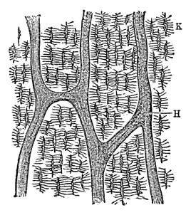
Scientists have discovered a system of small vessels that cross every part of a bone's cortical layers to supply most of the bone's blood—over 80% of the arterial supply and 59% of venous drainage. The proposed name is trans-cortical vessels (TCVs).
  • We’ve discovered a new type of blood vessel in our bones (brief article summarizing the discovery) my-ap.us/2B6tHce
  • A network of trans-cortical capillaries as mainstay for blood circulation in long bones (full research article) my-ap.us/2B6ttSq

 bone vessels


3 | Big Year: Content Updates & Applications

6 minutes
The first of several segments in a "debriefing" of the first year of this podcast.
This year brought many content updates and brief teaching snippets, from using green pens for grading to the role of platelets in immunity. Too many to mention, but Kevin gives his best shot at listing some of those that seem to be most memorable.

 Animated BP Categories


4 | Big Year: Evolution of This Podcast

9.5 minutes
What are show notes and how do they differ from the episode page? How best to listen and share? What's the episode list (theAPprofessor.org/podlist)? Preview episodes! And a word about our sponsors:
HAPS logo
The Human Anatomy & Physiology Society (HAPS) is a sponsor of this podcast. Did you know there's a one-day regional HAPS conference in March? Check it out. You can help appreciate their support by clicking the link below and checking out the many resources and benefits found there.

American Association of Anatomists
The searchable transcript for this episode, as well as the captioned audiogram of this episode, are sponsored by The American Association of Anatomists (AAA) at anatomy.org


5 | Big Year: The Big Ideas of the Past Year

 11 minutes
Each full episode usually has a "featured topic." This segment reviews some of the major topics and themes that emerged during the first year of the podcast.  From caring-empathy-compassion to integrity to storytelling, and from testing-as-teaching to elephant skin to learning science, a lot was covered! And what about those guest interviews--the Learning Scientists, media expert Barbara Waxer, anatomy educator Aaron Fried, and artist-professor Paul Krieger? And let's not forget those "lucky numbers"!

 one year

5 | Big Year: Wrapping Up

2 minutes
What do you want me to cover? I have some ideas, of course. But I want to hear  your ideas, too!
1-833-LION-DEN
1-833-546-6336
podcast@theAPprofessor.org
@theAPprofessor (Facebook, Twitter, Instagram, Pinterest, and beyond)

If the hyperlinks here are not active, go to TAPPradio.org to find the episode page.

Amazon referrals help defray podcasting expenses.
Transcript and captions for this episode are supported by the
American Association of Anatomists.anatomy.org


The Human Anatomy & Physiology Societyalso provides support for this podcast. theAPprofessor.org/haps

(Clicking on sponsor links 
helps let them know you appreciatetheir support of this podcast!)

Click here to listen to this episode—or access the detailed notes and transcript.

Wednesday, April 15, 2015

Mechanism of Cracking Knuckles


It never fails—somebody always asks about what's going on when knuckles crack when we get to the topic of articulations.

Recently, researchers used modern MRI techniques to demonstrate exactly what is happening—a process first described in the 1940s.  As joint surfaces separate, the changing tension in the synovial fluid causes a vapor pocket or "air bubble" to form in a process called cavitation.

The recent research proved that the popping sound typical of cracking a knuckle is produced as the vapor pocket forms. It also disproved the theory that the popping occurs when the bubble collapses.

What can we use from this in teaching undergraduate A&P?

  • You'll now always be ready for that inevitable "what causes knuckles to crack?" question when discussing joints in your A&P course.

    • Consider having a slide containing the media offered below to show students.

  • Discussing joint-cracking mechanisms engages students in a subject that may not immediately interest them.

  • Joint-cracking allows us to reinforce concepts of fluid dynamics in the context of synovial joints.

  • Consider stimulating some critical thinking by asking "what could cause a cracking sound in a cartilaginous or fibrous joint?"   (Hint: think "fracture" as cavitation would not occur where there are no fluids)
  • Do a demonstration by hitting a glass bottle full of water with a rubber mallet (see the embedded video below)
    • I've done this with an unopened glass bottle of ketchup by upturning it and hitting the base with my palm

Want to know more?

  • Real-Time Visualization of Joint Cavitation
    • GN Kawchuk, et al. PLoS|ONE. April 15, 2015 DOI: 10.1371/journal.pone.0119470
    • The original journal article. Includes downloadable images and PowerPoint slides, plus a link to a video showing the MRI results.
  • Researchers pull fingers to solve why knuckles crack | Finding bursts bubble of popular theory
    • Tina Hesman Saey Science News April 15, 2015
    • A plain-English summary of the new work on knuckle cracking with an embedded video (see below)
    • my-ap.us/1J4cGKP
Check out this video from the research article


Here's a video of the glass bottle demo of cavitation


Photo credit: Kawchuk et al.

Monday, August 4, 2014

Forensic Anthropologist Clyde Snow Passes


I just learned that the renowned forensic anthropologist Clyde Snow passed away this past May at the age of 86.

Famous for identifying the remains of Nazi war criminal Josef Mengele and victims of mass murderer John Wayne Gacy, Snow was one of a generation of pioneers who sparked the current popular interest in forensic sciences.

What can we use from this in teaching undergraduate A&P?  Some of stories of his achievements (see links below) can help spark interest in studying the human skeleton and other systems.  And some of his familiar quotes are worth using in the lab or classroom:

  • "There are 206 bones and 32 teeth in the human body, and each has a story to tell."
  • "Bones can be puzzles, but they never lie, and they don't smell bad."
  • "The ground is like a beautiful woman—if you treat her gently, she'll tell you all her secrets."
  • "If you can make people feel they're not going to get away with it—that's all we're asking." (regarding using forensic science to solve crimes)

Want to know more?

Clyde Snow, forensic anthropologist who helped ID victims of John Wayne Gacy, dies at 86
  • M. Weil Washington Post May 18, 2014
  • Article summarizing Snow's work, including several stories that could be used in an A&P class discussion.
  • my-ap.us/1qDQMVV

Science Detectives: The Masters Case
  • Discovery Channel January 22, 1997
  • Reenactment of forensic fieldwork in a killing that occurred decades before the victim's bones were discovered.
  • my-ap.us/U7DKoO

Image credit: Toony

Wednesday, November 6, 2013

New knee ligament confirmed

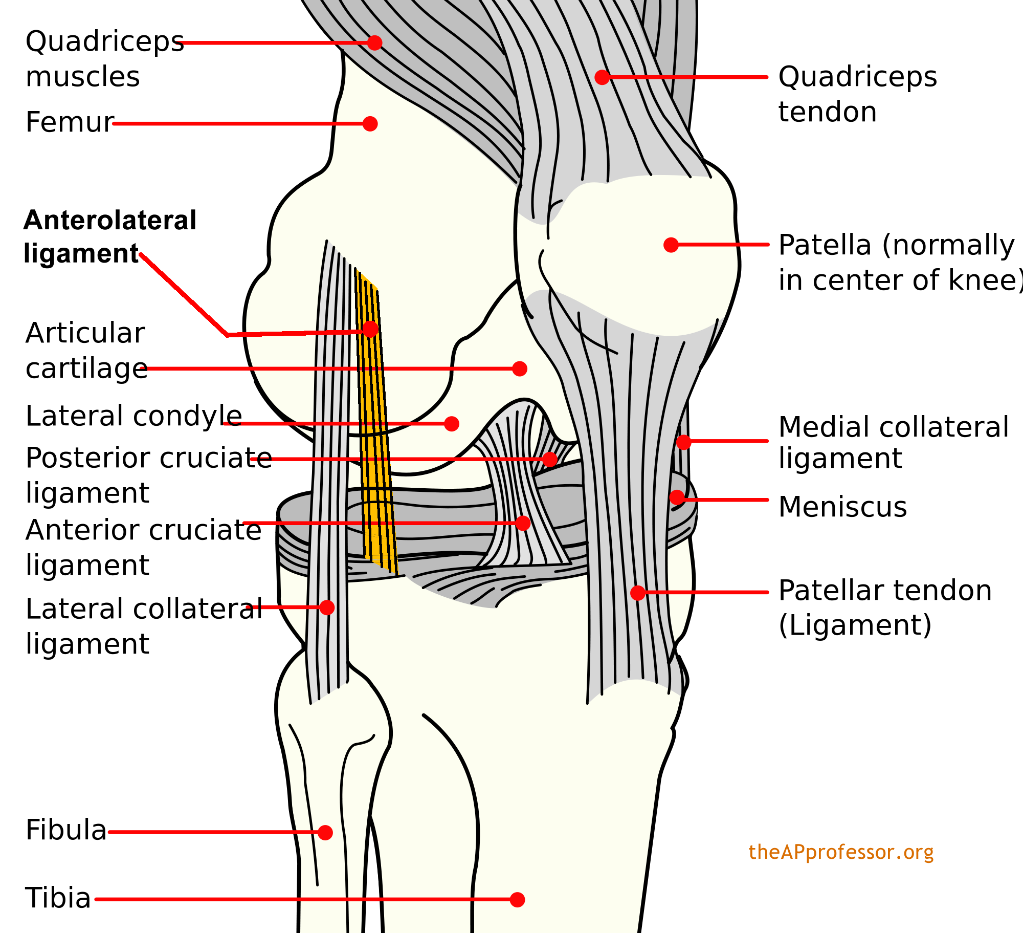
It's been there all along.  In most of us, at least.  Back in 1879, French surgeon Paul Ferdinand Segond first described it as it related to a particular type of avulsion fracture of the knee—the Segond fracture. But it was never really confirmed as separate from the joint capsule and named as a normal ligament of the human knee.  Until now.

Dubbed the anterolateral ligament (ALL), it originates at the prominence of the lateral femoral epicondyle (just anterior to the lateral collateral ligament) and running obliquely to the anterolateral part of the tibia (attached at the lateral meniscus).

Segond
So maybe we should pencil the ALL into our anatomic atlases, eh?  And wait for some research to confirm its biomechanical function—probably related to controlling internal rotation of the tibia.

This might also provide a good opportunity to talk about the dynamic nature of anatomical science—and the fact that human anatomy is not "finished."


Want to know more?

Anatomy of the anterolateral ligament of the knee
  • Steven Claes et al. Journal of Anatomy. Volume 223, Issue 4, pages 321–328, October 2013 (First published online: 1 AUG 2013) DOI: 10.1111/joa.12087
  • This the original journal article (free abstract).
  • my-ap.us/1aFUVV3

The Anterolateral Ligament of the Knee: Anatomy, Radiology, Biomechanics and Clinical Implications
  • Steven Claes, et al. American Academy of Orthopedic Surgeons (AAOS) Annual Meeting, SE73, 20 March 2013
  • This is an abstract (with image) of a preliminary presentation giving prior to journal publication.
  • my-ap.us/1b7bU0Q

Photo of the ALL
  • Steven Claes, et al. American Academy of Orthopedic Surgeons (AAOS) Annual Meeting, SE73, 20 March 2013
  • my-ap.us/1cF4sgC

Diagram of ALL


Monday, September 16, 2013

Teaching bones & bone features

I've found that one of the things that stand in the way of student success in learning the anatomy of the skeleton is that they don't know the underlying language.

If they already knew that a foramen is a hole and a condyle is a bump, then they could make short work of identifying and learning a long list of bones and bone features. And they'd have a better understanding of the concepts, plus an easier time remembering each structure for the long term.

We can't do anything about the fact that they just don't teach Latin as a required subject  in grade schools and high schools anymore.  But that doesn't mean there's nothing we can do to give them a head start on learning the skull!

For a long time, I've started out my students learning the general types of bone features before starting on the specific skeletal structures.  I find that they have a much easier time identifying structures because they have a better understanding of what the bone names mean.  They know they're looking for a hole when they get to the foramen ovale.  That oblong dark spot in the skull diagram or photo could be a bump, for all they know.

I use the list of general types of bone features found in my textbooks, lab manuals, and other publications (see list below).  I've also provided you with a downloadable version of this list you can link to in your course or distribute as a handout (see links below).

I've just uploaded two videos to my YouTube channel that help students get started this way.  You can link to the videos in an email, syllabus, or LMS; embed them in your course website or LMS; or embed them in a PowerPoint slide.  Or you could use the slides I used in the video to make your own video or live presentationperhaps even customize it for your course.  This links to all of these resources are below.

I'll be posting these videos on my study-tip blog for students, The A&P Student, soon.  If your students are not yet using this blog, you can distribute my FREE bookmarks to get them started.  See below for details.


Want to know more?

Bone Names Have Meaning - Part 1 - Get Started (video)
  • by Kevin Patton, Kevin's YouTube Channel,  uploaded September 2013
  • Video briefly explains the importance of knowing the meaning of bone names and how to get started doing that.  Includes link to list of bone features.
  • Link URL: youtu.be/heBjZIZP328
  • Embed code is available once you link to the above URL and click "Share"
Bone Names Have Meaning - Part 2 - Types of Bone Markings (video)
  • by Kevin Patton, Kevin's YouTube Channel, uploaded September 2013
  • Brief video runs through each of the major bone feature types, showing one or more examples. Includes link to list of bone features.
  • Link URL: youtu.be/NRR-t93jyFw
  • Embed code is available once you link to the above URL and click "Share"
Bone-Names-1-2 (PowerPoint slides)
  • by Kevin Patton, Lion Den Slide Collection,  September 2013
  • Download these animated slides I used to make the videos and use them in your recorded or live presentation.  The slide notes include the narration, which you could customize (or ignore).  You can use some or all of the slides.
  • To get the download password, fill out the form at lionden.com/slides-form.htm
Bone Names (web page)
  • by Kevin Patton, Lion Den Study Tips & Tools
  • Web page for students briefly outlining how to get started learning bone structures. The videos above are embedded in the page, so you could link to this page in your course instead of linking to the individual videos.  Links to related, downloadable lists. Part of the Field Guide to the Body.
  • lionden.com/tips-lab-anatomy-bone-names.htm
List of Bone Markings
Lion Den Slide Collection
  • by Kevin Patton, Lion Den Slide Collection
  • Download all kinds of  animated slides you can use in your own course to supplement your own slides and those provided with your textbook or school. Most slides are animated.
  • To get access to the whole collection, fill out the form at lionden.com/slides-form.htm
Free Bookmarks
  • Get packs of 50 bookmarks to distribute to students.  The bookmarks tell students how to access The A&P Student blog, which has a collection of study tips for A&P.
  • theapprofessor.org/free-bookmarks.html

Sunday, September 6, 2009

Inflammation images


Some interesting images of the inflammation seen in early stages of rheumatoid arthritis (RA) were released recently.

These images use a special type of fluorescent dye that is activated by the infrared radiation released at the site of inflammation. The intent of the project is to develop a way to detect such inflammation at an early stage of RA, when there's still time to help avert some of the damage produced by this condition.

However, the images can be useful in your A&P course when discussing the process of inflammation. The images are a dramatic example that contrasts the heat present in a normal hand versus the heat produced by hands that are inflamed.

To access the images go to:
Rheumatism Video Discloses Center Of Inflammation At An Early Stage.
Physikalisch-Technische Bundesanstalt (PTB) (2009, August 29).
ScienceDaily. Retrieved August 30, 2009
[Press release with associated images. Be sure to click the images for the larger view.]

Wednesday, August 5, 2009

FREE herniated disc video

There's a new FREE video available from one of my favorite medical/anatomical animation producers Nucleus Medical Art.

Even if you don't care to discuss the herniation aspect, it's a nice animation showing the location and basic structure of an intervertebral disc.

Check out the other videos from Nucleus, many of which you might be able to use in your A&P course.


[If you don't see the video viewer in your newsletter or feed version of this article, please go to The A&P Professor blog site to view it. Want to learn how to embed YouTube videos in your blog, website, or Powerpoint? Check it out at The A&P Professor website.]