Showing posts with label images. Show all posts

Friday, January 5, 2018

Platelets vs. Bacteria

Platelets as potent scavengers of bacteria? Really?

Something like 750 billion tiny cell fragments called platelets circulate in the human blood stream. When an injury to a blood vessel occurs, they stick to the exposed collagen in groups—forming platelet plug. And trigger additional reactions that eventually result in a blood clot.

But did you know that they have other helpful jobs, too? Like rounding up bacteria and feeding them up to immune cells, which devour them to make us safe.

This innate immune function of platelets has recently been outlined by researchers, as the information below summarizes.

Read through the quick points below to get an overview of some immune functions of platelets. Then read the full articles if you want to know more about these discoveries—including some great diagrams, micrographs, and videos.

[A short item on this topic also appears in today's edition of my daily Nuzzel newsletter of curated headlines for A&P professors.]

Quick points about platelets as bacterial scavengers


  • At sites of vessel injury/inflammation, platelets that contact intact collagen stick together—but platelets that do not contact collagen are motile.

  • Motile platelets change shape from a "fried egg" to a polarized "half moon" to better navigate the shearing forces of blood flow.

    • They can even navigate "upstream" against the flow of blood.

  • Platelets can use mechanical force to pull particles—including bacteria—from surrounding substrates.

  • Platelets collect and bundle bacteria, which facilitates neutrophil activation and subsequent phagocytosis. 

Migration pattern of motile platelet (left). Platelets collecting bacteria into bundles.

What can we use from this in teaching undergraduate A&P?


  • Yeah, okay we don't have time to go into all the ins and outs of platelets in a typical A&P course, but we can mention that platelets are now known to have immune functions.

    • Consider circling back to this mention later, when (if) you cover innate immune mechanisms a bit later in the course.

  • Consider calling attention to the sensory functions needed for platelets to analyze their microenvironment within the bloodstream.

  • Consider pointing out the specialized structure and function of the platelet's plasma membrane.

    • Integrins (integral membrane proteins) have a role in detecting particles for adhesion, binding to them, and sorting them.

    • Invaginations of the plasma membrane facilitate bundling of bacteria.

  • The shape changes needed for migration and handling of bacteria require actin-myosin reactions to power them. As in muscle fibers, these contractions are triggered by influx of extracellular calcium. In case you want to circle back to that.

  • Perhaps we should make a stronger point in reminding students that although they are "cell fragments" without a nucleus, they're more than just bags of hemostatic chemicals.

  • All these opportunities to "circle back" to previously studied concepts helps students make connections in their developing conceptual framework. And help them form a better understanding of the "big picture."

Want to know more?


Platelets, On Your Marks, Get Set, Migrate!

  • Bambach S, Lämmermann T. Cell. 2017 vol: 171 (6) pp: 1256-1258
  • Introduction to the Gaertner, et. al., paper below—giving background and overview to enhance understanding of the new discoveries. Great diagram, too! Click "Supplemental information" in the article to access video clips. 
  • my-ap.us/2EbU6Eu


Migrating Platelets Are Mechano-scavengers that Collect and Bundle Bacteria.

  • Gaertner F et. al. Cell. 2017 vol: 171 (6) pp: 1368-1382.e23
  • Journal article describing the scavenger role of platelets. Includes a few very nice, simple diagrams—and some cool micrographs and data graphs. These can also be downloaded as PowerPoint slides. Click "Supplemental information" in the article to access video clips. 
  • my-ap.us/2F5iM2U


Platelets Mediate Host Defense against Staphylococcus aureus through Direct Bactericidal Activity and by Enhancing Macrophage Activities.

  • Ali R et. al. Journal of immunology. 2017 vol: 198 (1) pp: 344-351
  • Journal article that supports the concept that platelets can kill MRSA bacteria and enhance their phagocytosis by macrophages.
  • my-ap.us/2EaO5be


[NOTE: If you can't access the full text of any resource, ask your school's reference librarian for help. If they can't provide direct access, they'll probably know how to get a copy of the resource for you. Quickly.]


Sketch: パタゴニア
Photos: LMU

Wednesday, October 4, 2017

Biomolecule Imaging Pioneers Share Nobel Prize

Today, the Royal Swedish Academy of Sciences has decided to award the Nobel Prize in Chemistry 2017 to Jacques Dubochet (University of Lausanne, Switzerland) and Joachim Frank (Columbia University, New York, USA), and Richard Henderson (MRC Laboratory of Molecular Biology, Cambridge, UK). The award is given "for developing cryo-electron microscopy for the high-resolution structure determination of biomolecules in solution"

Cool microscope technology revolutionises biochemistry

We may soon have detailed images of life’s complex machineries in atomic resolution. The Nobel Prize in Chemistry 2017 is awarded to Jacques Dubochet, Joachim Frank and Richard Henderson for the development of cryo-electron microscopy, which both simplifies and improves the imaging of biomolecules. This method has moved biochemistry into a new era.

A picture is a key to understanding. Scientific breakthroughs often build upon the successful visualization of objects invisible to the human eye. However, biochemical maps have long been filled with blank spaces because the available technology has had difficulty generating images of much of life’s molecular machinery. Cryo-electron microscopy changes all of this. Researchers can now freeze biomolecules mid-movement and visualize processes they have never previously seen, which is decisive for both the basic understanding of life’s chemistry and for the development of pharmaceuticals.

Electron microscopes were long believed to only be suitable for imaging dead matter, because the powerful electron beam destroys biological material. But in 1990, Richard Henderson succeeded in using an electron microscope to generate a three-dimensional image of a protein at atomic resolution. This breakthrough proved the technology’s potential.

Joachim Frank made the technology generally applicable. Between 1975 and 1986 he developed an image processing method in which the electron microscope’s fuzzy two-dimensional images are analysed and merged to reveal a sharp three-dimensional structure.

Jacques Dubochet added water to electron microscopy. Liquid water evaporates in the electron microscope’s vacuum, which makes the biomolecules collapse. In the early 1980s, Dubochet succeeded in vitrifying water – he cooled water so rapidly that it solidified in its liquid form around a biological sample, allowing the biomolecules to retain their natural shape even in a vacuum.

Following these discoveries, the electron microscope’s every nut and bolt have been optimised. The desired atomic resolution was reached in 2013, and researchers can now routinely produce three-dimensional structures of biomolecules. In the past few years, scientific literature has been filled with images of everything from proteins that cause antibiotic resistance, to the surface of the Zika virus. Biochemistry is now facing an explosive development and is all set for an exciting future.

About the Nobel Laureates

Jacques Dubochet, born 1942 in Aigle, Switzerland. Ph.D. 1973, University of Geneva and University of Basel, Switzerland. Honorary Professor of Biophysics, University of Lausanne, Switzerland.
www.unil.ch/dee/en/home/menuinst/people/honorary-professors/prof-jacques-dubochet.html

Joachim Frank, born 1940 in Siegen, Germany. Ph.D. 1970, Technical University of Munich, Germany. Professor of Biochemistry and Molecular Biophysics and of Biological Sciences, Columbia University, New York, USA.
http://franklab.cpmc.columbia.edu/franklab/

Richard Henderson, born 1945 in Edinburgh, Scotland. Ph.D. 1969, Cambridge University, UK. Programme Leader, MRC Laboratory of Molecular Biology, Cambridge, UK.
www2.mrc-lmb.cam.ac.uk/groups/rh15/


What can we use from this in teaching undergraduate A&P?


  • If you talk about imaging molecules in your course, this could be a way to garner student interest—considering that this is a current and ongoing effort in science. I always have a brief "shape is important in biological chemistry and here's what we can see with current tools" because they're going to see all those little odd-shaped rutabaga blobs in illustrations in their textbooks.

  • If you bring up microscopy in your course, perhaps describing the types of microscopy, adding a bit of info on this could help show students that microscopy is still evolving—in exciting ways.

  • Consider using the annual Nobel Prize announcements as a springboard to discuss the process of scientific discovery. 

  • Consider mentioning the other major awards for scientific achievement and discuss what the judges seem to value most about scientific discoveries. The Nobel Prize is the one everyone has heard of, so it's a great place to start.

  • Use the Nobel Prizes (and other awards) over time as a way to keep students aware of the history of, and progress, of human biology. One could also address the global diversity of laureates.  Or the lack of other kinds of diversity among laureates.

Want to know more?

Popular Information 


Scientific Background


Images

Image - 3D structures (pdf 1.4 MB)


Image - Blobology (pdf 8.5 MB)


Image - Dubochet's preparation method (948 kB)


Image - Frank's image analysis (pdf 1 MB)


Cool Animations (literally)

Structure and gating of the nuclear pore complex

Ion gating in the sarcoplasmic reticulum membrane

Antibody structure

Native LDL particles
  • Kumar V, Butcher S, Öörni K, Engelhardt P, Heikkonen J, Kaski K, Ala-Korpela M, Kovanen P
  • my-ap.us/2hO4Qms

Changes in the water and ion contents of organelles during apoptosis
  • Nolin F, Michel J, Wortham L, Tchelidze P, Banchet V, Lalun N, Terryn C, Ploton D
  • my-ap.us/2hMTYW4
Adapted from press release at nobelprize.org
Click each image for its source/attribution


Wednesday, February 17, 2016

What About That Negative Blood?

Every once in a while, I get an A&P student who expresses the concept of a negative Rh blood type as "having negative blood"—along with the connotation that having this blood type has a negative health impact.

We do not ordinarily think about red blood cell types such as A, B, AB, O, Rh+/-, or others, as being "bad for you" or even "good for you" healthwise. We most often think of them simply as different "flavors" of RBCs present in the human population.

Oh yeah, there are specific situations in which have a particular blood type can have significant health consequences. If you need an organ or tissue transplant—especially a blood donation—having the same RBC type as the available donor supply is "good for you." The lack thereof, then, is "bad for you" to at least some degree. Just like being tall can be bad for you when going through a low doorway.

Likewise, we all know there are health risks associated with a Rh- mother carrying an Rh+ fetus—especially the situation is not identified or if precautions are not taken.  But it's not like the Rh- type itself has a direct health impact on the person with that type.

However, such a view may be a bit more complex than it first seems.  Research continues to confirm that having a particular RBC type may affect your risk for certain health conditions.

For example, a little over a year ago, research published in the journal Neurology found that adults with type AB blood were at an increased risk of cognitive impairment compared to type O.  Of course, much more work needs to be done to establish a potential mechanism for this phenomenon. But it does give some evidence that the idea of certain blood types having health consequences may be true.

Other studies have suggested these links:

  • Type O may be linked to depression, anxiety, low (female) fertility

  • Type O and/or A may be linked to attention-deficit disorder (ADD) in children

  • Type B may be linked to a lower risk of ADD in children

  • Type A may be linked to obsessive-compulsive disorder and stomach cancer

  • Type A, B, and AB may be linked to heart disease and abnormal blood clotting


What can we use from this in teaching undergraduate A&P?


  • Another interesting aside to throw into an exploration of blood types to "liven up" the conversation a bit to motivate students.

  • Consider using a diagram of the actual ABO markers to show what's involved at the cellular level—and their similarity to each other.

  • One may want to mention that blood types may become a factor health professionals may look at when assessing health risks in patients

  • A classroom discussion on possible mechanisms of a blood-type—health risk could be interesting and useful. This could lead to some great insights about methods of scientific discovery. For example, what's the difference between correlation and cause? How confident should we be in one study?

    • Consider leading the discussion toward exactly what you want your students to know about blood types and health (e.g., blood donors and recipients, erythroblastosis fetalis, etc.)

Want to know more?


Blood Type Matters for Brain Health

  • A. Anderson and V. Stern. Scientific American MIND January 1, 2015
  • Brief article explains discovery that people with AB blood type are at higher risk for age-related cognitive decline. Also lists some of the other blood-type links I mentioned above.
  • my-ap.us/1KohroF


ABO blood type, factor VIII, and incident cognitive impairment in the REGARDS cohort

  • K. S. Alexander, et al. Neurology September 30, 2014 vol. 83 no. 14 1271-1276 
  • doi:http://dx.doi.org/ 10.1212/WNL.0000000000000844
  • Original research article about the discovery about AB blood type and brain health.
  • my-ap.us/1Koiq8j


Type O blood may be a fertility barrier

  • New Scientist. 10:15 26 October 2010
  • Brief article in plain English.
  • my-ap.us/1KolWzn


Yes, your blood group DOES affect your health

  • J. Naish Daily Mail 22 February 2011
  • Brief article includes some interesting historical facts.
  • my-ap.us/1Kompl7


Your Blood Type May Boost Your Heart Risk, Study Finds

  • S. Reinberg HealthDay Aug. 14, 2012
  • Brief article on link between blood type and heart disease
  • my-ap.us/1KomIfW

Blood photo: M. Osuchowicz
Diagram: InvictaHOG

Tuesday, July 14, 2015

Dissolving Microneedle Vaccinations

Researchers recently demonstrated that a flu vaccine delivered using microneedles that dissolve in the skin can protect people against infection even better than the standard needle-delivered vaccine.


The new microneedle patch is made of dissolvable material, eliminating needle-related risks. Not to mention the sea change it may mean for patients with severe needle anxiety!  I suspect this approach may also be more tolerable for many patients than oral and nasal vaccination methods. It is also easy to use without the need for trained medical personnel—making it ideal for use where healthcare resources are limited.

“Our novel transcutaneous vaccination using a dissolving microneedle patch is the only application vaccination system that is readily adaptable for widespread practical use,” said Professor Shinsaku Nakagawa, one of the authors of the study from Osaka University. “Because the new patch is so easy to use, we believe it will be particularly effective in supporting vaccination in developing countries.”

The new microneedle patch – MicroHyala – is dissolvable in water. The tiny needles are made of hyaluronic acid, a naturally occurring substance in tissue matrix and the synovial fluid that cushions the joints. When the patch is applied sort of like a Band-Aid, the needles pierce the epidermis of skin and dissolve into the body, taking the vaccine with them.

The researchers compared the new system to traditional needle delivery by vaccinating two groups of people against three strains of influenza: A/H1N1, A/H3N2 and B. None of the subjects had a bad reaction to the vaccine, showing that it is safe to use in humans. The patch was also effective: people given the vaccine using the microneedles had an immune reaction that was equal to or stronger than those given the vaccine by injection.

“We were excited to see that our new microneedle patch is just as effective as the needle-delivered flu vaccines, and in some cases even more effective,” said Professor Nakagawa.

Previous research has evaluated the use of microneedles made of silicon or metal, but they were not shown to be safe. Microneedles made from these materials also run the risk of breaking off in the skin, leaving tiny fragments behind. The new dissolvable patch eliminates this risk because the microneedles are designed to dissolve in the skin.

What can we use from this in teaching undergraduate A&P?


  • Consider mentioning this advance when discussing the layers of the skin, this giving a clinical application to pique student interest.

  • When discussing immunity and vaccination, consider mentioning this discovery.

  • If you discuss hyaluronic acid when covering histology, this information may help students realize the importance of knowing such details because of clinical applications of materials science.

Want to know more?


  • Clinical study and stability assessment of a novel transcutaneous influenza vaccination using a dissolving microneedle patch.
    • Sachiko Hirobe, et al. Biomaterials. Vol 57 (July 2015), Elsevier. doi: 10.1016/j.biomaterials.2015.04.007
    • The original research article.
    • my-ap.us/1eXzAud


Microneedle image courtesy of S. Nakagawa
Some content adapted from an Elsevier newsroom release

Wednesday, April 15, 2015

Mechanism of Cracking Knuckles


It never fails—somebody always asks about what's going on when knuckles crack when we get to the topic of articulations.

Recently, researchers used modern MRI techniques to demonstrate exactly what is happening—a process first described in the 1940s.  As joint surfaces separate, the changing tension in the synovial fluid causes a vapor pocket or "air bubble" to form in a process called cavitation.

The recent research proved that the popping sound typical of cracking a knuckle is produced as the vapor pocket forms. It also disproved the theory that the popping occurs when the bubble collapses.

What can we use from this in teaching undergraduate A&P?

  • You'll now always be ready for that inevitable "what causes knuckles to crack?" question when discussing joints in your A&P course.

    • Consider having a slide containing the media offered below to show students.

  • Discussing joint-cracking mechanisms engages students in a subject that may not immediately interest them.

  • Joint-cracking allows us to reinforce concepts of fluid dynamics in the context of synovial joints.

  • Consider stimulating some critical thinking by asking "what could cause a cracking sound in a cartilaginous or fibrous joint?"   (Hint: think "fracture" as cavitation would not occur where there are no fluids)
  • Do a demonstration by hitting a glass bottle full of water with a rubber mallet (see the embedded video below)
    • I've done this with an unopened glass bottle of ketchup by upturning it and hitting the base with my palm

Want to know more?

  • Real-Time Visualization of Joint Cavitation
    • GN Kawchuk, et al. PLoS|ONE. April 15, 2015 DOI: 10.1371/journal.pone.0119470
    • The original journal article. Includes downloadable images and PowerPoint slides, plus a link to a video showing the MRI results.
  • Researchers pull fingers to solve why knuckles crack | Finding bursts bubble of popular theory
    • Tina Hesman Saey Science News April 15, 2015
    • A plain-English summary of the new work on knuckle cracking with an embedded video (see below)
    • my-ap.us/1J4cGKP
Check out this video from the research article


Here's a video of the glass bottle demo of cavitation


Photo credit: Kawchuk et al.

Friday, January 2, 2015

Fat Cells in Skin Kill Bacteria


Scientists reported today that adipocytes in mouse and human skin produce an antimicrobial peptide (AMP) called cathelicidin is response to Staph aureus infections, including MRSA. Experimental animals that were deficient in the AMP were more susceptible to skin infections.

Adipocytes may recognize S. aureus by detecting bacterial peptides with toll-like receptors (TLRs), but more work is needed to fully understand the mechanisms.

This finding adds more to our understanding of human skin as a vital part of our body's defenses against infection. It also opens the door to understanding how diabetes, metabolic syndrome, and other conditions can reduce resistance to skin infections by altering the availability of AMPs in the fat associated with skin.

All of this may eventually lead to additional—perhaps more effective—strategies in preventing or curing serious skin infections such as MRSA.

I realize that we generally think of fat cells as belonging to the hypodermis, not the dermis, as described in the research. However, recent evidence shows the presence of adipocytes in the dermis that are distinct from those in the hypodermis. These adipocytes derive from a common precursor cell that produces both dermal fibroblasts and intradermal adipocytes. These dermal adipocytes have been shown to have a role in wound healing and the regeneration of hair follicles. And the research summarized here suggests that they also have a role in immunity.


What can we use from this in teaching undergraduate A&P?

  • Mention this discovery when discussing the roles of adipose tissue and adipocytes in your coverage of tissues of the body.

    • Consider clarifying that dermal adipocytes are distinct from fat cells in the hypdermis. And perhaps mention that it's a detail often left out of introductory discussions of skin.

  • This is a good point to mention when discussing the protective functions of the skin when covering the integumentary system.

  • When discussing the immune system, this concept helps illustrate several important principles:

    • The role of the skin as the first line of defense against infection

      • The variety of mechanisms available in the skin to act defensively

    • The role of TLRs and pattern recognition in immunity

    • The fact that immunity is a role for many tissues—not just lymphocytes and other WBCs

  • Take a moment NOW to add this to your course notes!

Want to know more?


Killer Fat
  • J. Alcorn and J. Kolls. Science 2 January 2015: Science Vol. 347 no. 6217 pp. 26-27 DOI: 10.1126/science.aaa4567
  • Editorial summary of the research in plain English. Includes a really nice, simple illustration of the concept (includes FREE teaching slide)
  • my-ap.us/1vBWbNP

Dermal adipocytes protect against invasive Staphylococcus aureus skin infection
  • L. Zhang1, et al. Science 2 January 2015: Vol. 347 no. 6217 pp. 67-71 DOI: 10.1126/science.126097
  • Original research article. Additional images available here, including some nice micrographs showing increase in adipocytes in response to S. aureus infection
  • my-ap.us/1xePNS4

Defining dermal adipose tissue.
  • Driskell RR, et al. Exp Dermatol. 2014. Exp Dermatol. 2014 Sep;23(9):629-31. doi: 10.1111/exd.12450.
  • Review article describing dermal adipocytes.
  • my-ap.us/1GuH3bL

FREE teaching slide
Click the image
to download a


Adipose image credit: my-ap.us/13MYGWO
This post was updated 6 OCT 2015

Monday, December 8, 2014

5 Useful Features of The A&P Professor Blog That You May Have Missed


Okay, the title is a bit misleading because a some of the five features you may have missed are new, so it may just be too early to have noticed them yet.  But there are some that have been around a while and you may not have given them much thought until now.

  1. Top menu bar.  

    I recently added a menu bar to the top of the blog page, just under the blog description.   You can link to the Home Page, Site Map, Subscription and various off-blog resources.  Explore each one to see if it's useful to you.

  2. Site Map.

    This new feature is a table of contents that lists blog posts in alphabetical lists by topic.  It's pretty big because I've been doing this blog for many years, and some posts are listed under more than one topic.  But if you are "into" the teaching of A&P, this might be a fun treasure hunt to find things you may have missed—or have simply forgotten about.

  3. Email Newsletter.

    The email newsletter for this blog, powered by Feedblitz, is a way to subscribe to this blog and be notified by email each time a new article is posted in blog.  It's free and you can safely unsubscribe at any time.  Then re-subscribe later when you find that you missed getting updates!  You may not realize that by subscribing, you also get additional content.  For example, once a month, I send out a Throwback Thursday newsletter reprising a popular article from the past that remain relevant.  I occasionally send out other "extra issues" that do not appear in the blog itself.

  4. Social Media.

    As more and more of you join the rest of the world on Facebook, Twitter, and other social media networks, you may want to follow The A&P Professor there, too.  As with the email newsletters, my Twitter and Facebook followers get additional content not found on the blog or other channels.  You can even follow my YouTube channel, where I post videos with study tips for A&P that you can share with your students.

  5. Family of Blogs and Websites.

    I have other blogs that are focused on particular topics that overlap your interest in teaching A&P.  For example, The A&P Student blog is directed at, er, A&P students.  Teachers who follow that blog pick up ideas for sharing with students who are having difficulty in your class, or link to blog posts in their course pages or syllabus.  My o-log-y blog relates specifically to the terminology of human science and medicine.  There, you may find tips for helping your students learn the language of A&P or you may find information about some of the tricky aspects of terminology that you may not already know. Check out each one to see if any of them interest you.

    Oh, and don't forget the separate website theAPprofessor.org, where you can find my free image library, sign up to get free bookmarks for your students, participate in teaching seminars, and gain access to my free library of teaching slides.

Play around the edges of this blog and see what other helpful little tools you may have missed.

Monday, October 13, 2014

RNA Interference. Again.


Five years ago, I extolled the virtues of teaching a little bit about RNA interference (RNAi) in undergraduate A&P courses.  But for a while it looked like the promise of RNAi in basic and clinical research might be sputtering.  However, a recent article by Eric Bender called The Second Coming of RNAi shows that RNAi "the gene-silencing technique [now] begins to fulfill some of its promises."

I recommend reading the entire article at my-ap.us/1BbxvB9  Before you read it, allow me to reprise my reasons of five years ago supporting my proposal to include RNAi in your course.

What can we use from this in teaching undergraduate A&P?


  • RNAi plays a role in defending our cells against viruses by stopping viral genetic code from being translated in host cells

  • RNAi likely plays a role in regulating gene activity in a cell by preventing translation of the gene product(s)

  • RNAi is increasingly used as method for "knocking out" a particular gene's effects in research animals in order to study the gene's functions

  • RNAi is being used to treat genetic disease. . . an application that will likely expand greatly over the next few decades
I'll add two more items to my previous list:
  • RNA interference is a mechanism of human disease, as has been demonstrated in some cases of inherited progressive hearing loss (for example).

  • Learning about RNAi helps clarify a general understanding of the many roles played by RNA in our lives—some perhaps still undiscovered.

I'm not sure that it's useful to expect beginning undergraduate students to learn the nitty-gritty details of RNAi mechanisms.  But I do think it's valuable to be exposed to the general concept of RNA interference and gene silencing.  A&P students are going to run up against these eventually as they learn about and then administer RNAi-based therapies, after all.  And perhaps we should prepare them.

Want to know more?


The Second Coming of RNAi
  • Eric Bender. The Scientist. September 1, 2014
  • Article mentioned above. In plain English, it shows that clinical progress in RNAi therapy against liver diseases, the gene-silencing technique begins to fulfill some of its promises. Includes useful illustrations and links to other resources.
  • my-ap.us/1BbxvB9

Why do we need to know about RNA interference?
  • Kevin Patton. The A&P Professor. 14 April 2009
  • My first article promoting the idea of teaching RNAi in the A&P course.  It links to an expanded article with additional teaching resources.
  • my-ap.us/1xbq4v6

RNA interference revisited
  • Kevin Patton. The A&P Professor. 9 June 2009
  • Brief follow-up article that references the role of RNA interference as a mechanism of human disease.  Links to other resources.
  • my-ap.us/1oDflzw

RNA Interference Animation and Slideshow
  • Nature Reviews Genetics. Accessed 3 September 2014
  • FREE animation, slideshow, and poster on RNAi, as well as a link to more details.
  • my-ap.us/1roNGYm

RNA Interference BioInteractive
  • Howard Hughes Medical Institute. Accessed 3 September 2014
  • FREE slideshow with worksheet that students fill out as they view the slideshow.  Links to FREE DVD from HMMI called The Double Life of RNA.
  • my-ap.us/1pJ9r5V


Wednesday, October 8, 2014

Nobel Prize 2014: Super-resolved fluorescence microscopy


The Royal Swedish Academy of Sciences has decided to award the Nobel Prize in Chemistry for 2014 to

Eric Betzig
Janelia Farm Research Campus, Howard Hughes Medical Institute, Ashburn, VA, USA,

Stefan W. Hell
Max Planck Institute for Biophysical Chemistry, Göttingen, and German Cancer Research Center, Heidelberg, Germany

and

William E. Moerner
Stanford University, Stanford, CA, USA

“for the development of
super-resolved fluorescence microscopy”


Surpassing the limitations of the light microscope


For a long time optical microscopy was held back by a presumed limitation: that it would never obtain a better resolution than half the wavelength of light. Helped by fluorescent molecules the Nobel Laureates in Chemistry 2014 ingeniously circumvented this limitation. Their ground-breaking work has brought optical microscopy into the nanodimension.

In what has become known as nanoscopy, scientists visualize the pathways of individual molecules inside living cells. They can see how molecules create synapses between nerve cells in the brain; they can track proteins involved in Parkinson’s, Alzheimer’s and Huntington’s diseases as they aggregate; they follow individual proteins in fertilized eggs as these divide into embryos.

It was all but obvious that scientists should ever be able to study living cells in the tiniest molecular detail. In 1873, the microscopist Ernst Abbe stipulated a physical limit for the maximum resolution of traditional optical microscopy: it could never become better than 0.2 micrometres. Eric Betzig, Stefan W. Hell and William E. Moerner are awarded the Nobel Prize in Chemistry 2014 for having bypassed this limit. Due to their achievements the optical microscope can now peer into the nanoworld.

Two separate principles are rewarded. 


One enables the method stimulated emission depletion (STED) microscopy, developed by Stefan Hell in 2000. Two laser beams are utilized; one stimulates fluorescent molecules to glow, another cancels out all fluorescence except for that in a nanometre-sized volume. Scanning over the sample, nanometre for nanometre, yields an image with a resolution better than Abbe’s stipulated limit.

Eric Betzig and William Moerner, working separately, laid the foundation for the second method, single-molecule microscopy. The method relies upon the possibility to turn the fluorescence of individual molecules on and off. Scientists image the same area multiple times, letting just a few interspersed molecules glow each time. Superimposing these images yields a dense super-image resolved at the nanolevel. In 2006 Eric Betzig utilized this method for the first time.

Today, nanoscopy is used world-wide and new knowledge of greatest benefit to mankind is produced on a daily basis.


This video is a brief animation of how STED works and how it improves resolution of individual particles.


This video is a longer, more detailed presentation by one of the Nobel laureates (Hell).


What can we use from this in teaching undergraduate A&P?

  • Discuss how this technology has enabled us to better visualize the chemicals and structures within our cells, enabling scientists to better understand the structure and function of cell, organelles, microbiome constituents, and other structures of the human body.

  • If you do a brief run-through of the theory of microscopy—perhaps in your A&P lab—you can add a mention of this technology.  

  • Your textbook or other learning resource may already have an example of this type of microscopy.

  • A discussion of this  Nobel Prize could evolve into a meaningful example of how science works, including how incremental improvements in classical tools for observation expand the number of questions that can be answered.

  • Use the links below (and images above) to use for a handout and/or teaching slides.


Want to know more?


Resources from Nobelprize.org

  • Popular Information 
  • Scientific Background
    • Handout: More detailed information includes references to original research articles
    • my-ap.us/ZdLJ69
  • Advanced Information
  • Images
  • Biographies
    • Eric Betzig, 
      • U.S. citizen. Born 1960 in Ann Arbor, MI, USA. Ph.D. 1988 from Cornell University, Ithaca, NY, USA. Group Leader at Janelia Farm Research Campus, Howard Hughes Medical Institute, Ashburn, VA, USA.
      • http://janelia.org/lab/betzig-lab
    • Stefan W. Hell, German citizen. 
      • Born 1962 in Arad, Romania. Ph.D. 1990 from the University of Heidelberg, Germany. Director at the Max Planck Institute for Biophysical Chemistry, Göttingen, and Division head at the German Cancer Research Center, Heidelberg, Germany.
      • http://www3.mpibpc.mpg.de/groups/hell
    • William E. Moerner, U.S. citizen. 
      • Born 1953 in Pleasanton, CA, USA. Ph.D. 1982 from Cornell University, Ithaca, NY, USA. Harry S. Mosher Professor in Chemistry and Professor, by courtesy, of Applied Physics at Stanford University, Stanford, CA, USA.
      • http://web.stanford.edu/group/moerner


Diagrram cretit: Ganbaatar
Micrograph credit: Tesselkaffee
Text adapted from press release from Nobel Media

Monday, September 15, 2014

Updated Cell Transport Slides


Many longtime readers of this blog know that I have a set of animated PowerPoint-compatible slides available for you to use FREE in your A&P classes.  These slides—the Lion Den Slide Collection— supplement the publisher-supplied slides or homegrown slides that you are already using.

I recently updated, improved, and expanded the set of slides that animate several key cell transport processes such as diffusion, osmosis, endocytosis, etc.

If you've accessed the Lion Den Slide Collection in the last several months, you've already registered in the system and probably have already received an email notification of the update.

If you're new to the collection (or accessed it before June 2014) then you need to go to the Lion Den Slide Collection page and click on the link to the form.  It takes a few steps, but by filling out the brief forms you register for a service that will notify you (if you want it) of any updates or additions to the collection.

Here is a preview the newly updated Membrane Transport Animations
Click image for download

When you download the linked file, you'll be able to play it in PowerPoint or any other program that plays "show" or PPTSX files.  The slides can only be viewed, not edited or added to your own slide deck.  To do that, you must download the fully editable PPTX files from the Lion Den Slide Collection

Once you download any slide deck from the Lion Den Slide Collection, you can mix and match them to blend them with your existing presentations.  You can also alter the content or timings to suit your needs.  

I'm hoping you'll also get some ideas from them to create new additional slides!  If you modify, create, or already have any slides (for which you own the content) that you want to place in the collection for sharing, please contact me directly. 

Want to know more?


Presentation Zen
  • Kevin Patton The Electronic Professor 6 August 2014
  • The zen approach to presentations.  Includes a video and links to resources.
  • my-ap.us/1mZjY7e

Are your students dodging bullets?
  • Kevin Patton. The Electronic Professor. 28 July 2011
  • My blog article on improving slide presentations.  With illustrated examples.
  • my-ap.us/1ni8c72

Handling bullets safely
  • Kevin Patton. The Electronic Professor.  3 August 2012
  • Another of my blog articles on improving slide presentations. Includes my own video on how to trim down those wordy slides to something that actually works in a slide.
  • my-ap.us/1ooEced

Monday, August 18, 2014

Human Microbial System


A recent article in The Scientist once again reminds us of the ongoing explosion in the scientific understanding of the human microbial system.  In a few short years, this area of exploration has moved to the forefront of medical and basic science research in human biology.

I think it's becoming clear that the most useful way to think of human body function is to recognize that an "organism" is really a sort of "habitat."  And like any habitat, it functions best when all the inhabitants are within a limited range of balanced relationships.

Who are the inhabitants?  Besides our own cells?  Well, one could think of mitochondria and cilia and other organelles as symbiotic internal inhabitants of our cells.  They're not that literally, of course, but I think its a useful metaphor for understanding the human body.  Then there are the many microbes and animals that cover our internal and external surfaces, burrow into some of our pores and glands, and inhabit our body fluids.

I call the balanced functional relationship among the various microbomes of the body and our own tissues the human microbial system.  And I am certain that it won't be long before we will be discussing this system alongside the major organ systems of the body.  That is if we truly want to understand how the body really works.

The article in The Scientist I mention is a great summary of some of the major roles that the human microbial system plays in the human body—and a good survey of some of the areas of the body where the human-microbial functional relationships play out.  See the link to the article below.


What can we use from this in teaching undergraduate A&P?


  • Why not introduce the concept of the human microbial system at the beginning of our A&P course, when we set the stage by explain how scientists understand the body and its functions as an integrated system of different parts?
  • We can mention the different microbiomes of the body when we explore each organ system where they play an important role—which is pretty much all of them!
  • Consider discussing what happens to normal human function when microbiomes get out of balance.  For example, in the gut a microbial imbalance can lead to ulcers, diarrhea, and other dysfunctions.  On the skin a pathogenic microbe may become dominant and cause a rash.
  • Promote a discussion of what kinds of wellness strategies might be employed to prevent microbial imbalances.
  • Our students can leave our A&P course with an up-to-date understanding of human biology that will help them understand new clinical concepts and treatment strategies.

Want to Know More?

The Body’s Ecosystem

  • By The Scientist Staff.  The Scientist. August 1, 2014
  • Plain-English article (cited above) on how research on the human microbiome is booming, and scientists have moved from simply taking stock of gut flora to understanding the influence of microbes throughout the body.
  • my-ap.us/1vgOu5y

Articles from The A&P Professor


Moving pictures of the human microbiome

  • J Gregory Caporaso et al. Genome Biology 2011, 12:R50  doi:10.1186/gb-2011-12-5-r50
  • Open-access journal article that includes FREE videos that show how dynamic the human microbial system is
  • my-ap.us/V7St3Q

Human Microbiome Project


The Microbiome and Disease

  • List of diseases associated with microbiome imbalances from Genetic Science Learning Center
  • my-ap.us/1nDpxr1

Audio

  • Radio stories from National Public Radio on human microbiomes and their role in health and disease.  The growing number of these stories tells us something as A&P teachers: maybe we better be covering this!
  • my-ap.us/1uA7Qyg

Thursday, July 24, 2014

Number of Human Genes Revised Downward. Again.

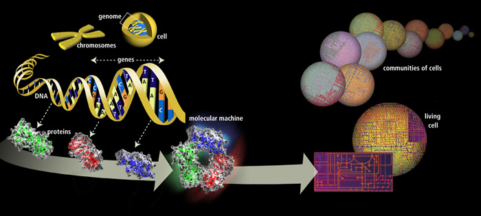
Genomic researchers in Spain have recently proposed a new, lower number of protein-coding genes in the human genome.  Previously, the number of coding genes was estimated by some at nearly 22,000 in the human genome.  The new estimate is approximately 19,000 protein-coding genes.

How can we use this new information in teaching undergraduate A&P?  Well, first we can update the numbers we use when discussing the role of genes in protein synthesis.  If it fits with our course objectives, we can use this as a way to transition to a discussion of coding vs. noncoding genes.

We also have an opportunity to discuss how science works—we are constantly checking our facts and revising our conclusions to improve the accuracy of our knowledge.  And that the story of genomics is far from complete.

I often tell students that I'm trying to tell them "the last, best story" of the human body's structure and function.  So if my story changes over time, that's a good thing!

FREE image you can use in your course


Want to know more?

Size of the human genome reduced to 19,000 genes

  • Science Daily. July 3, 2014
  • Press release in plain English based on information provided by researchers.
  • my-ap.us/1qrtHWf


Multiple evidence strands suggest that there may be as few as 19 000 human protein-coding genes. 

  • I. Ezkurdia, et al. Human Molecular Genetics, 2014; DOI: 10.1093/hmg/ddu309
  • Research article proposing the newly revised number.  Open access to full text of article.
  • my-ap.us/1oL7aTh

Want a FREE digital image of the nuclear genome that you can use in your presentation, handout, or other course material?  The image above is in the public domain and can be used in your course materials.


Monday, July 21, 2014

Biological Pacemaker Using Gene Therapy


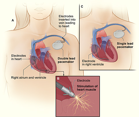
Researchers recently induced ordinary cardiac muscle fibers into becoming functioning pacemaker cells by injecting a therapeutic gene.

Working with pigs, a common model for human cardiovascular research, researchers first destroyed the natural pacemaker cells in each subject's heart and installed an electronic pacemaker. They then inserted a gene for transcription factor TBX18 into cardiac muscle tissue using an adenovirus.  Using adenovirous vectors for inserting genes is a common strategy in gene therapy.

Within a couple of days, ordinary myocardial fibers had developed the structure and function of pacemaker cells.  In about 5 days, the electronic pacemakers were no longer needed.

However, this biological pacemaking peaked at about 8 days, then eventually disappeared.  This may occur because the virus-infected cells are probably destroyed by the body's immune defenses.  So researchers are thinking that perhaps, at the very least, this could eventually lead to a temporary treatment for certain arrythmias in humans.


What can we use from this in teaching undergraduate A&P?  


  • This is an interesting bit of news that helps illustrate the frontiers of human biomedical sciences.
  • This story provides a good case to provoke a discussion of the nature of gene therapy.  
    • Why did the effect last only 8 or so day?  
    • What does this tell us about transcription factor TBX18?  
    • What benefit might this treatment have if developed for humans?
  • This may add interest to an discussion of the function of the electrical system of the heart in general, and artificial pacemakers in particular.
  • The case also provides a scenario in which the body attacks and destroys virus-infected cells.
FREE image you can use in your course

Want to know more?


Next Generation: Biological Pacemakers

  • R Williams, The Scientist (the-scientist.com) July 16, 2014
  • Plain-English article summarizing the discovery.  Includes quotes from the researchers.
  • my-ap.us/1ruocYu


Biological pacemaker created by minimally invasive somatic reprogramming in pigs with complete heart block
  • Y-F. Hu et al., Science Translational Medicine, 6:245ra94, 2014. DOI:10.1126/scitranslmed.3008681
  • Original research report.
  • my-ap.us/1nfErcA
FREE image you can use in your course

Photo credit: vfdbsn

Thursday, July 10, 2014

Mutations in Mitochondrial DNA

A new study suggests that DNA mutations in some of the mitochondria of healthy people may be a lot more common than scientists thought.

The term heteroplasmy describes a situation in which some mitochondria of a cell have mutant mitochondrial DNA (mtDNA) and other mitochondria have normal mtDNA.  Cell function can become disordered, perhaps producing disease, when the balance of mutant vs. normal mtDNA crosses a certain threshold.

The recent study shows that about 90% of healthy people studied in the 1000 Genomes Project had at least one heteroplasmy.  Some of these (about 20%) have been shown to correlate to disease.  That's a lot more than we were thinking prior to the study (25%-65% heteroplasmy rate).

We don't  know the significance of this finding yet, but it could influence how likely it is for mitochondrial diseases to develop over time—or to be inherited.  Could the mutant/normal mtDNA balance get skewed as oocytes form, thus giving different offspring different probabilities of inheriting mitochondrial disorders?  Or change the probabilities from one generation to the next?  Mitochondrial dysfunction is thought to be a mechanism of agingcould the rate of heteroplasmy be part of the aging mechanism?

What can we use from this in teaching undergraduate A&P?  The fact that we now know that mtDNA mutations are common in healthy people will be interesting and useful to students.


Want to know more?

Mutations Pervade Mitochondrial DNA

  • Jyoti Madhusoodanan. The Scientist (the-scientist.com) July 7, 2014
  • This is a plain-English article summarizing the new findings; includes quotes from the researchers.
  • my-ap.us/1oo18X8 


Extensive pathogenicity of mitochondrial heteroplasmy in healthy human individuals

  • K. Ye et al., Proceedings of the National Academy of Science (PNAS), doi:10.1073/pnas.1403521111, 2014. 
  • This is the original research report.
  • my-ap.us/1sDPV8S


Want a FREE labeled image of mtDNA that you can use in your presentation, handout, or other course material?
my-ap.us/1ncmAxl


Wednesday, November 6, 2013

New knee ligament confirmed

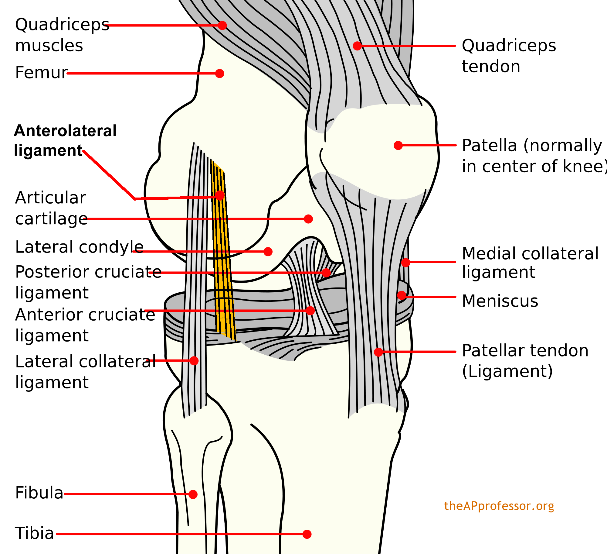
It's been there all along.  In most of us, at least.  Back in 1879, French surgeon Paul Ferdinand Segond first described it as it related to a particular type of avulsion fracture of the knee—the Segond fracture. But it was never really confirmed as separate from the joint capsule and named as a normal ligament of the human knee.  Until now.

Dubbed the anterolateral ligament (ALL), it originates at the prominence of the lateral femoral epicondyle (just anterior to the lateral collateral ligament) and running obliquely to the anterolateral part of the tibia (attached at the lateral meniscus).

Segond
So maybe we should pencil the ALL into our anatomic atlases, eh?  And wait for some research to confirm its biomechanical function—probably related to controlling internal rotation of the tibia.

This might also provide a good opportunity to talk about the dynamic nature of anatomical science—and the fact that human anatomy is not "finished."


Want to know more?

Anatomy of the anterolateral ligament of the knee
  • Steven Claes et al. Journal of Anatomy. Volume 223, Issue 4, pages 321–328, October 2013 (First published online: 1 AUG 2013) DOI: 10.1111/joa.12087
  • This the original journal article (free abstract).
  • my-ap.us/1aFUVV3

The Anterolateral Ligament of the Knee: Anatomy, Radiology, Biomechanics and Clinical Implications
  • Steven Claes, et al. American Academy of Orthopedic Surgeons (AAOS) Annual Meeting, SE73, 20 March 2013
  • This is an abstract (with image) of a preliminary presentation giving prior to journal publication.
  • my-ap.us/1b7bU0Q

Photo of the ALL
  • Steven Claes, et al. American Academy of Orthopedic Surgeons (AAOS) Annual Meeting, SE73, 20 March 2013
  • my-ap.us/1cF4sgC

Diagram of ALL


Monday, February 25, 2013

Blood viscosity

Blood viscosity is a concept that is important in understanding blood flow.  It is, after all, one of the factors that affects peripheral resistance to blood flow.

One major factor influencing blood viscosity is hematocrit.  You may be interested in using the analogy of ketchup outlined a few months ago in my article for students Blood viscosity and peripheral resistance at theAPstudent.org

Recently, researchers also looked at the viscosity of the blood plasma alone (without the formed elements).  They found that blood plasma has unique characteristics of flow found only in non-Newtonian fluids, becoming less viscous with increasing pressure.  Again, just like ketchup. Plasma, unlike plain water, exhibits both viscous and elastic behaviors.

Researchers found in recent experiments that this characteristic of plasma may promote swirling where blood vessels diameters change—both at the beginning and end of a narrowed segment.  Thus, this could have an effect on formation of clots at stenoses or where a stent has been placed.

So, as you may have suspected all along, blood is not only thicker than water—it's weirder than water.

Want to know more?

  • Blood viscosity and peripheral resistance
    • Kevin Patton
    • The A&P Student 12 September 2012
    • [Analogy of ketchup flow for students.  Includes video.]
    • my-ap.us/XuR596
  • Blood Is Thicker Than Water – And Blood Plasma Is, Too
    • Science Daily Feb. 18, 2013
    • [Brief, plain-language article outlining the recent research.]
    • my-ap.us/YpGhas
  • Rheology of human blood plasma: Viscoelastic versus Newtonian behavior.
    • M. Brust, et al.
    • Phys. Rev. Lett, 110, 078305 (2013) DOI: 10.1103/PhysRevLett.110.078305
    • [Original journal article.  See photos from the experiments below.]
    • my-ap.us/15HVkle


Recording from one of the "drop-experiments": If blood plasma is placed between two plates and then they pulled apart, high-speed cameras show in conjunction with high-resolution microscope objectives that strands and droplets form. This demonstrates that plasma is elastic and viscous and does not behave like water.
Photo: Christof Schaefer, Phys. Rev. Lett. 110, 2013, 078305th Copyright (2013) by the American Physical Society

Plasma turbulence affects the blood. In one experiment, the researchers had plasma flow through a microfluidic constriction as in vasoconstriction. They showed turbulence at the end of the contraction, but also - as seen here in the pictures - sticking to its beginning. This turbulence is caused by the viscoelastic properties of blood plasma.
Photo: Mathias chest, Phys. Rev. Lett. 110, 2013, 078305th Copyright (2013) by the American Physical Society


Monday, November 19, 2012

Brain injury in high-def with fiber tracking

U. PITTSBURGH (US) — New imaging technology will allow doctors to clearly see for the first time neural connections broken by traumatic brain injury.

Called High Definition Fiber Tracking [1], the technology shows injuries much like X-rays show a fractured bone, according to researchers from the University of Pittsburgh [2] in a report published online in the Journal of Neurosurgery [3].

In the report [4], the researchers describe the case of a 32-year-old man who wasn’t wearing a helmet when his all-terrain vehicle crashed. Initially, his CT scans showed bleeding and swelling on the right side of the brain, which controls left-sided body movement.


High definition fiber tracking reveals loss of fibers, or connections, on the injured right side (yellow) and the intact, undamaged left side (green). The patient was injured in an ATV accident and lost function in his left leg, arm, and hand. (Credit: Walt Schneider Laboratory)


High definition fiber-tracking map of a million brain fibers. (Credit: Walt Schneider Laboratory)
Straight from the Source


A week later, while the man was still in a coma, a conventional MRI scan showed brain bruising and swelling in the same area. When he awoke three weeks later, the man couldn’t move his left leg, arm and hand.

“There are about 1.7 million cases of TBI in the country each year, and all too often conventional scans show no injury or show improvement over time even though the patient continues to struggle,” says co-senior author and neurosurgeon David O. Okonkwo, associate professor in the neurological surgery department.

“Until now, we have had no objective way of identifying how the injury damaged the patient’s brain tissue, predicting how the patient would fare, or planning rehabilitation to maximize the recovery.”

HDFT might be able to provide those answers, says co-senior author Walter Schneider, professor of psychology, who led the team that developed the technology.

Data from sophisticated MRI scanners is processed through computer algorithms to reveal the wiring of the brain in vivid detail and to pinpoint breaks in the cables, called fiber tracts. Each tract contains millions of neuronal connections.

“In our experiments, HDFT has been able to identify disruptions in neural pathways with a clarity that no other method can see,” Schneider says. “With it, we can virtually dissect 40 major fiber tracts in the brain to find damaged areas and quantify the proportion of fibers lost relative to the uninjured side of the brain or to the brains of healthy individuals. Now, we can clearly see breaks and identify which parts of the brain have lost connections.”

HDFT scans of the study patient’s brain were performed four and 10 months after he was injured; he also had another scan performed with current state-of the-art diffusion tensor imaging (DTI), an imaging modality that collects data points from 51 directions, while HDFT is based on data from 257 directions. For the latter, the injury site was compared to the healthy side of his brain, as well as to HDFT brain scans from six healthy individuals.

Only the HDFT scan identified a lesion in a motor fiber pathway of the brain that correlated with the patient’s symptoms of left-sided weakness, including mostly intact fibers in the region controlling his left leg and extensive breaks in the region controlling his left hand. The patient eventually recovered movement in his left leg and arm by six months after the accident, but still could not use his wrist and fingers effectively 10 months later.

Memory loss, language problems, personality changes and other brain changes occur with TBI, which the researchers are exploring with HDFT in other research protocols.

University of Pittsburgh neurosurgeons also have used the technology to supplement conventional imaging, noted Robert Friedlander, professor and chair in the neurological surgery department, who was not involved with the study.

“I have used HDFT scans to map my approach to removing certain tumors and vascular abnormalities that lie in areas of the brain that cannot be reached without going through normal tissue,” he says.

“It shows me where significant functional pathways are relative to the lesion, so that I can make better decisions about which fiber tracts must be avoided and what might be an acceptable sacrifice to maintain the patient’s best quality of life after surgery.”

Okonkwo notes that the patient and his family were relieved to learn that there was evidence of brain damage to explain his ongoing difficulties. The team continues to evaluate and validate HDFT’s utility as a brain imaging tool, so it is not yet routinely available.

“We have been wowed by the detailed, meaningful images we can get with this technology,” Okonkwo says. “HDFT has the potential to be a game-changer in the way we handle TBI and other brain disorders.”

The study was funded by the Defense Advanced Research Projects Agency.

More news from the University of Pittsburgh: www.news.pitt.edu/ [5]

Article reprinted by CC license from Futurity.org: http://www.futurity.org
Posted By Anita Srikameswaran-Pittsburgh On March 2, 2012 @ 12:58 pm

URL to original article: http://www.futurity.org/top-stories/brain-injury-in-high-def-with-fiber-tracking/

URLs in this post:
[1] High Definition Fiber Tracking: http://schneiderlab.lrdc.pitt.edu/projects/tbi
[2] University of Pittsburgh: http://www.upmc.com/MediaRelations/NewsReleases/2012/Pages/High-Definition-Fiber-Tracking-Traumatic-Brain-Injury.aspx
[3] Journal of Neurosurgery: http://thejns.org/doi/abs/10.3171/2012.1.JNS111282?prevSearch=%255BFulltext%253A%2BOkonkwo%255D&searchHistoryKey=
[4] Read the original study: http://thejns.org/doi/abs/10.3171/2012.1.JNS111282?prevSearch=%255BFulltext%253A%2BOkonkwo%255D&searchHistoryKey=
[5] www.news.pitt.edu/: http://www.news.pitt.edu/

---------------------------------------------------------
Want to know more?


Related textbook content
  • The Human Body in Health and Disease 5th ed. p. 246-254 my-ap.us/fNN00N 


Tuesday, July 10, 2012

Stem cells in the ovary

Beginning about 8 years ago, scientists began providing evidence that apparently overturns the dogma that adult ovaries do not contain stem cells capable of producing oocytes.  Jonathon Tilly found such cells first in mice, then later in middle-aged women. Other labs have replicated such findings.

However, a study by members of Kui Liu's lab published today disputes that the stem cells actually produce oocytes.  Needless to say, this is stirring up quite a bit of controversy.  Tilly believes Liu was looking at oocytes, not stem cells.  Liu doesn't agree.

What does this mean for the undergraduate A&P class?  I discuss the recent discoveries of stem cells in adult ovaries in my course.  I use it as an opportunity to point out that there is still much to learn about how the human body works . . . that we are continually surprised by new research findings.  This is part of a year-long subtext of "how science works."  I tell my A&P students that I'm telling them "the last, best story" about the human body.  But that my story changes from year to year as scientists tease out more information . . . and thus revise "the story."

How does this fit in?  Well, isn't this how science is supposed to work?  Wouldn't it seem logical that it is in the best interest of everyone to have vigorous debate and extensive re-examination before we throw out the last, best story in favor of a new version?

The first article below briefly summarizes the issues involved in today's publication.


Want to know more?


Ovarian Stem Cell Debate
Ed Yong
The Scientist
(online) July 9, 2012

[Brief article discusses  that opinion is divided on a new paper showing that adult ovaries do not contain egg-making stem cells, contrary to two recent studies that appeared to overturn longstanding dogma.]

my-ap.us/Mf1Y5w
Experimental evidence showing that no mitotically active female germline progenitors exist in postnatal mouse ovaries
H. Zhang et al.
Proceedings of the National Academy of Sciences doi:10.1073/pnas.1206600109
[Recent paper disputing presence of stem cells in adult ovaries]
my-ap.us/RX4McG

Ovarian Stem Cells in Humans?
Sabrina Richards
The Scientist (online) February 27, 2012
[Brief summary of recent paper by Tilly's lab stating that adult human ovaries contain a population of stem cells capable of generating immature egg cells.]
my-ap.us/OWGY8G

Oocyte formation by mitotically active germ cells purified from ovaries of reproductive-age women
Y.A.R. White et al.
Nature Medicine 18:413–21, 2012.
[Original research paper by Tilly's lab showing presence of stem cells in human ovaries.]
my-ap.us/LFaYqX

The image above is copyright free and can be used in your course!