Kevin shares his experience of using cumulative testing to strengthen long-term learning.
Use video walk-throughs to help students navigate your digital course platforms.
Sometimes other people's genes influence an individual's biological traits.
(0:50) Things will get a lot more interesting if listeners start calling in with questions, comments, teaching tips, programming suggestions, updates, ...or anything else on your mind!
(2:20) A recent article in Science explores the idea the genomes of parents and others can affect traits—not just the genes within the individual's genome. This concept of "genetic nurture" expands the notion of the measurable biological influences of genes.
(7:34) You can help your colleagues and others find episodes of this podcast by influencing the search algorithms that guide the process. How? By subscribing to The A&P Professor podcast in iTunes or your favorite podcast app. Even better, consider giving a rating and leaving a brief review. Unless you really hate my podcast, in which case, pleaseclick here. Fans, please click on one (or all of these):
(8:14) In this age of digital teaching and learning, we must be able to help our students navigate their digital platforms: learning management systems, adaptive learning platforms, college/department/course websites, and more. Brief narrated video walkthroughs (screencasts) are simple and quick ways to provide clear guidance to individuals and whole classes.
Short Video Walk-Throughs Help Your Students(brief blog post clarifies the point of this segment, providing links to tools you can use and an example of how Kevin uses it in his courses).
(13:13) In the featured segment, Kevin shares his case story of using cumulative testing to strengthen long-term learning in his course. Using an easy method of adding a few questions from prior tests to each test and exam, Kevin was able to better prepare his students for the comprehensive final exam. And hopefully carry the essential concepts of A&P forward into future courses and careers.
Platelets as potent scavengers of bacteria? Really?
Something like 750 billion tiny cell fragments called platelets circulate in the human blood stream. When an injury to a blood vessel occurs, they stick to the exposed collagen in groups—forming platelet plug. And trigger additional reactions that eventually result in a blood clot.
But did you know that they have other helpful jobs, too? Like rounding up bacteria and feeding them up to immune cells, which devour them to make us safe.
This innate immune function of platelets has recently been outlined by researchers, as the information below summarizes.
Read through the quick points below to get an overview of some immune functions of platelets. Then read the full articles if you want to know more about these discoveries—including some great diagrams, micrographs, and videos.
Quick points about platelets as bacterial scavengers
At sites of vessel injury/inflammation, platelets that contact intact collagen stick together—but platelets that do not contact collagen are motile.
Motile platelets change shape from a "fried egg" to a polarized "half moon" to better navigate the shearing forces of blood flow.
They can even navigate "upstream" against the flow of blood.
Platelets can use mechanical force to pull particles—including bacteria—from surrounding substrates.
Platelets collect and bundle bacteria, which facilitates neutrophil activation and subsequent phagocytosis.
Migration pattern of motile platelet (left). Platelets collecting bacteria into bundles.
What can we use from this in teaching undergraduate A&P?
Yeah, okay we don't have time to go into all the ins and outs of platelets in a typical A&P course, but we canmention that platelets are now known to have immune functions.
Consider circling back to this mention later, when (if) you cover innate immune mechanisms a bit later in the course.
Consider calling attention to the sensory functions needed for platelets to analyze their microenvironment within the bloodstream.
Consider pointing out the specialized structure and function of the platelet's plasma membrane.
Integrins (integral membrane proteins) have a role in detecting particles for adhesion, binding to them, and sorting them.
Invaginations of the plasma membrane facilitate bundling of bacteria.
The shape changes needed for migration and handling of bacteria require actin-myosin reactions to power them. As in muscle fibers, these contractions are triggered by influx of extracellular calcium. In case you want to circle back to that.
Perhaps we should make a stronger point in reminding students that although they are "cell fragments" without a nucleus, they're more than just bags of hemostatic chemicals.
All these opportunities to "circle back" to previously studied concepts helps students make connections in their developing conceptual framework. And help them form a better understanding of the "big picture."
Introduction to the Gaertner, et. al., paper below—giving background and overview to enhance understanding of the new discoveries. Great diagram, too! Click "Supplemental information" in the article to access video clips.
Journal article describing the scavenger role of platelets. Includes a few very nice, simple diagrams—and some cool micrographs and data graphs. These can also be downloaded as PowerPoint slides. Click "Supplemental information" in the article to access video clips.
[NOTE: If you can't access the full text of any resource, ask your school's reference librarian for help. If they can't provide direct access, they'll probably know how to get a copy of the resource for you. Quickly.]
With the new guidelines for high blood pressure popping up all over the news recently, we may wonder what we need to know when this comes up in our A&P classrooms. And we know it will—students love, love, love to connect what they are learning in A&P with what they are experiencing in their lives.
It turns out that although the new 2017 Guideline For the Prevention, Detection, Evaluation, and Management of High Blood Pressure in Adults is focused on how physicians should make diagnoses and manage patient care, the definitions of exactly what constitutes high blood pressure (hypertension or HTN) are important learning points in the undergrad A&P course.
I'll outline the main things for us A&P professors to know here, but do check out the resources I've linked below to deepen your understanding of current thinking regarding approaches to blood pressure (BP) health.
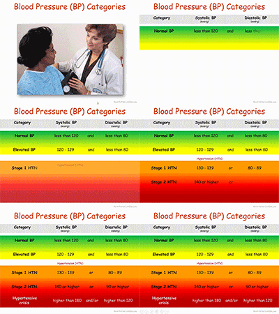
First, there are revised guidelines as to what constitutes high blood pressure or HTN:
Normal BP: Less than 120/80 mm Hg;
Elevated BP: Systolic between 120-129 and diastolic less than 80;
Stage 1 HTN: Systolic between 130-139 or diastolic between 80-89;
Stage 2 HTN: Systolic at least 140 or diastolic at least 90 mm Hg;
Hypertensive crisis: Systolic over 180 and/or diastolic over 120, with patients needing prompt changes in medication if there are no other indications of problems, or immediate hospitalization if there are signs of organ damage.
Regardless of the precise cutoffs listed above, in an interview discussing the new guidelines, the main author states that, "120/80 is normal, the same as we had before" the new guidelines. So I think we're safe in using 120/80 as an example of BP when discussing the normal science, even though technically it could be designated as "elevated." Not that we can't use an elevated variable measurement as an example when discussing the physiology of anything. The fact that even the main author of the guidelines uses 120/80 as the starting point of discussion makes me feel more confident in using it as the starting point of my course discussions, too.
The main thing to note in the categories above is that the cutoffs for HTN categories have been lowered. This puts many more people in an HTN category that were not there before. The main goal is for those folks to have conversations with their physicians to evaluate their risk for complications and develop a personalized prevention and care plan.
Note also that the category of prehypertension has been eliminated.
The new guidelines also recommend prescribing medication for Stage 1 HTN if the patient already had a cardiovascular event—or is at a high risk for such an event. They also recognize that many patients will need more than one medication to manage BP and that combining meds into one pill is likely to help folks take them consistently.
There are a lot of other recommendations, so reviewing the Executive Summary or similar resource (see below) may be a good idea.
What can we use from this in teaching undergraduate A&P?
If you discuss hypertension, or use case studies in teaching, you need to update the cutoff BPs you are using.
A BP of 120/80 is still considered the starting point for discussing blood pressure.
Consider discussing the impact of the changes in the new guidelines for ordinary people.
Discuss why such diagnosis, prevention, and treatment recommendations often change over time. Consider discussion other recent clinical updates.
Consider discussing specific changes suggested in the new guidelines.
Consider having students explore the Executive Summary and/or other documents and write their own summary or interpretation of key points. Perhaps they can create their own chart or concept map.
Need some free teaching materials?
SLIDE SET: High Blood Pressure
Kevin Patton. Lion Den Slide Collection. 18 Nov 2017
Small slide deck that includes an animated version of the BP Category chart pictured above. Part of the Lion Den Slide Collection (requires free registration to download). You can also download a static PNG image file of the chart in the slide collection set.
VIDEO: AHA 2017 | New High Blood Pressure Guidelines
America Heart Association. 13 Nov 2017.
Free video (viewable in the player above) features a chat with the main author of the new guidelines and summarizes the main points. Very practical and easy to understand.
SLIDE SET: 2017 Guideline For the Prevention, Detection, Evaluation and Management of High Blood Pressure in Adults
American College of Cardiology. 13 Nov 2017
Free set of almost 100 PowerPoint slides to use in teaching. And it has a decided focus on clinical applications, rather than the basic science. These are way, way beyond the coverage desirable in an undergrad A&P course. But some slides may be useful to you.
New blood pressure guidelines put half of U.S. adults in unhealthy range
A. Cunningham Science News. 13 Nov 2017
Plain-English article summarizing the first major update since 2003 aims to spur heart-healthy lifestyle changes. Has a useful graph and links to other articles and resources.
2017 ACC/AHA/AAPA/ABC/ACPM/AGS/APhA/ASH/ASPC/NMA/PCNA Guideline for the Prevention, Detection, Evaluation, and Management of High Blood Pressure in Adults: Executive Summary A Report of the American College of Cardiology/American Heart Association Task Force on Clinical Practice Guidelines
PK Whelton et al. Hypertension, Dec 2017, Volume 70, Issue 6. DOI: 10.1161/HYP.0000000000000066
Free PDF of the Executive Summary of the larger report. I recommend reading this first, then decide if you need to read the whole report.
2017 ACC/AHA/AAPA/ABC/ACPM/AGS/APhA/ASH/ASPC/NMA/PCNA Guideline for the Prevention, Detection, Evaluation, and Management of High Blood Pressure in Adults A Report of the American College of Cardiology/American Heart Association Task Force on Clinical Practice Guidelines
PK Whelton et al. Hypertension. Dec 2017, Volume 70, Issue 6. DOI: 10.1161/HYP.0000000000000065
Free PDF of the entire report. It's huge, so make a whole pot of tea before starting it.
Today, the Royal Swedish Academy of Sciences has decided to award the Nobel Prize in Chemistry 2017 to Jacques Dubochet (University of Lausanne, Switzerland) and Joachim Frank (Columbia University, New York, USA), and Richard Henderson (MRC Laboratory of Molecular Biology, Cambridge, UK). The award is given "for developing cryo-electron microscopy for the high-resolution structure determination of biomolecules in solution"
We may soon have detailed images of life’s complex machineries in atomic resolution. The Nobel Prize in Chemistry 2017 is awarded to Jacques Dubochet, Joachim Frank and Richard Henderson for the development of cryo-electron microscopy, which both simplifies and improves the imaging of biomolecules. This method has moved biochemistry into a new era.
A picture is a key to understanding. Scientific breakthroughs often build upon the successful visualization of objects invisible to the human eye. However, biochemical maps have long been filled with blank spaces because the available technology has had difficulty generating images of much of life’s molecular machinery. Cryo-electron microscopy changes all of this. Researchers can now freeze biomolecules mid-movement and visualize processes they have never previously seen, which is decisive for both the basic understanding of life’s chemistry and for the development of pharmaceuticals.
Electron microscopes were long believed to only be suitable for imaging dead matter, because the powerful electron beam destroys biological material. But in 1990, Richard Henderson succeeded in using an electron microscope to generate a three-dimensional image of a protein at atomic resolution. This breakthrough proved the technology’s potential.
Joachim Frank made the technology generally applicable. Between 1975 and 1986 he developed an image processing method in which the electron microscope’s fuzzy two-dimensional images are analysed and merged to reveal a sharp three-dimensional structure.
Jacques Dubochet added water to electron microscopy. Liquid water evaporates in the electron microscope’s vacuum, which makes the biomolecules collapse. In the early 1980s, Dubochet succeeded in vitrifying water – he cooled water so rapidly that it solidified in its liquid form around a biological sample, allowing the biomolecules to retain their natural shape even in a vacuum.
Following these discoveries, the electron microscope’s every nut and bolt have been optimised. The desired atomic resolution was reached in 2013, and researchers can now routinely produce three-dimensional structures of biomolecules. In the past few years, scientific literature has been filled with images of everything from proteins that cause antibiotic resistance, to the surface of the Zika virus. Biochemistry is now facing an explosive development and is all set for an exciting future.
Joachim Frank, born 1940 in Siegen, Germany. Ph.D. 1970, Technical University of Munich, Germany. Professor of Biochemistry and Molecular Biophysics and of Biological Sciences, Columbia University, New York, USA. http://franklab.cpmc.columbia.edu/franklab/ Richard Henderson, born 1945 in Edinburgh, Scotland. Ph.D. 1969, Cambridge University, UK. Programme Leader, MRC Laboratory of Molecular Biology, Cambridge, UK. www2.mrc-lmb.cam.ac.uk/groups/rh15/
What can we use from this in teaching undergraduate A&P?
If you talk about imaging molecules in your course, this could be a way to garner student interest—considering that this is a current and ongoing effort in science. I always have a brief "shape is important in biological chemistry and here's what we can see with current tools" because they're going to see all those little odd-shaped rutabaga blobs in illustrations in their textbooks.
If you bring up microscopy in your course, perhaps describing the types of microscopy, adding a bit of info on this could help show students that microscopy is still evolving—in exciting ways.
Consider using the annual Nobel Prize announcements as a springboard to discuss the process of scientific discovery.
Consider mentioning the other major awards for scientific achievement and discuss what the judges seem to value most about scientific discoveries. The Nobel Prize is the one everyone has heard of, so it's a great place to start.
Use the Nobel Prizes (and other awards) over time as a way to keep students aware of the history of, and progress, of human biology. One could also address the global diversity of laureates. Or the lack of other kinds of diversity among laureates.
The Nobel Assembly at Karolinska Institutet has today decided to award the 2016 Nobel Prize in Physiology or Medicine to Yoshinori Ohsumi for his discoveries of mechanisms for autophagy.
Overview
This year's Nobel Laureate discovered and elucidated mechanisms underlying autophagy, a fundamental process for degrading and recycling cellular components.
The word autophagy (aw-toh-FAY-jee) originates from the Greek words auto-, meaning "self", and phagein, meaning "to eat". Thus, autophagy denotes "self eating".
This concept emerged during the 1960's, when researchers first observed that the cell could destroy its own contents by enclosing it in membranes, forming sack-like vesicles that were transported to a recycling compartment, called the lysosome, for degradation.
Difficulties in studying the phenomenon meant that little was known until, in a series of brilliant experiments in the early 1990's, Yoshinori Ohsumi used baker's yeast to identify genes essential for autophagy. He then went on to elucidate the underlying mechanisms for autophagy in yeast and showed that similar sophisticated machinery is used in our cells.
Ohsumi's discoveries led to a new paradigm in our understanding of how the cell recycles its content. His discoveries opened the path to understanding the fundamental importance of autophagy in many physiological processes, such as in the adaptation to starvation or response to infection. Mutations in autophagy genes can cause disease, and the autophagic process is involved in several conditions including cancer and neurological disease.
Degradation – a central function in all living cells
In the mid 1950's scientists observed a new specialized cellular compartment, called an organelle, containing enzymes that digest proteins, carbohydrates and lipids. This specialized compartment is referred to as a "lysosome" and functions as a workstation for degradation of cellular constituents. The Belgian scientist Christian de Duve was awarded the Nobel Prize in Physiology or Medicine in 1974 for the discovery of the lysosome.
New observations during the 1960's showed that large amounts of cellular content, and even whole organelles, could sometimes be found inside lysosomes. The cell therefore appeared to have a strategy for delivering large cargo to the lysosome. Further biochemical and microscopic analysis revealed a new type of vesicle transporting cellular cargo to the lysosome for degradation (Figure 1).
Christian de Duve, the scientist behind the discovery of the lysosome, coined the term autophagy, "self-eating", to describe this process. The new vesicles were named autophagosomes.
Figure 1: Autophagosome. Our cells have different specialized compartments. Lysosomes constitute one such compartment and contain enzymes for digestion of cellular contents. A new type of vesicle called autophagosome was observed within the cell. As the autophagosome forms, it engulfs cellular contents, such as damaged proteins and organelles. Finally, it fuses with the lysosome, where the contents are degraded into smaller constituents. This process provides the cell with nutrients and building blocks for renewal.
During the 1970's and 1980's researchers focused on elucidating another system used to degrade proteins, namely the "proteasome". Within this research field Aaron Ciechanover, Avram Hershko and Irwin Rose were awarded the 2004 Nobel Prize in Chemistry for "the discovery of ubiquitin-mediated protein degradation". The proteasome efficiently degrades proteins one-by-one, but this mechanism did not explain how the cell got rid of larger protein complexes and worn-out organelles. Could the process of autophagy be the answer and, if so, what were the mechanisms?
A groundbreaking experiment
Yoshinori Ohsumi had been active in various research areas, but upon starting his own lab in 1988, he focused his efforts on protein degradation in the vacuole, an organelle that corresponds to the lysosome in human cells.
Yeast cells are relatively easy to study and consequently they are often used as a model for human cells. They are particularly useful for the identification of genes that are important in complex cellular pathways. But Ohsumi faced a major challenge; yeast cells are small and their inner structures are not easily distinguished under the microscope and thus he was uncertain whether autophagy even existed in this organism.
Ohsumi reasoned that if he could disrupt the degradation process in the vacuole while the process of autophagy was active, then autophagosomes should accumulate within the vacuole and become visible under the microscope. He therefore cultured mutated yeast lacking vacuolar degradation enzymes and simultaneously stimulated autophagy by starving the cells.
The results were striking! Within hours, the vacuoles were filled with small vesicles that had not been degraded (Figure 2). The vesicles were autophagosomes and Ohsumi's experiment proved that authophagy exists in yeast cells. But even more importantly, he now had a method to identify and characterize key genes involved this process. This was a major break-through and Ohsumi published the results in 1992.
Figure 2: Yeast. In yeast (left panel) a large compartment called the vacuole corresponds to the lysosome in mammalian cells. Ohsumi generated yeast lacking vacuolar degradation enzymes. When these yeast cells were starved, autophagosomes rapidly accumulated in the vacuole (middle panel). His experiment demonstrated that autophagy exists in yeast. As a next step, Ohsumi studied thousands of yeast mutants (right panel) and identified 15 genes that are essential for autophagy.
Autophagy genes are discovered
Ohsumi now took advantage of his engineered yeast strains in which autophagosomes accumulated during starvation. This accumulation should not occur if genes important for autophagy were inactivated. Ohsumi exposed the yeast cells to a chemical that randomly introduced mutations in many genes, and then he induced autophagy.
His strategy worked! Within a year of his discovery of autophagy in yeast, Ohsumi had identified the first genes essential for autophagy. In his subsequent series of elegant studies, the proteins encoded by these genes were functionally characterized. The results showed that autophagy is controlled by a cascade of proteins and protein complexes, each regulating a distinct stage of autophagosome initiation and formation (Figure 3).
Figure 3: Stages of autophagosome formation. Ohsumi studied the function of the proteins encoded by key autophagy genes. He delineated how stress signals initiate autophagy and the mechanism by which proteins and protein complexes promote distinct stages of autophagosome formation.
Autophagy – an essential mechanism in our cells
After the identification of the machinery for autophagy in yeast, a key question remained. Was there a corresponding mechanism to control this process in other organisms? Soon it became clear that virtually identical mechanisms operate in our own cells. The research tools required to investigate the importance of autophagy in humans were now available.
Thanks to Ohsumi and others following in his footsteps, we now know that autophagy controls important physiological functions where cellular components need to be degraded and recycled.
Autophagy can rapidly provide fuel for energy and building blocks for renewal of cellular components, and is therefore essential for the cellular response to starvation and other types of stress.
After infection, autophagy can eliminate invading intracellular bacteria and viruses. Autophagy contributes to embryo development and cell differentiation. Cells also use autophagy to eliminate damaged proteins and organelles, a quality control mechanism that is critical for counteracting the negative consequences of aging.
Disrupted autophagy has been linked to Parkinson's disease, type 2 diabetes and other disorders that appear in the elderly. Mutations in autophagy genes can cause genetic disease. Disturbances in the autophagic machinery have also been linked to cancer. Intense research is now ongoing to develop drugs that can target autophagy in various diseases.
Autophagy has been known for over 50 years but its fundamental importance in physiology and medicine was only recognized after Yoshinori Ohsumi's paradigm-shifting research in the 1990's. For
Yoshinori Ohsumi was born 1945 in Fukuoka, Japan. He received a Ph.D. from University of Tokyo in 1974. After spending three years at Rockefeller University, New York, USA, he returned to the University of Tokyo where he established his research group in 1988. He is since 2009 a professor at the Tokyo Institute of Technology.
More background on the winner and the prize
Yoshinori Ohsumi was born in Fukuoka, Japan, in 1945. He is affiliated with the Tokyo Institute of Technology in Tokyo, Japan. His monetary award will be nearly one million dollars.
The Nobel Assembly, consisting of 50 professors at Karolinska Institutet, awards the Nobel Prize in Physiology or Medicine. Its Nobel Committee evaluates the nominations. Since 1901 the Nobel Prize has been awarded to scientists who have made the most important discoveries for the benefit of mankind.his discoveries, he is awarded this year's Nobel Prize in physiology or medicine.
Nobel Prize® is the registered trademark of the Nobel Foundation
What can we use from this in teaching undergraduate A&P?
Consider using the Nobel Prizes as a discussion-starter in your class about
How science influences society
How society influences science
How science progresses
Rewarding of science discoveries
What makes a discovery "important"
Relate this discovery to prior (or upcoming) discussions of
Cell function
Organelle specialization
How cells handle protein
How autophagosomes work with lysosomes
Compare/contrast with phagocytosis
Compare/contrast with proteasome function and protein "quality control
Relate this discovery to the general idea of cellular mechanisms of disease
Consider taking this opportunity to emphasize "why we need to know all this" detail about cellular structure and function.
Want to know more?
Scientific Background Discoveries of Mechanisms for Autophagy
Larsson, N-G, Msucci, M. G. Nobelprize.org accessed 8 October 2016
A more advanced summary of the prizewinning discovery, including a handy glossary of terms.
A ubiquitin-like system mediates protein lipidation.
Ichimura, Y., Kirisako T., Takao, T., Satomi, Y., Shimonishi, Y., Ishihara, N., Mizushima, N., Tanida, I., Kominami, E., Ohsumi, M., Noda, T. and Ohsumi, Y. (2000). Nature, 408, 488-492
Honoring the 2016 Nobel laureates with free access to selections of their research
Elisa Nelissen Elsevier Connect. October 3, 2016
This blog post provides links to download the most cited papers the laureates published with Elsevier, a major publisher of scientific journals and references.
A new study published in Advances in Physiology Education adds additional evidence of the effectiveness that students do better when they take the time to analyze their tests immediately after taking them.
In my blog The A&P Student, I published an article in October of 2009 that outlines an easy and effective way for A&P students to "debrief" after each test and exam so that they can both clarify misconceptions and gain insights into possible weakness in test preparation.
By having students focus on missed questions coupled with addressing deficiencies in their test preparation strategies and behaviors, they likely engage in more self-regulated learning to better prepare for exams and avoid repeating past mistakes. (Favero & Hendricks 2016).
What can we use from this in teaching undergraduate A&P?
Encouraging students to debrief after every test and exam—individually or in study groups—can be more effective than whole-class reviews of exams or tests.
Test self-analysis can help students learn (or re-learn) concepts they are unable to retrieve on a test.
Test self-analysis can help students identify patterns of misconceptions, poor test preparation, and poor test-taking skills that make them better aware of their own thinking (metacognition) and thus more likely to succeed in later testing.
Consider encouraging your students to debrief after every test by providing them with the tools needed (see the links below).
In my many years of using this technique, I've found that the process described in the links below works equally well for either/both online tests (including adaptive quizzing) and traditional paper tests and exams
Want to know more?
Student exam analysis (debriefing) promotes positive changes in exam preparation and learning
Terence G. Favero, Nora Hendricks. Advances in Physiology Education Published 1 September 2016 Vol. 40 no. 3, 323-328 DOI: 10.1152/advan.00060.2016
The new research confirming the value of self-analysis of recent tests in an A&P course.
Brief blog post directed at A&P students giving them resources to perform this on their own, including an instructional video (see below), and access to a sample analysis form (see below). Consider linking to this post in your syllabus (and/or the other embedded resources).
Entry from Kevin's library of Study Tips & Tools for A&P students, briefly runs through the advantages of debriefing after a test and provides an instructional video (see below) and sample analysis form that students can download and use (or adapt). Consider adding a link from your syllabus or course website/LMS.
Brief instructional video that teaches students how to perform a test analysis (and why it's important). Link to this video in your course, or embed the video.
Kevin Patton. 2014. Mosby, Inc., an affiliate of Elsevier Inc. St. Louis MO ISBN: 978-0-323-11280-2
Brief paperback book that you can make available in your school or classroom library or require/suggest for purchase in your college bookstore. Covers the process of test debriefing and gives tips on how to resolves inefficient patterns of test preparation and test taking. Also contains A&P specific content tips and analogies.
It never fails—somebody always asks about what's going on when knuckles crack when we get to the topic of articulations.
Recently, researchers used modern MRI techniques to demonstrate exactly what is happening—a process first described in the 1940s. As joint surfaces separate, the changing tension in the synovial fluid causes a vapor pocket or "air bubble" to form in a process called cavitation.
The recent research proved that the popping sound typical of cracking a knuckle is produced as the vapor pocket forms. It also disproved the theory that the popping occurs when the bubble collapses.
What can we use from this in teaching undergraduate A&P?
You'll now always be ready for that inevitable "what causes knuckles to crack?" question when discussing joints in your A&P course.
Consider having a slide containing the media offered below to show students.
Discussing joint-cracking mechanisms engages students in a subject that may not immediately interest them.
Joint-cracking allows us to reinforce concepts of fluid dynamics in the context of synovial joints.
Consider stimulating some critical thinking by asking "what could cause a cracking sound in a cartilaginous or fibrous joint?" (Hint: think "fracture" as cavitation would not occur where there are no fluids)
Do a demonstration by hitting a glass bottle full of water with a rubber mallet (see the embedded video below)
I've done this with an unopened glass bottle of ketchup by upturning it and hitting the base with my palm
Want to know more?
Real-Time Visualization of Joint Cavitation
GN Kawchuk, et al. PLoS|ONE. April 15, 2015 DOI: 10.1371/journal.pone.0119470
The original journal article. Includes downloadable images and PowerPoint slides, plus a link to a video showing the MRI results.
Liven up your A&P class with a great video showing a gruesome attack by a killer T cell on a cancer cell. It's a fantastic bit of video microscopy produced by Cambridge University.
Okay, with the oddly soothing music score instead of a more appropriate score for the graphic violence shown in this video, it's not much of a horror flick. Especially when you consider that it's the "bad guy" cell getting whacked. But it is graphic and dramatic and impressive.
Just the thing to liven up a discussion of adaptive immunity, which (let's face it) can often cause a catatonic state in many students.
Five years ago, I extolled the virtues of teaching a little bit about RNA interference (RNAi) in undergraduate A&P courses. But for a while it looked like the promise of RNAi in basic and clinical research might be sputtering. However, a recent article by Eric Bender called The Second Coming of RNAi shows that RNAi "the gene-silencing technique [now] begins to fulfill some of its promises."
I recommend reading the entire article at my-ap.us/1BbxvB9 Before you read it, allow me to reprise my reasons of five years ago supporting my proposal to include RNAi in your course.
What can we use from this in teaching undergraduate A&P?
RNAi plays a role in defending our cells against viruses by stopping viral genetic code from being translated in host cells
RNAi likely plays a role in regulating gene activity in a cell by preventing translation of the gene product(s)
RNAi is increasingly used as method for "knocking out" a particular gene's effects in research animals in order to study the gene's functions
RNAi is being used to treat genetic disease. . . an application that will likely expand greatly over the next few decades
I'll add two more items to my previous list:
RNA interference is a mechanism of human disease, as has been demonstrated in some cases of inherited progressive hearing loss (for example).
Learning about RNAi helps clarify a general understanding of the many roles played by RNA in our lives—some perhaps still undiscovered.
I'm not sure that it's useful to expect beginning undergraduate students to learn the nitty-gritty details of RNAi mechanisms. But I do think it's valuable to be exposed to the general concept of RNA interference and gene silencing.A&P students are going to run up against these eventually as they learn about and then administer RNAi-based therapies, after all. And perhaps we should prepare them.
Want to know more?
The Second Coming of RNAi
Eric Bender. The Scientist. September 1, 2014
Article mentioned above. In plain English, it shows that clinical progress in RNAi therapy against liver diseases, the gene-silencing technique begins to fulfill some of its promises. Includes useful illustrations and links to other resources.
The Royal Swedish Academy of Sciences has decided to award the Nobel Prize in Chemistry for 2014 to
Eric Betzig
Janelia Farm Research Campus, Howard Hughes Medical Institute, Ashburn, VA, USA,
Stefan W. Hell
Max Planck Institute for Biophysical Chemistry, Göttingen, and German Cancer Research Center, Heidelberg, Germany
and
William E. Moerner
Stanford University, Stanford, CA, USA
“for the development of super-resolved fluorescence microscopy”
Surpassing the limitations of the light microscope
For a long time optical microscopy was held back by a presumed limitation: that it would never obtain a better resolution than half the wavelength of light. Helped by fluorescent molecules the Nobel Laureates in Chemistry 2014 ingeniously circumvented this limitation. Their ground-breaking work has brought optical microscopy into the nanodimension.
In what has become known as nanoscopy, scientists visualize the pathways of individual molecules inside living cells. They can see how molecules create synapses between nerve cells in the brain; they can track proteins involved in Parkinson’s, Alzheimer’s and Huntington’s diseases as they aggregate; they follow individual proteins in fertilized eggs as these divide into embryos.
It was all but obvious that scientists should ever be able to study living cells in the tiniest molecular detail. In 1873, the microscopist Ernst Abbe stipulated a physical limit for the maximum resolution of traditional optical microscopy: it could never become better than 0.2 micrometres. Eric Betzig, Stefan W. Hell and William E. Moerner are awarded the Nobel Prize in Chemistry 2014 for having bypassed this limit. Due to their achievements the optical microscope can now peer into the nanoworld.
Two separate principles are rewarded.
One enables the method stimulated emission depletion (STED) microscopy, developed by Stefan Hell in 2000. Two laser beams are utilized; one stimulates fluorescent molecules to glow, another cancels out all fluorescence except for that in a nanometre-sized volume. Scanning over the sample, nanometre for nanometre, yields an image with a resolution better than Abbe’s stipulated limit.
Eric Betzig and William Moerner, working separately, laid the foundation for the second method, single-molecule microscopy. The method relies upon the possibility to turn the fluorescence of individual molecules on and off. Scientists image the same area multiple times, letting just a few interspersed molecules glow each time. Superimposing these images yields a dense super-image resolved at the nanolevel. In 2006 Eric Betzig utilized this method for the first time.
Today, nanoscopy is used world-wide and new knowledge of greatest benefit to mankind is produced on a daily basis.
This video is a brief animation of how STED works and how it improves resolution of individual particles.
This video is a longer, more detailed presentation by one of the Nobel laureates (Hell).
What can we use from this in teaching undergraduate A&P?
Discuss how this technology has enabled us to better visualize the chemicals and structures within our cells, enabling scientists to better understand the structure and function of cell, organelles, microbiome constituents, and other structures of the human body.
If you do a brief run-through of the theory of microscopy—perhaps in your A&P lab—you can add a mention of this technology.
Your textbook or other learning resource may already have an example of this type of microscopy.
A discussion of this Nobel Prize could evolve into a meaningful example of how science works, including how incremental improvements in classical tools for observation expand the number of questions that can be answered.
Use the links below (and images above) to use for a handout and/or teaching slides.
U.S. citizen. Born 1960 in Ann Arbor, MI, USA. Ph.D. 1988 from Cornell University, Ithaca, NY, USA. Group Leader at Janelia Farm Research Campus, Howard Hughes Medical Institute, Ashburn, VA, USA.
Born 1962 in Arad, Romania. Ph.D. 1990 from the University of Heidelberg, Germany. Director at the Max Planck Institute for Biophysical Chemistry, Göttingen, and Division head at the German Cancer Research Center, Heidelberg, Germany.
Born 1953 in Pleasanton, CA, USA. Ph.D. 1982 from Cornell University, Ithaca, NY, USA. Harry S. Mosher Professor in Chemistry and Professor, by courtesy, of Applied Physics at Stanford University, Stanford, CA, USA.
The Nobel Assembly at Karolinska Institutet has today decided to award
The 2014 Nobel Prize in Physiology or Medicine
with one half to
John O´Keefe
and the other half jointly to
May-Britt Moser and Edvard I. Moser
for their discoveries of cells that constitute a positioning
system in the brain.
How do we know where we are? How can we find the way from one place to another? And how can we store this information in such a way that we can immediately find the way the next time we trace the same path? This year´s Nobel Laureates have discovered a positioning system, an “inner GPS” in the brain that makes it possible to orient ourselves in space, demonstrating a cellular basis for higher cognitive function.
In 1971, John O´Keefe discovered the first component of this positioning system. He found that a type of nerve cell in an area of the brain called the hippocampus that was always activated when a rat was at a certain place in a room. Other nerve cells were activated when the rat was at other places. O´Keefe concluded that these “place cells” formed a map of the room.
More than three decades later, in 2005, May-Britt and Edvard Moser discovered another key component of the brain’s positioning system. They identified another type of nerve cell, which they called “grid cells”, that generate a coordinate system and allow for precise positioning and pathfinding.
Their subsequent research showed how place and grid cells make it possible to determine position and to navigate.
The discoveries of John O´Keefe, May-Britt Moser and Edvard Moser have solved a problem that has occupied philosophers and scientists for centuries – how does the brain create a map of the space surrounding us and how can we navigate our way through a complex environment?
How do we experience our environment?
The sense of place and the ability to navigate are fundamental to our existence. The sense of place gives a perception of position in the environment. During navigation, it is interlinked with a sense of distance that is based on motion and knowledge of previous positions.
Questions about place and navigation have engaged philosophers and scientists for a long time. More than 200 years ago, the German philosopher Immanuel Kant argued that some mental abilities exist as a priori knowledge, independent of experience. He considered the concept of space as an inbuilt principle of the mind, one through which the world is and must be perceived. With the advent of behavioural psychology in the mid-20th century, these questions could be addressed experimentally. When Edward Tolman examined rats moving through labyrinths, he found that they could learn how to navigate, and proposed that a “cognitive map” formed in the brain allowed them to find their way. But questions still lingered - how would such a map be represented in the brain?
John O´Keefe and the place in space
John O´Keefe was fascinated by the problem of how the brain controls behaviour and decided, in the late 1960s, to attack this question with neurophysiological methods. When recording signals from individual nerve cells in a part of the brain called the hippocampus, in rats moving freely in a room, O’Keefe discovered that certain nerve cells were activated when the animal assumed a particular place in the environment (Figure 1). He could demonstrate that these “place cells” were not merely registering visual input, but were building up an inner map of the environment. O’Keefe concluded that the hippocampus generates numerous maps, represented by the collective activity of place cells that are activated in different environments. Therefore, the memory of an environment can be stored as a specific combination of place cell activities in the hippocampus.
May-Britt and Edvard Moser find the coordinates
May-Britt and Edvard Moser were mapping the connections to the hippocampus in rats moving in a room when they discovered an astonishing pattern of activity in a nearby part of the brain called the entorhinal cortex. Here, certain cells were activated when the rat passed multiple locations arranged in a hexagonal grid (Figure 2). Each of these cells was activated in a unique spatial pattern and collectively these “grid cells” constitute a coordinate system that allows for spatial navigation. Together with other cells of the entorhinal cortex that recognize the direction of the head and the border of the room, they form circuits with the place cells in the hippocampus. This circuitry constitutes a comprehensive positioning system, an inner GPS, in the brain (Figure 3).
A place for maps in the human brain
Recent investigations with brain imaging techniques, as well as studies of patients undergoing neurosurgery, have provided evidence that place and grid cells exist also in humans. In patients with Alzheimer´s disease, the hippocampus and entorhinal cortex are frequently affected at an early stage, and these individuals often lose their way and cannot recognize the environment. Knowledge about the brain´s positioning system may, therefore, help us understand the mechanism underpinning the devastating spatial memory loss that affects people with this disease.
The discovery of the brain’s positioning system represents a paradigm shift in our understanding of how ensembles of specialized cells work together to execute higher cognitive functions. It has opened new avenues for understanding other cognitive processes, such as memory, thinking and planning.
What can we use from this in teaching undergraduate A&P?
The role of the hippocampus and it's place cells could be briefly discussed as you explore the cognitive functions of the brain.
A discussion of this year's Nobel Prize could evolve into a meaningful example of how science works, including the use of discoveries in animals that can later be applied to learning more about human structure and function.
Information revealed by this discovery could be discussed when discussing human disorders, such as Alzheimer disease, that involve impairments of spatial orientation and/or place memory.
Want to know more?
Here's a video in which May-Britt Moser and Edvard I. Moser explain their research, which you can use in your course discussion.
2014 Nobel Prize announcement
Official announcement, which includes photos of the laureates and links to related information.
A story broadcast recently on National Public Radio (NPR) highlighted the role of a Pre-A&P course in student success.
Listen to the story yourself (link below) and tell me you don't recognize the issues brought up there. Students failing A&P because they just don't know how to read a science textbook, don't know how to study, and don't have higher-order thinking skills.
The story mentions a course to help students through these difficulties at West Kentucky Community and Technical College. But I know of a lot of colleges that are taking this approach to improving student success, including my own.
Some of you have already participated in one of my past seminars on how we did it. It involves a two-phase approach:
Pre-A&P Foundations in Science
An elective, developmental-level one-credit course.
Completely online, self-paced course.
Ten modules reviewing basic concepts needed for success in A&P:
Science Basics
Introductory Chemistry
Biological Chemistry
Introduction to Cells
Cell Transport
Getting Energy
Making Proteins
Introductory Genetics
Tissues
The Human Body
Each module is comprehensive, reviewing all prior modules.
Mastery-based: students proceed to the next module only if they pass the current module by 85% or better.
Passing the comprehensive final exam at mastery level earns a "Pass" grade.
A&P 1 Supplement
An elective, 200-level one-credit course.
Runs concurrently with A&P 1 lecture/lab sequence.
Addresses basic study skills, as applied to what students are studying in A&P
Specifically addresses "trouble spots" typically encountered
Provides coaching and support of students in real time
What can we use from this in teaching undergraduate A&P?
Look at what others have done and consider trying your own version of Pre-A&P and/or supplemental A&P study-skills courses!
If you are interested in our take on this, then listen to my seminar (link below). But I've helped several other colleges get their own version of this strategy off the ground, and it's working.
Want to know more?
The Toughest Class In Nursing School Is The First One
Zoe Chace. Planet Money (NPR). September 02, 2014
Radio story of one example of Pre-A&P helping students (and increasing A&P success rates).
A recent article in The Scientist once again reminds us of the ongoing explosion in the scientific understanding of the human microbial system. In a few short years, this area of exploration has moved to the forefront of medical and basic science research in human biology.
I think it's becoming clear that the most useful way to think of human body function is to recognize that an "organism" is really a sort of "habitat." And like any habitat, it functions best when all the inhabitants are within a limited range of balanced relationships.
Who are the inhabitants? Besides our own cells? Well, one could think of mitochondria and cilia and other organelles as symbiotic internal inhabitants of our cells. They're not that literally, of course, but I think its a useful metaphor for understanding the human body. Then there are the many microbes and animals that cover our internal and external surfaces, burrow into some of our pores and glands, and inhabit our body fluids.
I call the balanced functional relationship among the various microbomes of the body and our own tissues the human microbial system. And I am certain that it won't be long before we will be discussing this system alongside the major organ systems of the body. That is if we truly want to understand how the body really works.
The article in The Scientist I mention is a great summary of some of the major roles that the human microbial system plays in the human body—and a good survey of some of the areas of the body where the human-microbial functional relationships play out. See the link to the article below.
What can we use from this in teaching undergraduate A&P?
Why not introduce the concept of the human microbial system at the beginning of our A&P course, when we set the stage by explain how scientists understand the body and its functions as an integrated system of different parts?
We can mention the different microbiomes of the body when we explore each organ system where they play an important role—which is pretty much all of them!
Consider discussing what happens to normal human function when microbiomes get out of balance. For example, in the gut a microbial imbalance can lead to ulcers, diarrhea, and other dysfunctions. On the skin a pathogenic microbe may become dominant and cause a rash.
Promote a discussion of what kinds of wellness strategies might be employed to prevent microbial imbalances.
Our students can leave our A&P course with an up-to-date understanding of human biology that will help them understand new clinical concepts and treatment strategies.
Want to Know More?
The Body’s Ecosystem
By The Scientist Staff. The Scientist. August 1, 2014
Plain-English article (cited above) on how research on the human microbiome is booming, and scientists have moved from simply taking stock of gut flora to understanding the influence of microbes throughout the body.
Radio stories from National Public Radio on human microbiomes and their role in health and disease. The growing number of these stories tells us something as A&P teachers: maybe we better be covering this!

.jpeg)