Showing posts with label science history. Show all posts

Monday, February 11, 2019

The Last Best Story in Teaching Anatomy & Physiology | Episode 37


01:17 | Feedback in Online Tests
08:17 | The Anatomical Compass
14:47 | Sponsored by AAA
15:12 | Reserve hematopoiesis
18:09 | Sponsored by HAPS
18:54 | Featured: Last Best Story in Adult Neurogenesis & ANS Pathways
If you cannot see or activate the audio player click here.

Questions & Feedback: 1-833-LION-DEN (1-833-546-6336)
Follow The A&P Professor on Twitter, Facebook, Blogger, Nuzzel, Tumblr, or Instagram!

Scientific theories are tested every time someone makes an observation or conducts an experiment, so it is misleading to think of science as an edifice, built on foundations. Rather, scientific knowledge is more like a web. The difference couldn’t be more crucial. A tall edifice can collapse – if the foundations upon which it was built turn out to be shaky. But a web can be torn in several parts without causing the collapse of the whole. The damaged threads can be patiently replaced and re-connected with the rest – and the whole web can become stronger, and more intricate. (Massimo Pigliucci)

1 | Feedback in Online tests

7 minutes
In Episode 36, Adam Rich called in regarding how we can provide feedback to students taking online tests. I responded that I encourage students to get the correct response from their study buddies—or from me. After the episode aired, Krista Rompolski pointed out that this could be a challenge in very large courses. What do y'all think? Tell us. Really.


2 | The Anatomical Compass

6.5 minutes
Although you and I are comfortable in orienting ourselves to anatomical directions when looking at diagrams, photographs, and specimens in anatomy, our beginning student often are not. The simple process of adding an "anatomical rosette" reflecting the anatomical directions in each encountered diagram can  help students develop the skill of understanding anatomical perspective.
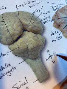
 anatomical rosette


3 | Sponsored by AAA

0.5 minutes
The searchable transcript for this episode, as well as the captioned audiogram of this episode, are sponsored by The American Association of Anatomists (AAA) at anatomy.org. Their big meeting is in April at the Experimental Biology (EB) meeting in Orlando FL. Check it out!
 American Association of Anatomists

4 | Reserve Hematopoiesis

3 minutes
Hematopoietic stem cells  (HSCs) may have a "back-up system" that helps out after damage to the working population. These "reserve" HSCs (rHSCs) may step up when the primed HSCs (pHSCs) cannot keep up with the demand for hematopoiesis.
  • Scientists have identified a bone marrow backup system (summary article) my-ap.us/2BmcoE0
  • N-Cadherin-Expressing Bone and Marrow Stromal Progenitor Cells Maintain Reserve Hematopoietic Stem Cells
    (report by Zhao, et al. in Cell Reports) my-ap.us/2Bk7vLN

 hematopoietic stem cell


5 | Sponsored by HAPS

0.5 minutes
The Human Anatomy & Physiology Society (HAPS) is a sponsor of this podcast. Did you know there's a one-day regional HAPS conference in March? Check it out. You can help appreciate their support by clicking the link below and checking out the many resources and benefits found there.

 HAPS logo

6 | Featured: Last Best Story in Adult Neurogenesis & ANS Pathways

12 minutes
The "last best story" is what I tell my students I'm providing to them. That approach emphasizes the evolving nature of scientific understanding. In this episode, I mention two stories that are evolving right now.
reading a book

If the hyperlinks here are not active, go to TAPPradio.org to find the episode page.

Amazon and TextExpander referrals help defray podcasting expenses.

Transcript and captions for this episode
are supported by theAmerican Association of Anatomists.anatomy.org


The Human Anatomy & Physiology Societyalso provides support for this podcast.
theAPprofessor.org/haps


(Clicking on sponsor links 
helps let them know you appreciatetheir support of this podcast!)

Click here to listen to this episode—or access the detailed notes and transcript.


Wednesday, October 4, 2017

Biomolecule Imaging Pioneers Share Nobel Prize

Today, the Royal Swedish Academy of Sciences has decided to award the Nobel Prize in Chemistry 2017 to Jacques Dubochet (University of Lausanne, Switzerland) and Joachim Frank (Columbia University, New York, USA), and Richard Henderson (MRC Laboratory of Molecular Biology, Cambridge, UK). The award is given "for developing cryo-electron microscopy for the high-resolution structure determination of biomolecules in solution"

Cool microscope technology revolutionises biochemistry

We may soon have detailed images of life’s complex machineries in atomic resolution. The Nobel Prize in Chemistry 2017 is awarded to Jacques Dubochet, Joachim Frank and Richard Henderson for the development of cryo-electron microscopy, which both simplifies and improves the imaging of biomolecules. This method has moved biochemistry into a new era.

A picture is a key to understanding. Scientific breakthroughs often build upon the successful visualization of objects invisible to the human eye. However, biochemical maps have long been filled with blank spaces because the available technology has had difficulty generating images of much of life’s molecular machinery. Cryo-electron microscopy changes all of this. Researchers can now freeze biomolecules mid-movement and visualize processes they have never previously seen, which is decisive for both the basic understanding of life’s chemistry and for the development of pharmaceuticals.

Electron microscopes were long believed to only be suitable for imaging dead matter, because the powerful electron beam destroys biological material. But in 1990, Richard Henderson succeeded in using an electron microscope to generate a three-dimensional image of a protein at atomic resolution. This breakthrough proved the technology’s potential.

Joachim Frank made the technology generally applicable. Between 1975 and 1986 he developed an image processing method in which the electron microscope’s fuzzy two-dimensional images are analysed and merged to reveal a sharp three-dimensional structure.

Jacques Dubochet added water to electron microscopy. Liquid water evaporates in the electron microscope’s vacuum, which makes the biomolecules collapse. In the early 1980s, Dubochet succeeded in vitrifying water – he cooled water so rapidly that it solidified in its liquid form around a biological sample, allowing the biomolecules to retain their natural shape even in a vacuum.

Following these discoveries, the electron microscope’s every nut and bolt have been optimised. The desired atomic resolution was reached in 2013, and researchers can now routinely produce three-dimensional structures of biomolecules. In the past few years, scientific literature has been filled with images of everything from proteins that cause antibiotic resistance, to the surface of the Zika virus. Biochemistry is now facing an explosive development and is all set for an exciting future.

About the Nobel Laureates

Jacques Dubochet, born 1942 in Aigle, Switzerland. Ph.D. 1973, University of Geneva and University of Basel, Switzerland. Honorary Professor of Biophysics, University of Lausanne, Switzerland.
www.unil.ch/dee/en/home/menuinst/people/honorary-professors/prof-jacques-dubochet.html

Joachim Frank, born 1940 in Siegen, Germany. Ph.D. 1970, Technical University of Munich, Germany. Professor of Biochemistry and Molecular Biophysics and of Biological Sciences, Columbia University, New York, USA.
http://franklab.cpmc.columbia.edu/franklab/

Richard Henderson, born 1945 in Edinburgh, Scotland. Ph.D. 1969, Cambridge University, UK. Programme Leader, MRC Laboratory of Molecular Biology, Cambridge, UK.
www2.mrc-lmb.cam.ac.uk/groups/rh15/


What can we use from this in teaching undergraduate A&P?


  • If you talk about imaging molecules in your course, this could be a way to garner student interest—considering that this is a current and ongoing effort in science. I always have a brief "shape is important in biological chemistry and here's what we can see with current tools" because they're going to see all those little odd-shaped rutabaga blobs in illustrations in their textbooks.

  • If you bring up microscopy in your course, perhaps describing the types of microscopy, adding a bit of info on this could help show students that microscopy is still evolving—in exciting ways.

  • Consider using the annual Nobel Prize announcements as a springboard to discuss the process of scientific discovery. 

  • Consider mentioning the other major awards for scientific achievement and discuss what the judges seem to value most about scientific discoveries. The Nobel Prize is the one everyone has heard of, so it's a great place to start.

  • Use the Nobel Prizes (and other awards) over time as a way to keep students aware of the history of, and progress, of human biology. One could also address the global diversity of laureates.  Or the lack of other kinds of diversity among laureates.

Want to know more?

Popular Information 


Scientific Background


Images

Image - 3D structures (pdf 1.4 MB)


Image - Blobology (pdf 8.5 MB)


Image - Dubochet's preparation method (948 kB)


Image - Frank's image analysis (pdf 1 MB)


Cool Animations (literally)

Structure and gating of the nuclear pore complex

Ion gating in the sarcoplasmic reticulum membrane

Antibody structure

Native LDL particles
  • Kumar V, Butcher S, Öörni K, Engelhardt P, Heikkonen J, Kaski K, Ala-Korpela M, Kovanen P
  • my-ap.us/2hO4Qms

Changes in the water and ion contents of organelles during apoptosis
  • Nolin F, Michel J, Wortham L, Tchelidze P, Banchet V, Lalun N, Terryn C, Ploton D
  • my-ap.us/2hMTYW4
Adapted from press release at nobelprize.org
Click each image for its source/attribution


Monday, October 2, 2017

Nobel Prize for Biological Clock Mechanisms

The Nobel Assembly at Karolinska Institutet has today decided to award the 2017 Nobel Prize in Physiology or Medicine jointly to Jeffrey C. Hall, Michael Rosbash, and Michael W. Young for their discoveries of molecular mechanisms controlling the circadian rhythm.

Summary

Life on Earth is adapted to the rotation of our planet. For many years we have known that living organisms, including humans, have an internal, biological clock that helps them anticipate and adapt to the regular rhythm of the day. But how does this clock actually work? Jeffrey C. Hall, Michael Rosbash and Michael W. Young were able to peek inside our biological clock and elucidate its inner workings. Their discoveries explain how plants, animals and humans adapt their biological rhythm so that it is synchronized with the Earth's revolutions.

Using fruit flies as a model organism, this year's Nobel laureates isolated a gene that controls the normal daily biological rhythm. They showed that this gene encodes a protein that accumulates in the cell during the night, and is then degraded during the day. Subsequently, they identified additional protein components of this machinery, exposing the mechanism governing the self-sustaining clockwork inside the cell. We now recognize that biological clocks function by the same principles in cells of other multicellular organisms, including humans.

With exquisite precision, our inner clock adapts our physiology to the dramatically different phases of the day. The clock regulates critical functions such as behavior, hormone levels, sleep, body temperature and metabolism. Our wellbeing is affected when there is a temporary mismatch between our external environment and this internal biological clock, for example when we travel across several time zones and experience "jet lag". There are also indications that chronic misalignment between our lifestyle and the rhythm dictated by our inner timekeeper is associated with increased risk for various diseases.

Our inner clock

Most living organisms anticipate and adapt to daily changes in the environment. During the 18th century, the astronomer Jean Jacques d'Ortous de Mairan studied mimosa plants, and found that the leaves opened towards the sun during daytime and closed at dusk. He wondered what would happen if the plant was placed in constant darkness. He found that independent of daily sunlight the leaves continued to follow their normal daily oscillation (Figure 1). Plants seemed to have their own biological clock.

Other researchers found that not only plants, but also animals and humans, have a biological clock that helps to prepare our physiology for the fluctuations of the day. This regular adaptation is referred to as the circadian rhythm, originating from the Latin words circa meaning "around" and dies meaning "day". But just how our internal circadian biological clock worked remained a mystery.

Figure 1. An internal biological clock. The leaves of the mimosa plant open towards the sun during day but close at dusk (upper part). Jean Jacques d'Ortous de Mairan placed the plant in constant darkness (lower part) and found that the leaves continue to follow their normal daily rhythm, even without any fluctuations in daily light.

Identification of a clock gene

During the 1970's, Seymour Benzer and his student Ronald Konopka asked whether it would be possible to identify genes that control the circadian rhythm in fruit flies. They demonstrated that mutations in an unknown gene disrupted the circadian clock of flies. They named this gene period. But how could this gene influence the circadian rhythm?

This year's Nobel Laureates, who were also studying fruit flies, aimed to discover how the clock actually works. In 1984, Jeffrey Hall and Michael Rosbash, working in close collaboration at Brandeis University in Boston, and Michael Young at the Rockefeller University in New York, succeeded in isolating the period gene. Jeffrey Hall and Michael Rosbash then went on to discover that PER, the protein encoded by period, accumulated during the night and was degraded during the day. Thus, PER protein levels oscillate over a 24-hour cycle, in synchrony with the circadian rhythm.

A self-regulating clockwork mechanism

The next key goal was to understand how such circadian oscillations could be generated and sustained. Jeffrey Hall and Michael Rosbash hypothesized that the PER protein blocked the activity of the period gene. They reasoned that by an inhibitory feedback loop, PER protein could prevent its own synthesis and thereby regulate its own level in a continuous, cyclic rhythm (Figure 2A).

Figure 2B. A simplified illustration of the molecular components of the circadian clock.
Such a regulatory feedback mechanism explained how this oscillation of cellular protein levels emerged, but questions lingered. What controlled the frequency of the oscillations? Michael Young identified yet another gene, doubletime, encoding the DBT protein that delayed the accumulation of the PER protein. This provided insight into how an oscillation is adjusted to more closely match a 24-hour cycle.

The paradigm-shifting discoveries by the laureates established key mechanistic principles for the biological clock. During the following years other molecular components of the clockwork mechanism were elucidated, explaining its stability and function. For example, this year's laureates identified additional proteins required for the activation of the period gene, as well as for the mechanism by which light can synchronize the clock.

Keeping time on our human physiology

The biological clock is involved in many aspects of our complex physiology. We now know that all multicellular organisms, including humans, utilize a similar mechanism to control circadian rhythms. A large proportion of our genes are regulated by the biological clock and, consequently, a carefully calibrated circadian rhythm adapts our physiology to the different phases of the day (Figure 3). Since the seminal discoveries by the three laureates, circadian biology has developed into a vast and highly dynamic research field, with implications for our health and wellbeing.
Figure 3. The circadian clock anticipates and adapts our physiology to the different phases of the day. Our biological clock helps to regulate sleep patterns, feeding behavior, hormone release, blood pressure, and body temperature.

About the Nobel Laureates

Jeffrey C. Hall was born 1945 in New York, USA. He received his doctoral degree in 1971 at the University of Washington in Seattle and was a postdoctoral fellow at the California Institute of Technology in Pasadena from 1971 to 1973. He joined the faculty at Brandeis University in Waltham in 1974. In 2002, he became associated with University of Maine.

Michael Rosbash was born in 1944 in Kansas City, USA. He received his doctoral degree in 1970 at the Massachusetts Institute of Technology in Cambridge. During the following three years, he was a postdoctoral fellow at the University of Edinburgh in Scotland. Since 1974, he has been on faculty at Brandeis University in Waltham, USA.

Michael W. Young was born in 1949 in Miami, USA. He received his doctoral degree at the University of Texas in Austin in 1975. Between 1975 and 1977, he was a postdoctoral fellow at Stanford University in Palo Alto. From 1978, he has been on faculty at the Rockefeller University in New York.

What can we use from this in teaching undergraduate A&P?

  • When you discuss biological clocks and rhythms in your course, this could be a way to garner student interest—considering that this is a current and ongoing effort in science. I begin discussing this at the beginning of the course—when covering  homeostasis.

  • Consider using the annual Nobel Prize announcements as a springboard to discuss the process of scientific discovery. 

  • Consider mentioning the other major awards for scientific achievement and discuss what the judges seem to value most about scientific discoveries. The Nobel Prize is the one everyone has heard of, so it's a great place to start.

  • Use the Nobel Prizes (and other awards) over time as a way to keep students aware of the history of, and progress, of human biology. One could also address the global diversity of laureates.  Or the lack of other kinds of diversity among laureates.


  • The sources below are great places to find media for teaching and for great, pithy explanations of complex topics for a "beginner" audience like our A&P students.

  • Want to know more?


    Advanced information


    P-element transformation with period locus DNA restores rhythmicity to mutant, arrhythmic Drosophila melanogaster.

    • Zehring, W.A., Wheeler, D.A., Reddy, P., Konopka, R.J., Kyriacou, C.P., Rosbash, M., and Hall, J.C. (1984).  Cell 39, 369–376.
    • my-ap.us/2kkb5ze


    Restoration of circadian behavioural rhythms by gene transfer in Drosophila. 

    • Bargiello, T.A., Jackson, F.R., and Young, M.W. (1984). Nature 312, 752–754.
    • my-ap.us/2kmvMux


    Antibodies to the period gene product of Drosophila reveal diverse tissue distribution and rhythmic changes in the visual system.

    • Siwicki, K.K., Eastman, C., Petersen, G., Rosbash, M., and Hall, J.C. (1988).  Neuron 1, 141–150.
    • my-ap.us/2klDMvI


    Feedback of the Drosophila period gene product on circadian cycling of its messenger RNA levels.

    • Hardin, P.E., Hall, J.C., and Rosbash, M. (1990).  Nature 343, 536–540.
    • my-ap.us/2knh2LS


    The period gene encodes a predominantly nuclear protein in adult Drosophila.

    • Liu, X., Zwiebel, L.J., Hinton, D., Benzer, S., Hall, J.C., and Rosbash, M. (1992).  J Neurosci 12, 2735–2744.
    • my-ap.us/2kngfuu


    Block in nuclear localization of period protein by a second clock mutation, timeless.

    • Vosshall, L.B., Price, J.L., Sehgal, A., Saez, L., and Young, M.W. (1994).  Science 263, 1606–1609.
    • my-ap.us/2kneqh8


    double-time is a novel Drosophila clock gene that regulates PERIOD protein accumulation. 

    • Price, J.L., Blau, J., Rothenfluh, A., Abodeely, M., Kloss, B., and Young, M.W. (1998). Cell 94, 83–95.
    • my-ap.us/2kocqVJ

    Content: Adapted from press release at nobelprize.org 
    Illustrations: © The Nobel Committee for Physiology or Medicine. Illustrator: Mattias Karlén


    Monday, November 28, 2016

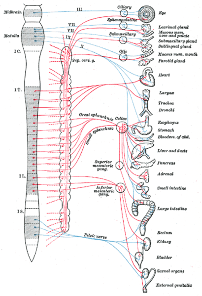
    Sacral Efferent Pathways are Sympathetic, Not Parasympathetic

    A recent report in the journal Science proposed a big change in how we understand the sympathetic and parasympathetic pathways of the autonomic nervous system (ANS).

    In a nutshell, the new model stipulates that the outflow (efferent pathways) are divided into a cranial division and spinal division—not the craniosacral and thoracolumbar divisions that we learned (and that exist in all A&P textbooks):

    Current model:
    • Craniosacral division (parasympathetic outflow)
    • Thoracolumbar division (sympathetic outflow)
    New model:
    • Cranial division (parasympathetic outflow)
    • Spinal division (sympathetic outflow)
    The authors lay out embryological and genetic phenotype evidence to show that the sacral components of the ANS outflow pathways are similar to sympathetic thoracic pathways—not to cranial parasympathetic pathways as we have long supposed. 

    But wait, you say, what about the parasympathetic control of the genitals, rectum, bladder? What about, well, all kinds of things that now seem to unravel? I suggest reading the rather brief and plainly written article in Science for the full answer. 

    However, a few quick points may reduce your blood pressure a bit—and perhaps pique your interest.

    Quick points about the new ANS model

    • Thoracic and sacral pathways share common embryologic development by location and when looking at transcriptional markers associated with neurotransmitters that differ from the developmental pattern of cranial pathways.

    • Thoracic and sacral pathways have a ventral exit point from the spinal cord; cranial pathways have a dorsal exit point.

    • The pelvic ganglion has been considered a "mixed" sympathetic/parasympathetic ganglion because it receives fibers from both the upper lumbar and sacral segments. But if the sacral pathways are sympathetic, the pelvic ganglion is clearly a sympathetic ganglion (not mixed). 

    • Analyses of transcription factors show that cells of the pelvic ganglia resemble those sympathetic ganglia and do not resemble cells in cranial ganglia.

    • The supposed lumbar vs. sacral antagonism in the urinary bladder's detrusor muscle does not seem to hold up, with the lumbar inhibitory effects either not demonstrable in experiments or of questionable functional relevance.

    • The effects on vessel dilation in genitals can be explained as a "continuity of action—rather than antagonism"

    • The sacral pathway to the rectum seems to resemble sympathetic structure, not cranial (parasympathetic) structure.

    What can we use from this in teaching undergraduate A&P?

    • When covering the craniosacral/thoracolumbar scheme, consider mentioning this newly proposed model.

    • Consider using this scenario to illustrate the dynamic nature of science. Perhaps discuss that long-held dogma is occasionally challenged using newer methods and ways of thinking.

    • Consider discussing pros and cons of adopting the new model. For example, can evidence from mice extend to all vertebrates? Which is stronger, evidence for the current model or the new model? Which model is most useful in understanding principles of ANS regulation? A little critical thinking never hurt anyone (at least not much).

    Want to know more?


    The sacral autonomic outflow is sympathetic
    • I. Espinosa-Medina, O. Saha, F. Boismoreau, Z. Chettouh, F. Rossi, W. D. Richardson, J.-F. Brunet. Science  18 Nov 2016: Vol. 354, Issue 6314, pp. 893-897 DOI: 10.1126/science.aah5454
    • Peer-reviewed research report describing this discovery, Includes an updated version of the classic diagram of sympathetic and parasympathetic pathways.
    • my-ap.us/2fNdcF3

    Neural circuitry gets rewired
    • Adameyko, I. Science 18 Nov 2016: Vol. 354, Issue 6314, pp. 833-834 DOI: 10.1126/science.aal2810
    • Companion article to the report cited above, stating that "This finding provokes a serious shift in textbook knowledge, and, as with any fundamental discovery, it brings important practical implications..." and goes on to mention of a few of the implications (e.g., how to treat bladder dysfunction).
    • my-ap.us/2gg9O8P

    The Autonomic Nervous System. Part I.
    • John Newport Langley. W. Heffer & Sons Ltd., Cambridge, 1921.80pp.
    • Classic "primary source" that codified the modern concept of the ANS. 
    • my-ap.us/2fYHt3M

    Gray's Anatomy ANS diagram
    • Henry Gray. 1918 (online edition at Bartleby)
    • Classic diagram by Henry Vandyke Carter of ANS pathways from an early edition of Gray's Anatomy.
    • my-ap.us/2fYGMaT or my-ap.us/2gcAmaW

    Monday, October 31, 2016

    Sex-Gender Differences in Medical Research

    We are only very slowly recognizing the many biological and medical differences between males and females (and masculine/feminine)—besides the obvious ones related to reproduction. There are divergent patterns in the anatomy and physiology of perhaps every body system. However, in medical research male and female subjects are often grouped together in a way that obscures those divergent patterns.

    Two "viewpoint" articles in the Journal of the American Medical Association (JAMA) today focus a light on this issue and point the way to improved—more clinically useful—medical research. Links to both articles are listed below.

    As one of the articles points out, women have been included in medical trials for only the past few decades. So there is still a lot of work to be done to shore up the database of male-female differences. But also a lot of work to be done in sorting out male-female patterns of health and disease. Then even more work in making this new knowledge part of the everyday practice medicine.

    Both articles are brief and relatively nontechnical, but when read together, they provide an important message for those of us teaching pre-clinical health professionals in A&P. That message is that we should consider introducing—then reinforcing—the notion of body-wide sex and gender differences.

    Both articles give examples of such differences, but many more are to be found elsewhere, as well. Not that we should teach every possible example in the undergraduate A&P course. However, the general concept of functional variation between males and females may be an important one to emphasize as a sub-theme in our story of the human body.


    What can we use from this in teaching undergraduate A&P? 

    • Consider making sex differences a sub-theme in your A&P course.
      • Occasionally point out examples of structural, functional, and clinical patterns of variation that differ between males and females.
        • Compare and contrast sex differences with other types of pattern variations.
        • Discuss "patterns of variability" in contrast to a strictly "binary" view.
      • Consider bringing up sex-difference research that is not yet fully supported.
        • Discuss whether more attention to sex differences across topics in scientific research might help advance this area of knowledge.
        • Discuss the opposing view that there are no clinically significant biological differences between males and females other than those related to reproduction.
      • Look for such examples in your textbook and other teaching/learning resources and point them out to your students.
      • Consider having a classroom or online discussion of this topic. 
      • Ask students to post links to articles that discuss male-female patterns of variation
        • Post to course discussion or course social media channel
        • Bring to class or email to instructor to share with class
        • Post on bulletin board

    • Bring up this issue when discussing how science is done.
      • Consider asking students what effects on public health a more thorough consideration of sex differences may produce.
      • Ask students to look at a study and ask whether sex differences were thoroughly accounted for in the methodology. Could this affect how the study is interpreted and applied in the clinic?


    Want to know more? 


    Consideration of Sex Differences in Medicine to Improve Health Care and Patient Outcomes

    • Marianne J. Legato, MD; Paula A. Johnson, MD, MPH; JoAnn E. Manson, MD, DrPH.
    • JAMA. Published online October 31, 2016. doi:10.1001/jama.2016.13995
    • One of the two articles cited in the post above.
    • my-ap.us/2f1pwEv


    Reporting Sex, Gender, or Both in Clinical Research? 

    • Janine Austin Clayton, MD; Cara Tannenbaum, MD, MS
    • JAMA. Published online October 31, 2016. doi:10.1001/jama.2016.16405
    • One of the two articles cited in the post above.
    • my-ap.us/2f1jcwM


    Let’s Talk About Sex…and Gender!

    • Amanda M. Rossi, PhD; Louise Pilote, MD, MPH, PhD
    • Circulation: Cardiovascular Quality and Outcomes. 2016; 9: S100-S101 doi: 10.1161/CIRCOUTCOMES.116.002660
    • Brief article that addresses the issue of terminology, specifically distinguishing between terms that address sex (male, female) and gender (masculine, feminine). Includes a solid list of references.
    • my-ap.us/2f1Y3Tr
    Q-angle image: OpenStax College

    Monday, October 3, 2016

    Autophagy Discovery Garners Nobel Prize

    The Nobel Assembly at Karolinska Institutet has today decided to award the 2016 Nobel Prize in Physiology or Medicine to Yoshinori Ohsumi for his discoveries of mechanisms for autophagy.

    Overview


    This year's Nobel Laureate discovered and elucidated mechanisms underlying autophagy, a fundamental process for degrading and recycling cellular components.

    The word autophagy (aw-toh-FAY-jee) originates from the Greek words auto-, meaning "self", and phagein, meaning "to eat". Thus, autophagy denotes "self eating".

    This concept emerged during the 1960's, when researchers first observed that the cell could destroy its own contents by enclosing it in membranes, forming sack-like vesicles that were transported to a recycling compartment, called the lysosome, for degradation.

    Difficulties in studying the phenomenon meant that little was known until, in a series of brilliant experiments in the early 1990's, Yoshinori Ohsumi used baker's yeast to identify genes essential for autophagy. He then went on to elucidate the underlying mechanisms for autophagy in yeast and showed that similar sophisticated machinery is used in our cells.

    Ohsumi's discoveries led to a new paradigm in our understanding of how the cell recycles its content. His discoveries opened the path to understanding the fundamental importance of autophagy in many physiological processes, such as in the adaptation to starvation or response to infection. Mutations in autophagy genes can cause disease, and the autophagic process is involved in several conditions including cancer and neurological disease.

    Degradation – a central function in all living cells


    In the mid 1950's scientists observed a new specialized cellular compartment, called an organelle, containing enzymes that digest proteins, carbohydrates and lipids. This specialized compartment is referred to as a "lysosome" and functions as a workstation for degradation of cellular constituents. The Belgian scientist Christian de Duve was awarded the Nobel Prize in Physiology or Medicine in 1974 for the discovery of the lysosome.

    New observations during the 1960's showed that large amounts of cellular content, and even whole organelles, could sometimes be found inside lysosomes. The cell therefore appeared to have a strategy for delivering large cargo to the lysosome. Further biochemical and microscopic analysis revealed a new type of vesicle transporting cellular cargo to the lysosome for degradation (Figure 1).

    Christian de Duve, the scientist behind the discovery of the lysosome, coined the term autophagy, "self-eating", to describe this process. The new vesicles were named autophagosomes.


    Figure 1: Autophagosome. Our cells have different specialized compartments. Lysosomes constitute one such compartment and contain enzymes for digestion of cellular contents. A new type of vesicle called autophagosome was observed within the cell. As the autophagosome forms, it engulfs cellular contents, such as damaged proteins and organelles. Finally, it fuses with the lysosome, where the contents are degraded into smaller constituents. This process provides the cell with nutrients and building blocks for renewal.

    During the 1970's and 1980's researchers focused on elucidating another system used to degrade proteins, namely the "proteasome". Within this research field Aaron Ciechanover, Avram Hershko and Irwin Rose were awarded the 2004 Nobel Prize in Chemistry for "the discovery of ubiquitin-mediated protein degradation". The proteasome efficiently degrades proteins one-by-one, but this mechanism did not explain how the cell got rid of larger protein complexes and worn-out organelles. Could the process of autophagy be the answer and, if so, what were the mechanisms?

    A groundbreaking experiment


    Yoshinori Ohsumi had been active in various research areas, but upon starting his own lab in 1988, he focused his efforts on protein degradation in the vacuole, an organelle that corresponds to the lysosome in human cells.

    Yeast cells are relatively easy to study and consequently they are often used as a model for human cells. They are particularly useful for the identification of genes that are important in complex cellular pathways. But Ohsumi faced a major challenge; yeast cells are small and their inner structures are not easily distinguished under the microscope and thus he was uncertain whether autophagy even existed in this organism.

    Ohsumi reasoned that if he could disrupt the degradation process in the vacuole while the process of autophagy was active, then autophagosomes should accumulate within the vacuole and become visible under the microscope. He therefore cultured mutated yeast lacking vacuolar degradation enzymes and simultaneously stimulated autophagy by starving the cells.

    The results were striking! Within hours, the vacuoles were filled with small vesicles that had not been degraded (Figure 2). The vesicles were autophagosomes and Ohsumi's experiment proved that authophagy exists in yeast cells. But even more importantly, he now had a method to identify and characterize key genes involved this process. This was a major break-through and Ohsumi published the results in 1992.


    Figure 2: Yeast. In yeast (left panel) a large compartment called the vacuole corresponds to the lysosome in mammalian cells. Ohsumi generated yeast lacking vacuolar degradation enzymes. When these yeast cells were starved, autophagosomes rapidly accumulated in the vacuole (middle panel). His experiment demonstrated that autophagy exists in yeast. As a next step, Ohsumi studied thousands of yeast mutants (right panel) and identified 15 genes that are essential for autophagy.


    Autophagy genes are discovered


    Ohsumi now took advantage of his engineered yeast strains in which autophagosomes accumulated during starvation. This accumulation should not occur if genes important for autophagy were inactivated. Ohsumi exposed the yeast cells to a chemical that randomly introduced mutations in many genes, and then he induced autophagy.

    His strategy worked! Within a year of his discovery of autophagy in yeast, Ohsumi had identified the first genes essential for autophagy. In his subsequent series of elegant studies, the proteins encoded by these genes were functionally characterized. The results showed that autophagy is controlled by a cascade of proteins and protein complexes, each regulating a distinct stage of autophagosome initiation and formation (Figure 3).


    Figure 3: Stages of autophagosome formation. Ohsumi studied the function of the proteins encoded by key autophagy genes. He delineated how stress signals initiate autophagy and the mechanism by which proteins and protein complexes promote distinct stages of autophagosome formation.


    Autophagy – an essential mechanism in our cells


    After the identification of the machinery for autophagy in yeast, a key question remained. Was there a corresponding mechanism to control this process in other organisms? Soon it became clear that virtually identical mechanisms operate in our own cells. The research tools required to investigate the importance of autophagy in humans were now available.

    Thanks to Ohsumi and others following in his footsteps, we now know that autophagy controls important physiological functions where cellular components need to be degraded and recycled.

    Autophagy can rapidly provide fuel for energy and building blocks for renewal of cellular components, and is therefore essential for the cellular response to starvation and other types of stress.

    After infection, autophagy can eliminate invading intracellular bacteria and viruses. Autophagy contributes to embryo development and cell differentiation. Cells also use autophagy to eliminate damaged proteins and organelles, a quality control mechanism that is critical for counteracting the negative consequences of aging.

    Disrupted autophagy has been linked to Parkinson's disease, type 2 diabetes and other disorders that appear in the elderly. Mutations in autophagy genes can cause genetic disease. Disturbances in the autophagic machinery have also been linked to cancer. Intense research is now ongoing to develop drugs that can target autophagy in various diseases.

    Autophagy has been known for over 50 years but its fundamental importance in physiology and medicine was only recognized after Yoshinori Ohsumi's paradigm-shifting research in the 1990's. For
    Yoshinori Ohsumi was born 1945 in Fukuoka, Japan. He received a Ph.D. from University of Tokyo in 1974. After spending three years at Rockefeller University, New York, USA, he returned to the University of Tokyo where he established his research group in 1988. He is since 2009 a professor at the Tokyo Institute of Technology.

    More background on the winner and the prize


    Yoshinori Ohsumi was born in Fukuoka, Japan, in 1945.  He is affiliated with the Tokyo Institute of Technology in Tokyo, Japan. His monetary award will be nearly one million dollars.

    The Nobel Assembly, consisting of 50 professors at Karolinska Institutet, awards the Nobel Prize in Physiology or Medicine. Its Nobel Committee evaluates the nominations. Since 1901 the Nobel Prize has been awarded to scientists who have made the most important discoveries for the benefit of mankind.his discoveries, he is awarded this year's Nobel Prize in physiology or medicine.

    Nobel Prize® is the registered trademark of the Nobel Foundation

    What can we use from this in teaching undergraduate A&P?


    • Consider using the Nobel Prizes as a discussion-starter in your class about 
      • How science influences society
      • How society influences science
      • How science progresses
      • Rewarding of science discoveries
      • What makes a discovery "important"

    • Relate this discovery to prior (or upcoming) discussions of 
      • Cell function
      • Organelle specialization
      • How cells handle protein
      • How autophagosomes work with lysosomes
      • Compare/contrast with phagocytosis
      • Compare/contrast with proteasome function and protein "quality control

    • Relate this discovery to the general idea of cellular mechanisms of disease
      • Consider taking this opportunity to emphasize "why we need to know all this" detail about cellular structure and function.

    Want to know more?


    Scientific Background Discoveries of Mechanisms for Autophagy

    • Larsson, N-G, Msucci, M. G. Nobelprize.org accessed 8 October 2016
    • A more advanced summary of the prizewinning discovery, including a handy glossary of terms.
    • my-ap.us/2dHaipm


    Honorary Professor Yoshinori Ohsumi wins Nobel Prize in Physiology or Medicine for 2016

    • Tokyo Tech News. 3 October 2016
    • Summary of biography and scientific work of the prizewinner.
    • my-ap.us/2dHbyc6


    Autophagy in yeast demonstrated with proteinase-deficient mutants and conditions for its induction.

    • Takeshige, K., Baba, M., Tsuboi, S., Noda, T. and Ohsumi, Y. (1992). Journal of Cell Biology 119, 301-311
    • One of the scientific reports of the discovery.
    • my-ap.us/2dHakO9


    Isolation and characterization of autophagy-defective mutants of Saccharomyces cervisiae. 

    • Tsukada, M. and Ohsumi, Y. (1993). FEBS Letters 333, 169-17
    • One of the scientific reports of the discovery.
    • my-ap.us/2dH9mkY


    A protein conjugation system essential for autophagy. 

    • Mizushima, N., Noda, T., Yoshimori, T., Tanaka, Y., Ishii, T., George, M.D., Klionsky, D.J., Ohsumi, M. and Ohsumi, Y. (1998). Nature 395, 395-398
    • One of the scientific reports of the discovery.
    • my-ap.us/2dHbd92

    A ubiquitin-like system mediates protein lipidation.

    • Ichimura, Y., Kirisako T., Takao, T., Satomi, Y., Shimonishi, Y., Ishihara, N., Mizushima, N., Tanida, I., Kominami, E., Ohsumi, M., Noda, T. and Ohsumi, Y. (2000).  Nature, 408, 488-492
    • One of the scientific reports of the discovery.
    • my-ap.us/2dH809y


    Honoring the 2016 Nobel laureates with free access to selections of their research

    • Elisa Nelissen Elsevier Connect. October 3, 2016
    • This blog post provides links to download the most cited papers the laureates published with Elsevier, a major publisher of scientific journals and references.
    • my-ap.us/2dH92T4

    Hot Topic in Biochemistry: Role of Autophagy in Human Health and Disease
    • Sharon Tooze. YouTube 20 December 2011
    • Brief video of webcast presentation at the Biochemical Society Hot Topic event.
    • youtu.be/0kiZdHhCtZQ


    Some content, including illustrations,
    is adapted from the press release
    and other resources at Nobelprize.org


    Wednesday, October 8, 2014

    Nobel Prize 2014: Super-resolved fluorescence microscopy


    The Royal Swedish Academy of Sciences has decided to award the Nobel Prize in Chemistry for 2014 to

    Eric Betzig
    Janelia Farm Research Campus, Howard Hughes Medical Institute, Ashburn, VA, USA,

    Stefan W. Hell
    Max Planck Institute for Biophysical Chemistry, Göttingen, and German Cancer Research Center, Heidelberg, Germany

    and

    William E. Moerner
    Stanford University, Stanford, CA, USA

    “for the development of
    super-resolved fluorescence microscopy”


    Surpassing the limitations of the light microscope


    For a long time optical microscopy was held back by a presumed limitation: that it would never obtain a better resolution than half the wavelength of light. Helped by fluorescent molecules the Nobel Laureates in Chemistry 2014 ingeniously circumvented this limitation. Their ground-breaking work has brought optical microscopy into the nanodimension.

    In what has become known as nanoscopy, scientists visualize the pathways of individual molecules inside living cells. They can see how molecules create synapses between nerve cells in the brain; they can track proteins involved in Parkinson’s, Alzheimer’s and Huntington’s diseases as they aggregate; they follow individual proteins in fertilized eggs as these divide into embryos.

    It was all but obvious that scientists should ever be able to study living cells in the tiniest molecular detail. In 1873, the microscopist Ernst Abbe stipulated a physical limit for the maximum resolution of traditional optical microscopy: it could never become better than 0.2 micrometres. Eric Betzig, Stefan W. Hell and William E. Moerner are awarded the Nobel Prize in Chemistry 2014 for having bypassed this limit. Due to their achievements the optical microscope can now peer into the nanoworld.

    Two separate principles are rewarded. 


    One enables the method stimulated emission depletion (STED) microscopy, developed by Stefan Hell in 2000. Two laser beams are utilized; one stimulates fluorescent molecules to glow, another cancels out all fluorescence except for that in a nanometre-sized volume. Scanning over the sample, nanometre for nanometre, yields an image with a resolution better than Abbe’s stipulated limit.

    Eric Betzig and William Moerner, working separately, laid the foundation for the second method, single-molecule microscopy. The method relies upon the possibility to turn the fluorescence of individual molecules on and off. Scientists image the same area multiple times, letting just a few interspersed molecules glow each time. Superimposing these images yields a dense super-image resolved at the nanolevel. In 2006 Eric Betzig utilized this method for the first time.

    Today, nanoscopy is used world-wide and new knowledge of greatest benefit to mankind is produced on a daily basis.


    This video is a brief animation of how STED works and how it improves resolution of individual particles.


    This video is a longer, more detailed presentation by one of the Nobel laureates (Hell).


    What can we use from this in teaching undergraduate A&P?

    • Discuss how this technology has enabled us to better visualize the chemicals and structures within our cells, enabling scientists to better understand the structure and function of cell, organelles, microbiome constituents, and other structures of the human body.

    • If you do a brief run-through of the theory of microscopy—perhaps in your A&P lab—you can add a mention of this technology.  

    • Your textbook or other learning resource may already have an example of this type of microscopy.

    • A discussion of this  Nobel Prize could evolve into a meaningful example of how science works, including how incremental improvements in classical tools for observation expand the number of questions that can be answered.

    • Use the links below (and images above) to use for a handout and/or teaching slides.


    Want to know more?


    Resources from Nobelprize.org

    • Popular Information 
    • Scientific Background
      • Handout: More detailed information includes references to original research articles
      • my-ap.us/ZdLJ69
    • Advanced Information
    • Images
    • Biographies
      • Eric Betzig, 
        • U.S. citizen. Born 1960 in Ann Arbor, MI, USA. Ph.D. 1988 from Cornell University, Ithaca, NY, USA. Group Leader at Janelia Farm Research Campus, Howard Hughes Medical Institute, Ashburn, VA, USA.
        • http://janelia.org/lab/betzig-lab
      • Stefan W. Hell, German citizen. 
        • Born 1962 in Arad, Romania. Ph.D. 1990 from the University of Heidelberg, Germany. Director at the Max Planck Institute for Biophysical Chemistry, Göttingen, and Division head at the German Cancer Research Center, Heidelberg, Germany.
        • http://www3.mpibpc.mpg.de/groups/hell
      • William E. Moerner, U.S. citizen. 
        • Born 1953 in Pleasanton, CA, USA. Ph.D. 1982 from Cornell University, Ithaca, NY, USA. Harry S. Mosher Professor in Chemistry and Professor, by courtesy, of Applied Physics at Stanford University, Stanford, CA, USA.
        • http://web.stanford.edu/group/moerner


    Diagrram cretit: Ganbaatar
    Micrograph credit: Tesselkaffee
    Text adapted from press release from Nobel Media

    Monday, October 6, 2014

    Nobel Prize 2014: The Brain's Positioning System


    The Nobel Assembly at Karolinska Institutet has today decided to award

    The 2014 Nobel Prize in Physiology or Medicine

    with one half to

    John O´Keefe

    and the other half jointly to

    May-Britt Moser and Edvard I. Moser

    for their discoveries of cells that constitute a positioning 
    system in the brain.


    How do we know where we are? How can we find the way from one place to another? And how can we store this information in such a way that we can immediately find the way the next time we trace the same path? This year´s Nobel Laureates have discovered a positioning system, an “inner GPS” in the brain that makes it possible to orient ourselves in space, demonstrating a cellular basis for higher cognitive function.

    In 1971, John O´Keefe discovered the first component of this positioning system. He found that a type of nerve cell in an area of the brain called the hippocampus that was always activated when a rat was at a certain place in a room. Other nerve cells were activated when the rat was at other places. O´Keefe concluded that these “place cells” formed a map of the room.

    More than three decades later, in 2005, May-Britt and Edvard Moser discovered another key component of the brain’s positioning system. They identified another type of nerve cell, which they called “grid cells”, that generate a coordinate system and allow for precise positioning and pathfinding.

    Their subsequent research showed how place and grid cells make it possible to determine position and to navigate.

    The discoveries of John O´Keefe, May-Britt Moser and Edvard Moser have solved a problem that has occupied philosophers and scientists for centuries – how does the brain create a map of the space surrounding us and how can we navigate our way through a complex environment?


    How do we experience our environment?


    The sense of place and the ability to navigate are fundamental to our existence. The sense of place gives a perception of position in the environment. During navigation, it is interlinked with a sense of distance that is based on motion and knowledge of previous positions.

    Questions about place and navigation have engaged philosophers and scientists for a long time. More than 200 years ago, the German philosopher Immanuel Kant argued that some mental abilities exist as a priori knowledge, independent of experience. He considered the concept of space as an inbuilt principle of the mind, one through which the world is and must be perceived. With the advent of behavioural psychology in the mid-20th century, these questions could be addressed experimentally. When Edward Tolman examined rats moving through labyrinths, he found that they could learn how to navigate, and proposed that a “cognitive map” formed in the brain allowed them to find their way. But questions still lingered - how would such a map be represented in the brain?


    John O´Keefe and the place in space


    John O´Keefe was fascinated by the problem of how the brain controls behaviour and decided, in the late 1960s, to attack this question with neurophysiological methods. When recording signals from individual nerve cells in a part of the brain called the hippocampus, in rats moving freely in a room, O’Keefe discovered that certain nerve cells were activated when the animal assumed a particular place in the environment (Figure 1). He could demonstrate that these “place cells” were not merely registering visual input, but were building up an inner map of the environment. O’Keefe concluded that the hippocampus generates numerous maps, represented by the collective activity of place cells that are activated in different environments. Therefore, the memory of an environment can be stored as a specific combination of place cell activities in the hippocampus.


    May-Britt and Edvard Moser find the coordinates


    May-Britt and Edvard Moser were mapping the connections to the hippocampus in rats moving in a room when they discovered an astonishing pattern of activity in a nearby part of the brain called the entorhinal cortex. Here, certain cells were activated when the rat passed multiple locations arranged in a hexagonal grid (Figure 2). Each of these cells was activated in a unique spatial pattern and collectively these “grid cells” constitute a coordinate system that allows for spatial navigation. Together with other cells of the entorhinal cortex that recognize the direction of the head and the border of the room, they form circuits with the place cells in the hippocampus. This circuitry constitutes a comprehensive positioning system, an inner GPS, in the brain (Figure 3).


    A place for maps in the human brain


    Recent investigations with brain imaging techniques, as well as studies of patients undergoing neurosurgery, have provided evidence that place and grid cells exist also in humans. In patients with Alzheimer´s disease, the hippocampus and entorhinal cortex are frequently affected at an early stage, and these individuals often lose their way and cannot recognize the environment. Knowledge about the brain´s positioning system may, therefore, help us understand the mechanism underpinning the devastating spatial memory loss that affects people with this disease.

    The discovery of the brain’s positioning system represents a paradigm shift in our understanding of how ensembles of specialized cells work together to execute higher cognitive functions. It has opened new avenues for understanding other cognitive processes, such as memory, thinking and planning.


    What can we use from this in teaching undergraduate A&P?

    • The role of the hippocampus and it's place cells could be briefly discussed as you explore the cognitive functions of the brain.

    • A discussion of this year's Nobel Prize could evolve into a meaningful example of how science works, including the use of discoveries in animals that can later be applied to learning more about human structure and function.

    • Information revealed by this discovery could be discussed when discussing human disorders, such as Alzheimer disease, that involve impairments of spatial orientation and/or place memory.


    Want to know more?

    Here's a video in which May-Britt Moser and Edvard I. Moser explain their research, which you can use in your course discussion.


    2014 Nobel Prize announcement

    • Official announcement, which includes photos of the laureates and links to related information.
    • my-ap.us/1oJH82y


    Scientific Background: The Brain’s Navigational Place and Grid Cell System

    • Nobelprize.org accessed 6 October 2014
    • Plain English hand-out that can be used with your students.  Illustrated with clear diagrams.
    • my-ap.us/1pGLVlm

    Nobel Prize in Medicine Is Awarded for Discovery of Brain’s ‘Inner GPS’

    • L. Altman The New York Times. OCT. 6, 2014
    • Plain English article summarizing the discoveries.
    • my-ap.us/10ELy5h

    The hippocampus as a spatial map. Preliminary evidence from unit activity in the freely‐moving rat.

    • O'Keefe, J., and Dostrovsky, J. (1971). Brain Research 34, 171-175.
    • Original research paper describing key discoveries for which this prize is given.
    • my-ap.us/1s4fi5Z


    Place units in the hippocampus of the freely moving rat.

    • O´Keefe, J. (1976). Experimental Neurology 51, 78-109.
    • Original research paper describing key discoveries for which this prize is given.
    • my-ap.us/1s2NYGd


    Spatial representation in the entorhinal cortex.

    • Fyhn, M., Molden, S., Witter, M.P., Moser, E.I., Moser, M.B. (2004)  Science 305, 1258-1264.
    • Original research paper describing key discoveries for which this prize is given.
    • my-ap.us/1BIvdI4


    Microstructure of spatial map in the entorhinal cortex.

    • Hafting, T., Fyhn, M., Molden, S., Moser, M.B., and Moser, E.I. (2005). Nature 436, 801-806.
    • Original research paper describing key discoveries for which this prize is given.
    • my-ap.us/1nX0jLe


    Article adapted from Nobel Media press release

    Monday, August 25, 2014

    Use Eponyms with Style!

    Paul Langerhans

    I love eponyms!

    I'm a bit sad that eponyms—terms that include a proper name—are going out of style in the world of human sciences, it seems.

    The international lists of anatomic terminology recommend against most eponyms, providing descriptive terms in their place.  For example, pancreatic islet is the term  preferred to the eponym islet of Langerhans.  Osteon is preferred over haversian system.

    Of course, I get that.  Descriptive terms are more intuitive and therefore easier to understand, learn, and remember. Related to that is that they are more accurate when it comes to medical applications.  And I'm all about accuracy in the medical professions.

    It's just kind of fun using eponyms.  And kind of sad to feel like I am leaving behind all those wonderful women and men who discovered our parts way back when.

    But it's not just the international lists of anatomy that are leaving eponyms behind—all the "authorities," including most A&P and medical textbooks are doing it. Professional societies, associations, and boards in the basic sciences and the health professions are doing it.  So if we want to be "in style" with our terminology—and more importantly, make sure our students are sporting the latest linguistic style—we'd better pay attention to the trends, eh?

    Let me give you some unasked-for fashion tips if you want to be a stylish A&P professor:

    Avoid eponyms

    If there's an accepted descriptive term, it's best to use that rather than the eponym.

    Be bilingual

    Some folks you'll encounter are old-fashioned or possibly don't know the newer descriptive term.  Or they know both and use them interchangeably.  Because we're on the cusp of a fashion revolution here, the most competent professionals will know both and be able to switch back and forth easily as the context requires. The goal is to understand and be understood, right?

    What to do if you have to use an eponym

    Then use an eponym! There are some commonly used terms for which there really isn't a great descriptive term to replace an eponym.  For example, Parkinson disease, Alzheimer disease, and other disorders often don't have a widely accepted alternative. So absolute avoidance of eponyms is not (yet) possible.

    Fashionable uses of eponyms

    If you must use an eponym, the trendy folks at AMA and elsewhere avoid the use of possessive forms.  For example, notice how I used the term Parkinson disease above and not Parkinson's disease?  It's better to use Down syndrome than Down's syndrome—and even better to use trisomy 21 syndrome.

    Likewise, the possessive loop of Henle is out of favor but using Henle loop may still get you into most of the trendy clubs.

    Gabriele Falloppio
    Illusory eponym styles

    When you try to get away with using a possessive form of an eponym and yet still avoid arrest by the fashion police, you have to be very cunning.  Here's a common way that's done: use the adjective form of a proper name.  So if you want to honor Gabriele Falloppio's work in describing uterine tubes, then use his Latinized name (Fallopius) in the form of an adjective and call them Fallopian tubes.

    But, you may say, that's not a very clever masking of the fact that it's an eponym. In fact, it's pretty obvious, right?  Well here's the sly part: cover it up by using a lowercase letter—thus obscuring the fact that it incorporates a proper noun. That's why many sources use fallopian tube instead of Fallopian tube.

    Terms like eustachian tube and haversian canal may not seem like well-hidden eponyms in our context here, where we're actually focusing on eponyms. But most grand stage illusions—like Blackstone's making an elephant appear on stage from thin air—rely on such subtle misdirection.

    So when you are using an adjective form of an eponym, it's best not to capitalize it and risk possible arrest by the fashion police.

    Fashion rules are not really rules

    With any fashion, the "rules" are not usually rules in the formal sense.  They are simple formulations of trends that, if heeded, will likely save you some embarrassment when you don't appear to be cool.  So if you have a good reason—or even a lame reason—to ignore these rules, I think you'll probably survive. People laugh (even hoot) at my disregard for current clothing fashion all the time—you get used to it.

    OK, sometimes fashion rules really are rules

    As with any professional communication, sometimes fashion rules get set in stone in a required style to which you must adhere in your work.  Sort of like a dress code for words. For example, students learn how to use professional styles when we require that they submit their assignments in APA, Chicago, or CBE style. Likewise, in publishing journal articles, books, and other works, there are rules established that provide consistency—and, therefore, also accuracy.  Each publisher, sometimes each journal or textbook, has it's own house style that defines such things. So if your journal editor insists on Eustachian tube instead of auditory tube or eustachian tube, then I recommend doing it.  Retro, in some contexts, can be cool.

    What can we use from this in teaching undergraduate A&P?


    • By modeling current styles in the use of  scientific terminology, we teach an important lesson when learning any language—how it's actually used out there on the streets.
    • Consider taking a moment every once in a while to explain why the eponym your are using—or avoiding—is widely used.  Or why it's different than what they may be reading or hearing elsewhere. This may make them more likely to listen (and think) more carefully in their professional communications.
    • Consider using this information to explain why you want your students to know more than one alternate term—you are preparing them to be nimble in their clinical experiences and professional reading.
    • Many students aren't competent in using different possessive forms and perhaps don't even know what an eponym is.  Perhaps we can help them gain such competence.


    Monday, August 4, 2014

    Forensic Anthropologist Clyde Snow Passes


    I just learned that the renowned forensic anthropologist Clyde Snow passed away this past May at the age of 86.

    Famous for identifying the remains of Nazi war criminal Josef Mengele and victims of mass murderer John Wayne Gacy, Snow was one of a generation of pioneers who sparked the current popular interest in forensic sciences.

    What can we use from this in teaching undergraduate A&P?  Some of stories of his achievements (see links below) can help spark interest in studying the human skeleton and other systems.  And some of his familiar quotes are worth using in the lab or classroom:

    • "There are 206 bones and 32 teeth in the human body, and each has a story to tell."
    • "Bones can be puzzles, but they never lie, and they don't smell bad."
    • "The ground is like a beautiful woman—if you treat her gently, she'll tell you all her secrets."
    • "If you can make people feel they're not going to get away with it—that's all we're asking." (regarding using forensic science to solve crimes)

    Want to know more?

    Clyde Snow, forensic anthropologist who helped ID victims of John Wayne Gacy, dies at 86
    • M. Weil Washington Post May 18, 2014
    • Article summarizing Snow's work, including several stories that could be used in an A&P class discussion.
    • my-ap.us/1qDQMVV

    Science Detectives: The Masters Case
    • Discovery Channel January 22, 1997
    • Reenactment of forensic fieldwork in a killing that occurred decades before the victim's bones were discovered.
    • my-ap.us/U7DKoO

    Image credit: Toony