Showing posts with label animation. Show all posts

Wednesday, October 4, 2017

Biomolecule Imaging Pioneers Share Nobel Prize

Today, the Royal Swedish Academy of Sciences has decided to award the Nobel Prize in Chemistry 2017 to Jacques Dubochet (University of Lausanne, Switzerland) and Joachim Frank (Columbia University, New York, USA), and Richard Henderson (MRC Laboratory of Molecular Biology, Cambridge, UK). The award is given "for developing cryo-electron microscopy for the high-resolution structure determination of biomolecules in solution"

Cool microscope technology revolutionises biochemistry

We may soon have detailed images of life’s complex machineries in atomic resolution. The Nobel Prize in Chemistry 2017 is awarded to Jacques Dubochet, Joachim Frank and Richard Henderson for the development of cryo-electron microscopy, which both simplifies and improves the imaging of biomolecules. This method has moved biochemistry into a new era.

A picture is a key to understanding. Scientific breakthroughs often build upon the successful visualization of objects invisible to the human eye. However, biochemical maps have long been filled with blank spaces because the available technology has had difficulty generating images of much of life’s molecular machinery. Cryo-electron microscopy changes all of this. Researchers can now freeze biomolecules mid-movement and visualize processes they have never previously seen, which is decisive for both the basic understanding of life’s chemistry and for the development of pharmaceuticals.

Electron microscopes were long believed to only be suitable for imaging dead matter, because the powerful electron beam destroys biological material. But in 1990, Richard Henderson succeeded in using an electron microscope to generate a three-dimensional image of a protein at atomic resolution. This breakthrough proved the technology’s potential.

Joachim Frank made the technology generally applicable. Between 1975 and 1986 he developed an image processing method in which the electron microscope’s fuzzy two-dimensional images are analysed and merged to reveal a sharp three-dimensional structure.

Jacques Dubochet added water to electron microscopy. Liquid water evaporates in the electron microscope’s vacuum, which makes the biomolecules collapse. In the early 1980s, Dubochet succeeded in vitrifying water – he cooled water so rapidly that it solidified in its liquid form around a biological sample, allowing the biomolecules to retain their natural shape even in a vacuum.

Following these discoveries, the electron microscope’s every nut and bolt have been optimised. The desired atomic resolution was reached in 2013, and researchers can now routinely produce three-dimensional structures of biomolecules. In the past few years, scientific literature has been filled with images of everything from proteins that cause antibiotic resistance, to the surface of the Zika virus. Biochemistry is now facing an explosive development and is all set for an exciting future.

About the Nobel Laureates

Jacques Dubochet, born 1942 in Aigle, Switzerland. Ph.D. 1973, University of Geneva and University of Basel, Switzerland. Honorary Professor of Biophysics, University of Lausanne, Switzerland.
www.unil.ch/dee/en/home/menuinst/people/honorary-professors/prof-jacques-dubochet.html

Joachim Frank, born 1940 in Siegen, Germany. Ph.D. 1970, Technical University of Munich, Germany. Professor of Biochemistry and Molecular Biophysics and of Biological Sciences, Columbia University, New York, USA.
http://franklab.cpmc.columbia.edu/franklab/

Richard Henderson, born 1945 in Edinburgh, Scotland. Ph.D. 1969, Cambridge University, UK. Programme Leader, MRC Laboratory of Molecular Biology, Cambridge, UK.
www2.mrc-lmb.cam.ac.uk/groups/rh15/


What can we use from this in teaching undergraduate A&P?


  • If you talk about imaging molecules in your course, this could be a way to garner student interest—considering that this is a current and ongoing effort in science. I always have a brief "shape is important in biological chemistry and here's what we can see with current tools" because they're going to see all those little odd-shaped rutabaga blobs in illustrations in their textbooks.

  • If you bring up microscopy in your course, perhaps describing the types of microscopy, adding a bit of info on this could help show students that microscopy is still evolving—in exciting ways.

  • Consider using the annual Nobel Prize announcements as a springboard to discuss the process of scientific discovery. 

  • Consider mentioning the other major awards for scientific achievement and discuss what the judges seem to value most about scientific discoveries. The Nobel Prize is the one everyone has heard of, so it's a great place to start.

  • Use the Nobel Prizes (and other awards) over time as a way to keep students aware of the history of, and progress, of human biology. One could also address the global diversity of laureates.  Or the lack of other kinds of diversity among laureates.

Want to know more?

Popular Information 


Scientific Background


Images

Image - 3D structures (pdf 1.4 MB)


Image - Blobology (pdf 8.5 MB)


Image - Dubochet's preparation method (948 kB)


Image - Frank's image analysis (pdf 1 MB)


Cool Animations (literally)

Structure and gating of the nuclear pore complex

Ion gating in the sarcoplasmic reticulum membrane

Antibody structure

Native LDL particles
  • Kumar V, Butcher S, Öörni K, Engelhardt P, Heikkonen J, Kaski K, Ala-Korpela M, Kovanen P
  • my-ap.us/2hO4Qms

Changes in the water and ion contents of organelles during apoptosis
  • Nolin F, Michel J, Wortham L, Tchelidze P, Banchet V, Lalun N, Terryn C, Ploton D
  • my-ap.us/2hMTYW4
Adapted from press release at nobelprize.org
Click each image for its source/attribution


Friday, June 26, 2015

There May Not Be a Single Language Comprehension Center in the Brain

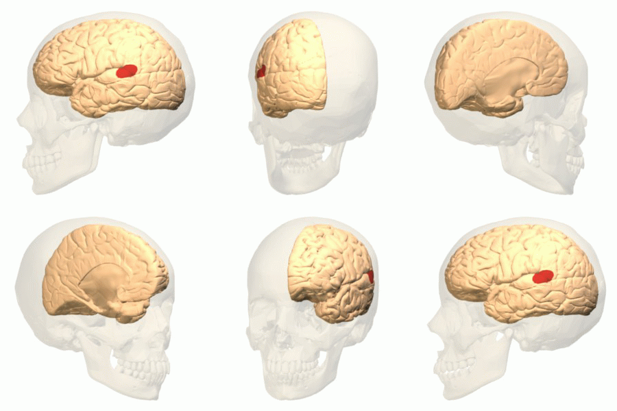
A recent paper in the journal Brain, a journal of neurology, challenges the long-held notion that the sensory speech area of the left temporal lobe of the cerebrum—often called the Wernicke area—acts as the center of language comprehension.

Wernicke area
The report describes research that leads one to the conclusion that although the Wernicke area (pictured) apparently has a role in understanding individual words, the task of sentence comprehension is accomplished by a complex network of diverse areas of the cerebrum. These include, "temporoparietal components of Wernicke’s area, Broca’s area, and [the] dorsal premotor cortex."


What can we use from this in teaching undergraduate A&P?


  • When discussing the sensory speech (Wernicke) area, one might bring up this discovery and how it may modify our description of its main function.

  • This discovery is a good example of how science works—how theories are formed and how they are modified as new information is discovered.  And how new research must be confirmed before it becomes widely established as fact.

  • When discussing cerebral localization of function, this story may be helpful in emphasizing that complex cerebral functions often involve integration of diverse cortical areas.

  • This story is a good example of our rapidly advancing knowledge of complex brain function.

  • You'll sound very "with it" when you can drop in casual asides about the latest brain research in your classroom discussions!

Want to know more?


New Human Brain Language Map | Researchers find that Wernicke’s area, thought to be the seat of language comprehension in the human brain for more than a century, is not.
  • Bob Grant. The Scientist  Published online June 26, 2015
  • Brief, plain-English article summarizing the recent findings.
  • my-ap.us/1QTTN5z 

The Wernicke conundrum and the anatomy of language comprehension in primary progressive aphasia 
  • M-Marsel Mesulam , et al. Brain. DOI: http://dx.doi.org/10.1093/brain/awv154 First published online: 25 June 2015
  • The original research article.
  • my-ap.us/1QTWPXw

Scientists redraw traditional brain map of language comprehension
  • Northwestern University press release. Published online June 25, 2015
  • my-ap.us/1QTYpsl


Cortical Areas Involved in Speech Processing


Image credits: Database Center for Life Science (Wernicke area)
Leuthardt, et al. (cortical speech areas)

Monday, June 15, 2015

Virtual Cardiology Lab


Looking for a supplemental hands-on activity with cardiology in your course?

Try the FREE online interactive Cardiology Virtual Lab from the Howard Hughes Medical Institute.

It covers these concepts:
  • Symptoms of a selection of heart diseases, to serve as examples of what kinds of things can go wrong with the heart. 
    • How are symptoms detected and why?
  • Tools and techniques used for diagnosis. 
    • What can the different techniques detect and how do they work?
  • Principles of pedigree analysis.


What can we use from this in teaching undergraduate A&P?


  • Link to this virtual lab activity from your online syllabus, course web page, or LMS (or in an email  or tweet to students)
  • If you want to give course points for the lab, consider an online quiz or lab report submitted through your learning management system (LMS) or emailed to you.
  • Gives students a "real life" clinical lab application for the concepts they are learning in A&P.
  • Provides a cardiology lab option for online/hybrid courses or wet labs that don't have funding for cardiology experiments.
  • Helps integrate principles of genetics with cardiology, so it can be used in your genetics unit.


Want to know more?


Cardiology Virtual Lab

  • BioInteractive. Howard Hughes Medical Institute. Accessed 16 Sep 2014.
  • This virtual lab will familiarize you with heritable diseases of the heart. Learn about the diagnostic tools used to examine and diagnose patients.
  • my-ap.us/1qKWiK7

BioInteractive Virtual Labs

  • BioInteractive. Howard Hughes Medical Institute. Accessed 16 Sep 2014.
  • List of all the FREE virtual labs offered by HHMI's BioInteractive project.
  • my-ap.us/1wAX92q

Cardiovascular Topics

  • Kevin Patton. The A&P Professor. Various dates.
  • List of previous blog posts on cardiovascular topics
  • my-ap.us/1uEGngu

Monday, October 13, 2014

RNA Interference. Again.


Five years ago, I extolled the virtues of teaching a little bit about RNA interference (RNAi) in undergraduate A&P courses.  But for a while it looked like the promise of RNAi in basic and clinical research might be sputtering.  However, a recent article by Eric Bender called The Second Coming of RNAi shows that RNAi "the gene-silencing technique [now] begins to fulfill some of its promises."

I recommend reading the entire article at my-ap.us/1BbxvB9  Before you read it, allow me to reprise my reasons of five years ago supporting my proposal to include RNAi in your course.

What can we use from this in teaching undergraduate A&P?


  • RNAi plays a role in defending our cells against viruses by stopping viral genetic code from being translated in host cells

  • RNAi likely plays a role in regulating gene activity in a cell by preventing translation of the gene product(s)

  • RNAi is increasingly used as method for "knocking out" a particular gene's effects in research animals in order to study the gene's functions

  • RNAi is being used to treat genetic disease. . . an application that will likely expand greatly over the next few decades
I'll add two more items to my previous list:
  • RNA interference is a mechanism of human disease, as has been demonstrated in some cases of inherited progressive hearing loss (for example).

  • Learning about RNAi helps clarify a general understanding of the many roles played by RNA in our lives—some perhaps still undiscovered.

I'm not sure that it's useful to expect beginning undergraduate students to learn the nitty-gritty details of RNAi mechanisms.  But I do think it's valuable to be exposed to the general concept of RNA interference and gene silencing.  A&P students are going to run up against these eventually as they learn about and then administer RNAi-based therapies, after all.  And perhaps we should prepare them.

Want to know more?


The Second Coming of RNAi
  • Eric Bender. The Scientist. September 1, 2014
  • Article mentioned above. In plain English, it shows that clinical progress in RNAi therapy against liver diseases, the gene-silencing technique begins to fulfill some of its promises. Includes useful illustrations and links to other resources.
  • my-ap.us/1BbxvB9

Why do we need to know about RNA interference?
  • Kevin Patton. The A&P Professor. 14 April 2009
  • My first article promoting the idea of teaching RNAi in the A&P course.  It links to an expanded article with additional teaching resources.
  • my-ap.us/1xbq4v6

RNA interference revisited
  • Kevin Patton. The A&P Professor. 9 June 2009
  • Brief follow-up article that references the role of RNA interference as a mechanism of human disease.  Links to other resources.
  • my-ap.us/1oDflzw

RNA Interference Animation and Slideshow
  • Nature Reviews Genetics. Accessed 3 September 2014
  • FREE animation, slideshow, and poster on RNAi, as well as a link to more details.
  • my-ap.us/1roNGYm

RNA Interference BioInteractive
  • Howard Hughes Medical Institute. Accessed 3 September 2014
  • FREE slideshow with worksheet that students fill out as they view the slideshow.  Links to FREE DVD from HMMI called The Double Life of RNA.
  • my-ap.us/1pJ9r5V


Monday, September 15, 2014

Updated Cell Transport Slides


Many longtime readers of this blog know that I have a set of animated PowerPoint-compatible slides available for you to use FREE in your A&P classes.  These slides—the Lion Den Slide Collection— supplement the publisher-supplied slides or homegrown slides that you are already using.

I recently updated, improved, and expanded the set of slides that animate several key cell transport processes such as diffusion, osmosis, endocytosis, etc.

If you've accessed the Lion Den Slide Collection in the last several months, you've already registered in the system and probably have already received an email notification of the update.

If you're new to the collection (or accessed it before June 2014) then you need to go to the Lion Den Slide Collection page and click on the link to the form.  It takes a few steps, but by filling out the brief forms you register for a service that will notify you (if you want it) of any updates or additions to the collection.

Here is a preview the newly updated Membrane Transport Animations
Click image for download

When you download the linked file, you'll be able to play it in PowerPoint or any other program that plays "show" or PPTSX files.  The slides can only be viewed, not edited or added to your own slide deck.  To do that, you must download the fully editable PPTX files from the Lion Den Slide Collection

Once you download any slide deck from the Lion Den Slide Collection, you can mix and match them to blend them with your existing presentations.  You can also alter the content or timings to suit your needs.  

I'm hoping you'll also get some ideas from them to create new additional slides!  If you modify, create, or already have any slides (for which you own the content) that you want to place in the collection for sharing, please contact me directly. 

Want to know more?


Presentation Zen
  • Kevin Patton The Electronic Professor 6 August 2014
  • The zen approach to presentations.  Includes a video and links to resources.
  • my-ap.us/1mZjY7e

Are your students dodging bullets?
  • Kevin Patton. The Electronic Professor. 28 July 2011
  • My blog article on improving slide presentations.  With illustrated examples.
  • my-ap.us/1ni8c72

Handling bullets safely
  • Kevin Patton. The Electronic Professor.  3 August 2012
  • Another of my blog articles on improving slide presentations. Includes my own video on how to trim down those wordy slides to something that actually works in a slide.
  • my-ap.us/1ooEced

Monday, August 18, 2014

Human Microbial System


A recent article in The Scientist once again reminds us of the ongoing explosion in the scientific understanding of the human microbial system.  In a few short years, this area of exploration has moved to the forefront of medical and basic science research in human biology.

I think it's becoming clear that the most useful way to think of human body function is to recognize that an "organism" is really a sort of "habitat."  And like any habitat, it functions best when all the inhabitants are within a limited range of balanced relationships.

Who are the inhabitants?  Besides our own cells?  Well, one could think of mitochondria and cilia and other organelles as symbiotic internal inhabitants of our cells.  They're not that literally, of course, but I think its a useful metaphor for understanding the human body.  Then there are the many microbes and animals that cover our internal and external surfaces, burrow into some of our pores and glands, and inhabit our body fluids.

I call the balanced functional relationship among the various microbomes of the body and our own tissues the human microbial system.  And I am certain that it won't be long before we will be discussing this system alongside the major organ systems of the body.  That is if we truly want to understand how the body really works.

The article in The Scientist I mention is a great summary of some of the major roles that the human microbial system plays in the human body—and a good survey of some of the areas of the body where the human-microbial functional relationships play out.  See the link to the article below.


What can we use from this in teaching undergraduate A&P?


  • Why not introduce the concept of the human microbial system at the beginning of our A&P course, when we set the stage by explain how scientists understand the body and its functions as an integrated system of different parts?
  • We can mention the different microbiomes of the body when we explore each organ system where they play an important role—which is pretty much all of them!
  • Consider discussing what happens to normal human function when microbiomes get out of balance.  For example, in the gut a microbial imbalance can lead to ulcers, diarrhea, and other dysfunctions.  On the skin a pathogenic microbe may become dominant and cause a rash.
  • Promote a discussion of what kinds of wellness strategies might be employed to prevent microbial imbalances.
  • Our students can leave our A&P course with an up-to-date understanding of human biology that will help them understand new clinical concepts and treatment strategies.

Want to Know More?

The Body’s Ecosystem

  • By The Scientist Staff.  The Scientist. August 1, 2014
  • Plain-English article (cited above) on how research on the human microbiome is booming, and scientists have moved from simply taking stock of gut flora to understanding the influence of microbes throughout the body.
  • my-ap.us/1vgOu5y

Articles from The A&P Professor


Moving pictures of the human microbiome

  • J Gregory Caporaso et al. Genome Biology 2011, 12:R50  doi:10.1186/gb-2011-12-5-r50
  • Open-access journal article that includes FREE videos that show how dynamic the human microbial system is
  • my-ap.us/V7St3Q

Human Microbiome Project


The Microbiome and Disease

  • List of diseases associated with microbiome imbalances from Genetic Science Learning Center
  • my-ap.us/1nDpxr1

Audio

  • Radio stories from National Public Radio on human microbiomes and their role in health and disease.  The growing number of these stories tells us something as A&P teachers: maybe we better be covering this!
  • my-ap.us/1uA7Qyg

Wednesday, June 13, 2012

Want to fold some proteins?

In a previous post, I proposed that A&P students should be aware of the basic elements of protein folding.  To follow up, I'd like to mention a interesting phenomenon related to protein folding and "citizen science" using an online game called Foldit.  


The Foldit game is an online puzzle game in which anybody can try their hand and finding which way a given protein folds most efficiently.  Interestingly, this has proven to yield useful results for biochemists not obtainable by traditional methods.

You may want to mention the Foldit game to students.  I've already posted it at my blog The A&P Student.

Want to know more?
Online Gamers Achieve First Crowd-Sourced Redesign of Protein
Jessica Marshall & Nature magazine
Scientific American Online January 22, 2012
[Brief article about recent redesign of a protein by online gamers using Foldit. Original paper published in Nature Biotechnology]
my-ap.us/wRK2bV

Foldit Online Protein Puzzle
Scientific American Citizen Science accessed 23 January 2012
[Brief description of the online game Foldit and the goals of the project.]
my-ap.us/zIV75F

Foldit - Solve Puzzles for Science
[Direct link to portal for the game Foldit]
my-ap.us/wfRQPF


Tuesday, May 22, 2012

Why bother with protein folding?

For those of you who use (or refer to) my textbooks, you may notice that I've been gradually adding more and more coverage of protein folding to most of them. My newest text (due out in March) adds a bit more to the story. Why bother?  Isn't that way more than beginning students need to know for an A&P course preparing students for health careers?

I submit that beginning A&P students should know a bit about protein folding.

Knowing the very basic principles of protein folding help students visualize the complex shape of proteins.  That, in turn, helps them understand that "it's all about shape" when trying to understand how proteins like enzymes, receptors, and most other proteins work—proteins that they'll encounter many times throughout their A&P course and beyond. 

Besides that, protein folding has become a key concept in understanding not only how the body functions, but how to intervene therapeutically in important diseases.  If a class of therapy based on protein folding is now being developed, a class of therapy that many of our students will likely encounter in their professions, don't we owe it to them to cover the basic ideas of protein folding? 

This latest idea was brought up at a recent meeting of the American Society of Cell Biology (ASCB).  You may want to read the article below, which briefly summarizes some current work being done in developing drugs that affect protein folding systems.  None of the specific  information in the article would be appropriate for A&P students to learn.  But reading it will give the A&P professor better insights about why the concept of protein folding is important for students to learn.  And it gives you a chance to say, "I was just reading about how scientists are now developing drugs based on protein folding . . . " to get their attention in class!

Want to know more?
Protein Folding and Disease: The Path from Bench to Bedside
V. Glaser
Genetic Engineering & Biotechnology News 15 Jan 2012. Vol. 32, No. 2
[Brief, illustrated article that clearly summarizes some recent work in applying principles of protein folding to drug therapies.]
my-ap.us/wFh6F2
Protein folding animation
[Interesting Quicktime animation that shows a protein folding]
my-ap.us/xzWnW4

GCSF Protein Folding Illustration Movie
[Another animation, a bit more complex than the previous one.  Clearly shows different types of models used in most A&P textbooks.]
my-ap.us/xpSisS


The Three-Dimensional Structure of Proteins
[Narrated animation showing four levels of protein structure, including visualization of protein folding.]
my-ap.us/yAS2fr

Monday, October 3, 2011

Dendritic cell pioneers win Nobel Prize

The Nobel Assembly at Karolinska Institutet has today decided that

The Nobel Prize in Physiology or Medicine 2011
shall be divided, with one half jointly to
Bruce A. Beutler and Jules A. Hoffmann
for their discoveries concerning the activation of innate immunity
and the other half to
Ralph M. Steinman
for his discovery of the dendritic cell and its role in adaptive immunity


Summary

This year's Nobel Laureates have revolutionized our understanding of the immune system by discovering key principles for its activation.
Scientists have long been searching for the gatekeepers of the immune response by which man and other animals defend themselves against attack by bacteria and other microorganisms. Bruce Beutler and Jules Hoffmann discovered receptor proteins that can recognize such microorganisms and activate innate immunity, the first step in the body's immune response. Ralph Steinman discovered the dendritic cells of the immune system and their unique capacity to activate and regulate adaptive immunity, the later stage of the immune response during which microorganisms are cleared from the body.
The discoveries of the three Nobel Laureates have revealed how the innate and adaptive phases of the immune response are activated and thereby provided novel insights into disease mechanisms. Their work has opened up new avenues for the development of prevention and therapy against infections, cancer, and inflammatory diseases.

Two lines of defense in the immune system

We live in a dangerous world. Pathogenic microorganisms (bacteria, virus, fungi, and parasites) threaten us continuously but we are equipped with powerful defense mechanisms (please see image below). The first line of defense, innate immunity, can destroy invading microorganisms and trigger inflammation that contributes to blocking their assault. If microorganisms break through this defense line, adaptive immunity is called into action. With its T and B cells, it produces antibodies and killer cells that destroy infected cells. After successfully combating the infectious assault, our adaptive immune system maintains an immunologic memory that allows a more rapid and powerful mobilization of defense forces next time the same microorganism attacks. These two defense lines of the immune system provide good protection against infections but they also pose a risk. If the activation threshold is too low, or if endogenous molecules can activate the system, inflammatory disease may follow.
The components of the immune system have been identified step by step during the 20th century. Thanks to a series of discoveries awarded the Nobel Prize, we know, for instance, how antibodies are constructed and how T cells recognize foreign substances. However, until the work of Beutler, Hoffmann and Steinman, the mechanisms triggering the activation of innate immunity and mediating the communication between innate and adaptive immunity remained enigmatic.

Discovering the sensors of innate immunity

Jules Hoffmann made his pioneering discovery in 1996, when he and his co-workers investigated how fruit flies combat infections. They had access to flies with mutations in several different genes including Toll, a gene previously found to be involved in embryonal development by Christiane Nüsslein-Volhard (Nobel Prize 1995). When Hoffmann infected his fruit flies with bacteria or fungi, he discovered that Toll mutants died because they could not mount an effective defense. He was also able to conclude that the product of the Toll gene was involved in sensing pathogenic microorganisms and Toll activation was needed for successful defense against them.
Bruce Beutler was searching for a receptor that could bind the bacterial product, lipopolysaccharide (LPS), which can cause septic shock, a life threatening condition that involves overstimulation of the immune system. In 1998, Beutler and his colleagues discovered that mice resistant to LPS had a mutation in a gene that was quite similar to the Toll gene of the fruit fly. This Toll-like receptor (TLR) turned out to be the elusive LPS receptor. When it binds LPS, signals are activated that cause inflammation and, when LPS doses are excessive, septic shock. These findings showed that mammals and fruit flies use similar molecules to activate innate immunity when encountering pathogenic microorganisms. The sensors of innate immunity had finally been discovered.
The discoveries of Hoffmann and Beutler triggered an explosion of research in innate immunity. Around a dozen different TLRs have now been identified in humans and mice. Each one of them recognizes certain types of molecules common in microorganisms. Individuals with certain mutations in these receptors carry an increased risk of infections while other genetic variants of TLR are associated with an increased risk for chronic inflammatory diseases.

A new cell type that controls adaptive immunity

Ralph Steinman discovered, in 1973, a new cell type that he called the dendritic cell. He speculated that it could be important in the immune system and went on to test whether dendritic cells could activate T cells, a cell type that has a key role in adaptive immunity and develops an immunologic memory against many different substances. In cell culture experiments, he showed that the presence of dendritic cells resulted in vivid responses of T cells to such substances. These findings were initially met with skepticism but subsequent work by Steinman demonstrated that dendritic cells have a unique capacity to activate T cells.
Further studies by Steinman and other scientists went on to address the question of how the adaptive immune system decides whether or not it should be activated when encountering various substances. Signals arising from the innate immune response and sensed by dendritic cells were shown to control T cell activation. This makes it possible for the immune system to react towards pathogenic microorganisms while avoiding an attack on the body's own endogenous molecules.

From fundamental research to medical use

The discoveries that are awarded the 2011 Nobel Prize have provided novel insights into the activation and regulation of our immune system. They have made possible the development of new methods for preventing and treating disease, for instance with improved vaccines against infections and in attempts to stimulate the immune system to attack tumors. These discoveries also help us understand why the immune system can attack our own tissues, thus providing clues for novel treatment of inflammatory diseases.


Bruce A. Beutler was born in 1957 in Chicago, USA. He received his MD from the University of Chicago in 1981 and worked as a scientist at Rockefeller University in New York and the University of Texas in Dallas, where he discovered the LPS receptor. Since 2000 he has been professor of genetics and immunology at The Scripps Research Institute, La Jolla, USA.
Jules A. Hoffmann was born in Echternach, Luxembourg in 1941. He studied at the University of Strasbourg in France, where he obtained his PhD in 1969. After postdoctoral training at the University of Marburg, Germany, he returned to Strasbourg, where he headed a research laboratory from 1974 to 2009. He has also served as director of the Institute for Molecular Cell Biology in Strasbourg and during 2007-2008 as President of the French National Academy of Sciences.
Ralph M. Steinman was born in 1943 in Montreal, Canada, where he studied biology and chemistry at McGill University. After studying medicine at Harvard Medical School in Boston, MA, USA, he received his MD in 1968. He has been affiliated with Rockefeller University in New York since 1970, has been professor of immunology at this institution since 1988, and is also director of its Center for Immunology and Immune Diseases.


Key publications:

Poltorak A, He X, Smirnova I, Liu MY, Van Huffel C, Du X, Birdwell D, Alejos E, Silva M, Galanos C, Freudenberg M, Ricciardi-Castagnoli P, Layton B, Beutler B. Defective LPS signaling in C3H/HeJ and C57BL/10ScCr mice: Mutations in Tlr4 gene. Science 1998;282:2085-2088.
Lemaitre B, Nicolas E, Michaut L, Reichhart JM, Hoffmann JA. The dorsoventral regulatory gene cassette spätzle/Toll/cactus controls the potent antifungal response in drosophila adults. Cell 1996;86:973-983.
Steinman RM, Cohn ZA. Identification of a novel cell type in peripheral lymphoid organs of mice. J Exp Med 1973;137:1142-1162.
Steinman RM, Witmer MD. Lymphoid dendritic cells are potent stimulators of the primary mixed leukocyte reaction in mice. Proc Natl Acad Sci USA 1978;75:5132-5136.
Schuler G, Steinman RM. Murine epidermal Langerhans cells mature into potent immunostimulatory dendritic cells in vitro. J Exp Med 1985;161:526-546.

illustration High resolution image (pdf 3,6 Mb) 

The Nobel Assembly, consisting of 50 professors at Karolinska Institutet, awards the Nobel Prize in Physiology or Medicine. Its Nobel Committee evaluates the nominations. Since 1901 the Nobel Prize has been awarded to scientists who have made the most important discoveries for the benefit of mankind.

Nobel Prize® is the registered trademark of the Nobel Foundation 

The information above is taken directly from 
The 2011 Nobel Prize in Physiology or Medicine - Press Release
Nobelprize.org. 3 Oct 2011 my-ap.us/pE7zzC

Want to know more?
Immune Responses
[An animated activity from the Nobel Prize folks.] 

Find a brief explanation of dendritic cells in these textbooks:
Find FREE images and videos you can use in your course
Dendritic cells
http://my-ap.us/pkQycM

Watch a brief video on dendritic cells.

Monday, May 9, 2011

FREE animated function of the appendix

When in comes to explaining the role of the human vermiform appendix, I must dispel students' belief that it has no role. And then I must try to convey that it has an immune function while at the same time promoting microbial growth.

Being a visual learner by preference, and therefore preferentially a visual teacher, I find any concept easier to teach if I can draw a picture of it.  Even better if I can put that picture into motion.

I recently uploaded a new set of animated PowerPoint slides that anyone can use to explain the immune function of the appendix.

To download and view the FREE set of slides, go to http://www.mediafire.com/file/jqgizrv1xr8s6kd/AppendixFunction-LionDen-KPatton.pptx

To access the file, you'll need a password.  If you don't already have a current password to the Lion Den Slide Collection, you can get one by filling out the form at http://my-ap.us/eOtyVq

Feel free to use it in your classroom or website (or both).

I'll be adding a narrated version to my YouTube channel soon at youtube.com/user/kevintpatton

Want to know more?
New "old" news about the appendix
K. Patton
The A&P Professor 24 Aug 2009
[From the archive of this blog, includes some interesting comments from our readers plus links to journal articles]
http://my-ap.us/eIHUKI

The Cecal Appendix: One More Immune Component With a Function Disturbed By Post-Industrial Culture
Michel Laurin et al.
The Anatomical Record. Article first published online: 2 MAR 2011 DOI: 10.1002/ar.21357
[Recent review article that outlines an interesting perspective on the function of the appendix.]
http://my-ap.us/fUeGPX

Monday, May 2, 2011

Video: Neutrophils to the Rescue

Have you seen this video from Science Videolab that shows fluorescent-stained neutrophils rushing toward the site of a tissue injury?

The clip actually strings together several videos showing bright green neutrophils rushing toward damaged cells in liver tissue (seen as bright red areas).  The narrator explains in simple terms what is going on and what it means in understanding what happens when tissue damage occurs.

This is a great FREE video to show your class when discussing any or all of these topics:
  • WBCs in general
  • Neutrophils
  • Immune response
  • Inflammation
  • Chemotaxis
Check out the video!
http://my-ap.us/fh1Exm
Want to know more?
Intravascular Danger Signals Guide Neutrophils to Sites of Sterile Inflammation
Braedon McDonald et al.

Science
15 October 2010: Vol. 330 no. 6002 pp. 362-366 DOI: 10.1126/science.1195491

[Research article that summarizes the discovery about how neutrophils use a multistep process to navigate toward noninfectious sites of tissue injury. ]

http://my-ap.us/f5Fua6
EDITORS' CHOICE: Immunology Inflammation Response in Living Color
Kristen L. Mueller
Sci. Signal., 19 October 2010 Vol. 3, Issue 144, p. ec324 DOI: 10.1126/scisignal.3144ec324
[Editor's summary of the processes described above]
http://my-ap.us/fFfwOq

Monday, April 18, 2011

First human brain map unveiled

Today NewScientist reported that the world's first computerized map of the brain was unveiled last week by neuroscientists at the Allen Institute for Brain Science.

The FREE interactive brain map must be downloaded and installed on your computer at http://my-ap.us/f8Rabf   It's fun . . . you should try it! 

You can see both of two brains used to produce the maps and check which parts of the brain you want to see.  Each is shown in a different color and you check and uncheck brain parts as you explore.  For example, you can visualize just the cerebral nuclei, then add in the cerebral cortex.  You can also click on each part of the cortex and it will highlight (and name) the particular gyrus or region that you are on.

There are far more features than I've had the time to explore . . . and far more than I'll need to use in the classroom to help my students visualize the brain's structure.

All these richer features are available because it's meant as a research tool rather than a teaching tool.  The new map can show the biochemistry and gene expression at various sites based on in depth studies done on two human brains, for example.  But you don't have to use any of the richer features.

One of many interesting and useful tidbits of information that has come out of the research end of the project is that there is a 94% similarity in the biochemistry of the two human brains used int he study.

Another interesting fact is that at least 82% of all human genes are expressed in the human brain.  (Except perhaps in mine, especially on Fridays.)

While exploring the website at Allen Institute for Brain Science I also stumbled upon a nifty, interactive tool that I'll also probably use in my A&P course.  This FREE tool allows you to view different planes of the brain simultaneously while navigating around the brain.  I imagine that this tool would be fun to use in class to visualize anatomical relationships of the brain as students themselves navigate around and answer their own questions about the general nature of brain structure.


Want to know more?

World's first human brain map unveiled
H. Crawford
NewScientist published online 15 April 2011
[Brief news synopsis with images of applications of the new brain map]
http://my-ap.us/dRY1Qy

Allen Institute's online MRI explorer
[FREE interactive tool that allows you to explore a human brain MRI to visualize brain structure at different levels that you control.] 
http://my-ap.us/hjGa7c

Allen Institute's download page for Brain Explorer 2
[FREE interactive tool that allows researchers to locate biochemistry and/or gene expression at specific brain locations.] 
http://my-ap.us/fDppYG

Sunday, April 3, 2011

Plaque-fighting bacteria

Have you noticed that the microbiome of the human body has taken off as one of the hottest areas?  Each month, new concepts of how our microbial partners keeps us healthy are revealed.  Last week, we were discussing teeth in my A&P 2 course and I wish I'd had this new tidbit to share with my students:

Researchers recently found that Streptococcus salivarius, one of the microbes in our mouth, can help fight the buildup of plaque on our teeth.  It does so by producing the enzyme FruA, which breaks down carbohydrates in our mouth more efficiently than can the bacteria that form plaque biofilms.  Thus, the plaque-forming bacteria are robbed of their nutrients.

I'll bet S. salivarius will become popular as an oral probiotic.  And its discovery may help us find better ways to manage our mouth's ecosystem to promote good health.

Want to know more?

Inhibition of Streptococcus mutans Biofilm Formation by Streptococcus salivarius FruA 
A. Ogawa, et al.
Applied and Environmental Microbiology Vol. 77, March 2011, p. 1572 doi:10.1128/AEM.02066-10, published online January 14, 2011
[Original research article]
http://my-ap.us/h7RuaA

Bacterial fight dental plaque
Tina Hesman Saey
Science News Published online April 1, 2011
[Brief summary of the discovery]
http://my-ap.us/hsj1nb
Click the image above to access a FREE animation of tooth decay you can use in your course as you explain the process.

Friday, March 11, 2011

Antibodies work INSIDE virus-infected cells

You already know that we use antibodies in several ways to combat infection in our immune system.  They bind to pathogens, they activate complement, you know the drill. Well, here's another bullet point to add to your antibody slide: we've found a new intracellular role for the antibody.

Researchers have recently shown that antibodies can attach to a virus, which then enters a host cell where a molecule called TRIM21 quickly binds at Fc on the IgG antibodies.  By ubiquitin ligase activity, TRIM21 targets the virus's proteins for destruction by the proteasome.

Click here for an awesome animation that shows all this violent destruction in a simple, dramatic way. Your students will love this animation, because the proteasomes' rapid and total destruction of the virus is so amazing to watch.  And it's a good opportunity to emphasize the importance of the proteasome in the cell. 

Not only does this observation give us a new intracellular role for antibodies, it also highlights a new and important strategic link between innate immunity (TRIM21/proteasome action) and adaptive immunity (antibodies).

Want to know more?

Antibodies mediate intracellular immunity through tripartite motif-containing 21 (TRIM21)
Donna L. Mallery, et al.
Proceedings of the National Academy of Science November 16, 2010 vol. 107 no. 46 19985-19990
Published online before print November 2, 2010, doi: 10.1073/pnas.1014074107
[This is the original paper, available open access]

Read this shortened evaluation highlighting the key findings:
Koch D, Sawtell N: 2010. F1000.com/6381958

Come Inside
R. Grant
The Scientist Volume 25 Issue 3 Page 58 2011-03-01
[Quick and easy summary, including that awesome video]

AntibodyBy the way, I love those movies showing miniaturized submarines exploring the inside of the body . . . and this reminds me of the SyFy thriller Antibody with Lance Henriksen and Robin Givens.  Yes, it's a hokey movie, but I like the scenes showing the immune cells attacking the miniaturized sub.