Showing posts with label development. Show all posts

Monday, November 28, 2016

Sacral Efferent Pathways are Sympathetic, Not Parasympathetic

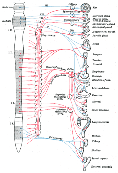
A recent report in the journal Science proposed a big change in how we understand the sympathetic and parasympathetic pathways of the autonomic nervous system (ANS).

In a nutshell, the new model stipulates that the outflow (efferent pathways) are divided into a cranial division and spinal division—not the craniosacral and thoracolumbar divisions that we learned (and that exist in all A&P textbooks):

Current model:
  • Craniosacral division (parasympathetic outflow)
  • Thoracolumbar division (sympathetic outflow)
New model:
  • Cranial division (parasympathetic outflow)
  • Spinal division (sympathetic outflow)
The authors lay out embryological and genetic phenotype evidence to show that the sacral components of the ANS outflow pathways are similar to sympathetic thoracic pathways—not to cranial parasympathetic pathways as we have long supposed. 

But wait, you say, what about the parasympathetic control of the genitals, rectum, bladder? What about, well, all kinds of things that now seem to unravel? I suggest reading the rather brief and plainly written article in Science for the full answer. 

However, a few quick points may reduce your blood pressure a bit—and perhaps pique your interest.

Quick points about the new ANS model

  • Thoracic and sacral pathways share common embryologic development by location and when looking at transcriptional markers associated with neurotransmitters that differ from the developmental pattern of cranial pathways.

  • Thoracic and sacral pathways have a ventral exit point from the spinal cord; cranial pathways have a dorsal exit point.

  • The pelvic ganglion has been considered a "mixed" sympathetic/parasympathetic ganglion because it receives fibers from both the upper lumbar and sacral segments. But if the sacral pathways are sympathetic, the pelvic ganglion is clearly a sympathetic ganglion (not mixed). 

  • Analyses of transcription factors show that cells of the pelvic ganglia resemble those sympathetic ganglia and do not resemble cells in cranial ganglia.

  • The supposed lumbar vs. sacral antagonism in the urinary bladder's detrusor muscle does not seem to hold up, with the lumbar inhibitory effects either not demonstrable in experiments or of questionable functional relevance.

  • The effects on vessel dilation in genitals can be explained as a "continuity of action—rather than antagonism"

  • The sacral pathway to the rectum seems to resemble sympathetic structure, not cranial (parasympathetic) structure.

What can we use from this in teaching undergraduate A&P?

  • When covering the craniosacral/thoracolumbar scheme, consider mentioning this newly proposed model.

  • Consider using this scenario to illustrate the dynamic nature of science. Perhaps discuss that long-held dogma is occasionally challenged using newer methods and ways of thinking.

  • Consider discussing pros and cons of adopting the new model. For example, can evidence from mice extend to all vertebrates? Which is stronger, evidence for the current model or the new model? Which model is most useful in understanding principles of ANS regulation? A little critical thinking never hurt anyone (at least not much).

Want to know more?


The sacral autonomic outflow is sympathetic
  • I. Espinosa-Medina, O. Saha, F. Boismoreau, Z. Chettouh, F. Rossi, W. D. Richardson, J.-F. Brunet. Science  18 Nov 2016: Vol. 354, Issue 6314, pp. 893-897 DOI: 10.1126/science.aah5454
  • Peer-reviewed research report describing this discovery, Includes an updated version of the classic diagram of sympathetic and parasympathetic pathways.
  • my-ap.us/2fNdcF3

Neural circuitry gets rewired
  • Adameyko, I. Science 18 Nov 2016: Vol. 354, Issue 6314, pp. 833-834 DOI: 10.1126/science.aal2810
  • Companion article to the report cited above, stating that "This finding provokes a serious shift in textbook knowledge, and, as with any fundamental discovery, it brings important practical implications..." and goes on to mention of a few of the implications (e.g., how to treat bladder dysfunction).
  • my-ap.us/2gg9O8P

The Autonomic Nervous System. Part I.
  • John Newport Langley. W. Heffer & Sons Ltd., Cambridge, 1921.80pp.
  • Classic "primary source" that codified the modern concept of the ANS. 
  • my-ap.us/2fYHt3M

Gray's Anatomy ANS diagram
  • Henry Gray. 1918 (online edition at Bartleby)
  • Classic diagram by Henry Vandyke Carter of ANS pathways from an early edition of Gray's Anatomy.
  • my-ap.us/2fYGMaT or my-ap.us/2gcAmaW

Thursday, July 10, 2014

Mutations in Mitochondrial DNA

A new study suggests that DNA mutations in some of the mitochondria of healthy people may be a lot more common than scientists thought.

The term heteroplasmy describes a situation in which some mitochondria of a cell have mutant mitochondrial DNA (mtDNA) and other mitochondria have normal mtDNA.  Cell function can become disordered, perhaps producing disease, when the balance of mutant vs. normal mtDNA crosses a certain threshold.

The recent study shows that about 90% of healthy people studied in the 1000 Genomes Project had at least one heteroplasmy.  Some of these (about 20%) have been shown to correlate to disease.  That's a lot more than we were thinking prior to the study (25%-65% heteroplasmy rate).

We don't  know the significance of this finding yet, but it could influence how likely it is for mitochondrial diseases to develop over time—or to be inherited.  Could the mutant/normal mtDNA balance get skewed as oocytes form, thus giving different offspring different probabilities of inheriting mitochondrial disorders?  Or change the probabilities from one generation to the next?  Mitochondrial dysfunction is thought to be a mechanism of agingcould the rate of heteroplasmy be part of the aging mechanism?

What can we use from this in teaching undergraduate A&P?  The fact that we now know that mtDNA mutations are common in healthy people will be interesting and useful to students.


Want to know more?

Mutations Pervade Mitochondrial DNA

  • Jyoti Madhusoodanan. The Scientist (the-scientist.com) July 7, 2014
  • This is a plain-English article summarizing the new findings; includes quotes from the researchers.
  • my-ap.us/1oo18X8 


Extensive pathogenicity of mitochondrial heteroplasmy in healthy human individuals

  • K. Ye et al., Proceedings of the National Academy of Science (PNAS), doi:10.1073/pnas.1403521111, 2014. 
  • This is the original research report.
  • my-ap.us/1sDPV8S


Want a FREE labeled image of mtDNA that you can use in your presentation, handout, or other course material?
my-ap.us/1ncmAxl


Monday, December 31, 2012

Rita Levi-Montalcini, growth factor pioneer

Yesterday, the scientific community lost another of its great people, Rita Levi-Montalcini.

In The Human Body in Health & Disease and Structure & Function of the Body, I wrote this about Levi-Montalcini:
Rita Levi-Montalcini had just finished a medical degree in her native Italy when in 1938 the Fascist government under Mussolini barred all “non-Aryans” from working in academic and professional careers. Being Jewish, Levi-Montalcini was forced to move to Belgium to work. But when Belgium was about to be invaded by the Nazis, she decided to return home to Italy and work in secret. Her home laboratory was very crude, but in it she made some important discoveries about how the nervous system develops during embryonic development. After World War II, she was invited to Washington University in St. Louis to work. There, she discovered the existence of nerve growth factor (NGF), for which she later won the 1986 Nobel Prize. Her discovery of a chemical that regulates the growth of new nerves during early brain development has led to many different paths of investigation. For example, by learning more about growth regulators we now know more about how the nervous system develops, as well as other tissues, organs, and systems of the body.
Note that I put in a little plug for my hometown of St. Louis, where we continue to be proud of this remarkable woman and her pioneering work.

As I said in a recent post about the passing of transplant pioneer Joseph Murray, I think the occasional story of a pioneer in the history of human science adds a lot to the A&P course.  Such stories give a human dimension to the pursuit of science and provide the context needed for students to understand how we know what we know.  Levi-Montalcini's story gives us the further opportunities to weave into our courses the themes of global collaboration among the scientific community as the role of women in science.

Want to know more?

  • Nobel Scientist Rita Levi-Montalcini Dies in Rome
  • Oldest Nobel winner Rita Levi-Montalcini dies at 103
  • Nobel Lecture by Rita Levi-Montalcini 
    • Media Player at Nobelprize.org
    • [Full video (in English) of Nobel lecture by Rita Levi-Montalcini in which she fully credits "good luck"; 57 minutes]
    • http://my-ap.us/Vf6sPz

  • Rita Levi-Montalcini Interview
    • Adam Smith, Editor-in-Chief of Nobelprize.org.
    • Nobel Interview, November 2008
    • [Video interview with Rita Levi-Montalcini, who talks about her daily work, why she had to make a laboratory in her bedroom to conduct research during World War II (3:06), the benefits of working in isolation (5:03), her post-war move to the United States (6:25), her work with Stanley Cohen and the discovery of nerve growth factor (7:15), the roles of intuition and chance in biological research (15:14), her current research (16:58), her advice to young scientists (17:41), and why this period of her life has been the best so far (28:10).]
    • http://my-ap.us/Ug6gA5

  • The Nobel Prize in Physiology or Medicine 1986 Press Release
    • Nobelprize.org
    • [Detailed news release that includes some simple diagrams that help illustrate the concepts involved.]
    • http://my-ap.us/ZPyacv
  • In Praise of Imperfection: My Life and Work
    • Rita Levi-Montalcini
    • Sloan Foundation Science Series, October 1989
    • [Her autobiography]
    • http://amzn.to/Wgjk7b


Related textbook content
  • Anatomy & Physiology 8th ed.  p. 409, 1111-1113, A&P Connect: The Nobel Legacy my-ap.us/QZTbK1
  • Essentials of Anatomy & Physiology p. 231-232, 241, 610-612 my-ap.us/SCfNlj  
  • The Human Body in Health and Disease 5th ed. p. 236-237, 644-645, 658 my-ap.us/fNN00N 
  • Structure & Function of the Body 14th ed. p. 168-169, 472-473 my-ap.us/X6QxqE


Photo: Presidenza della Repubblica Italiana

Monday, November 5, 2012

New method to avoid mitochondrial disorders

A recent paper in Nature describes a new approach to avoiding inherited mitochondrial disorders.

Although identified as "germline gene therapy," in the title of the paper the method is a lot like cloning. The method calls for transferring a healthy nucleus out of an egg with mutant mitochondria, then transferring that nucleus to a healthy donor egg.

In this technique, scientists are not making a genetic copy of an individual as an cloning — but the technique is very similar. The whole idea of this is an interesting one to bring up in an A&P course when discussing the topic of mitochondrial inheritance. It not only emphasizes and clarifies the central idea of mitochondrial inheritance, it's also a good way to connect students to "what's going on right now" in the world of science. 

Among the links below I have included an article from Science News that does a great job of summarizing the new research and pointing out some of the ethical concerns that the method poses.

The article also contains a sidebar listing some of the mitochondrial diseases that might be avoided using the technique. That sidebar complements the coverage of mitochondrial inheritance found in my textbooks. 

Check out this video that demonstrates how the method is carried out in the lab.  You can use this video in your course!


Want to know more?
  • Cloning-like method targets mitochondrial diseases: providing healthy ‘power plants’ in donor eggs appears feasible in humans
    • Tina Hesman Saey Science News Web edition: October 24, 2012 Print edition: November 17, 2012; Vol.182 #10 (p. 5)
    • A great feature article that summarizes the discovery in plain English.  Might be the best place to point your students if they want more information.
    • my-ap.us/SEPeg5
  • Towards germline gene therapy of inherited mitochondrial diseases. 
    • M. Tachibana et al. Nature. Published online 24 Oct 2012 doi:10.1038/nature11647
    • Abstract of original journal article describing the method. Includes images.
    • my-ap.us/RwC2cf

Related articles
  • Embryo transfer technique could prevent maternally inherited diseases | Body & Brain | Science News my-ap.us/VjPGSR
  • Mitochondrial DNA replacement successful in Rhesus monkeys | Genes & Cells | Science News my-ap.us/VInDbs

Related textbook content
  • The Human Body in Health and Disease 5th ed. p. 45, 672, A-13 my-ap.us/fNN00N 
Here's a related image you can use in your course: my-ap.us/QZVNaH

Monday, October 8, 2012

Reprogramming cells to be pluripotent


The Nobel Assembly at Karolinska Institutet has today decided to award

The Nobel Prize in Physiology or Medicine 2012
jointly to

John B. Gurdon and Shinya Yamanaka
for the discovery that mature cells can be reprogrammed to become pluripotent

Summary
The Nobel Prize recognizes two scientists who discovered that mature, specialised cells can be reprogrammed to become immature cells capable of developing into all tissues of the body. Their findings have revolutionised our understanding of how cells and organisms develop.

John B. Gurdon discovered in 1962 that the specialisation of cells is reversible. In a classic experiment, he replaced the immature cell nucleus in an egg cell of a frog with the nucleus from a mature intestinal cell. This modified egg cell developed into a normal tadpole. The DNA of the mature cell still had all the information needed to develop all cells in the frog.

Shinya Yamanaka discovered more than 40 years later, in 2006, how intact mature cells in mice could be reprogrammed to become immature stem cells. Surprisingly, by introducing only a few genes, he could reprogram mature cells to become pluripotent stem cells, i.e. immature cells that are able to develop into all types of cells in the body.

These groundbreaking discoveries have completely changed our view of the development and cellular specialisation. We now understand that the mature cell does not have to be confined forever to its specialised state. Textbooks have been rewritten and new research fields have been established. By reprogramming human cells, scientists have created new opportunities to study diseases and develop methods for diagnosis and therapy.

Life – a journey towards increasing specialisation

All of us developed from fertilized egg cells. During the first days after conception, the embryo consists of immature cells, each of which is capable of developing into all the cell types that form the adult organism. Such cells are called pluripotent stem cells. With further development of the embryo, these cells give rise to nerve cells, muscle cells, liver cells and all other cell types - each of them specialised to carry out a specific task in the adult body. This journey from immature to specialised cell was previously considered to be unidirectional. It was thought that the cell changes in such a way during maturation that it would no longer be possible for it to return to an immature, pluripotent stage.

Frogs jump backwards in development

John B. Gurdon challenged the dogma that the specialised cell is irreversibly committed to its fate. He hypothesised that its genome might still contain all the information needed to drive its development into all the different cell types of an organism. In 1962, he tested this hypothesis by replacing the cell nucleus of a frog's egg cell with a nucleus from a mature, specialised cell derived from the intestine of a tadpole. The egg developed into a fully functional, cloned tadpole and subsequent repeats of the experiment yielded adult frogs. The nucleus of the mature cell had not lost its capacity to drive development to a fully functional organism.

Gurdon's landmark discovery was initially met with scepticism but became accepted when it had been confirmed by other scientists. It initiated intense research and the technique was further developed, leading eventually to the cloning of mammals. Gurdon's research taught us that the nucleus of a mature, specialized cell can be returned to an immature, pluripotent state. But his experiment involved the removal of cell nuclei with pipettes followed by their introduction into other cells. Would it ever be possible to turn an intact cell back into a pluripotent stem cell?

A roundtrip journey – mature cells return to a stem cell state

Shinya Yamanaka was able to answer this question in a scientific breakthrough more than 40 years after Gurdon´s discovery. His research concerned embryonal stem cells, i.e. pluripotent stem cells that are isolated from the embryo and cultured in the laboratory. Such stem cells were initially isolated from mice by Martin Evans (Nobel Prize 2007) and Yamanaka tried to find the genes that kept them immature. When several of these genes had been identified, he tested whether any of them could reprogram mature cells to become pluripotent stem cells.

Yamanaka and his co-workers introduced these genes, in different combinations, into mature cells from connective tissue, fibroblasts, and examined the results under the microscope. They finally found a combination that worked, and the recipe was surprisingly simple. By introducing four genes together, they could reprogram their fibroblasts into immature stem cells!

The resulting induced pluripotent stem cells (iPS cells) could develop into mature cell types such as fibroblasts, nerve cells and gut cells. The discovery that intact, mature cells could be reprogrammed into pluripotent stem cells was published in 2006 and was immediately considered a major breakthrough.

From surprising discovery to medical use

The discoveries of Gurdon and Yamanaka have shown that specialised cells can turn back the developmental clock under certain circumstances. Although their genome undergoes modifications during development, these modifications are not irreversible. We have obtained a new view of the development of cells and organisms.

Research during recent years has shown that iPS cells can give rise to all the different cell types of the body. These discoveries have also provided new tools for scientists around the world and led to remarkable progress in many areas of medicine. iPS cells can also be prepared from human cells.

For instance, skin cells can be obtained from patients with various diseases, reprogrammed, and examined in the laboratory to determine how they differ from cells of healthy individuals. Such cells constitute invaluable tools for understanding disease mechanisms and so provide new opportunities to develop medical therapies.


Sir John B. Gurdon was born in 1933 in Dippenhall, UK. He received his Doctorate from the University of Oxford in 1960 and was a postdoctoral fellow at California Institute of Technology. He joined Cambridge University, UK, in 1972 and has served as Professor of Cell Biology and Master of Magdalene College. Gurdon is currently at the Gurdon Institute in Cambridge.

Shinya Yamanaka was born in Osaka, Japan in 1962. He obtained his MD in 1987 at Kobe University and trained as an orthopaedic surgeon before switching to basic research. Yamanaka received his PhD at Osaka City University in 1993, after which he worked at the Gladstone Institute in San Francisco and Nara Institute of Science and Technology in Japan. Yamanaka is currently Professor at Kyoto University and also affiliated with the Gladstone Institute.

Key publications:

Gurdon, J.B. (1962). The developmental capacity of nuclei taken from intestinal epithelium cells of feeding tadpoles. Journal of Embryology and Experimental Morphology 10:622-640.

Takahashi, K., Yamanaka, S. (2006). Induction of pluripotent stem cells from mouse embryonic and adult fibroblast cultures by defined factors. Cell 126:663-676.

Link to a FREE handout you can use in your A&P class: my-ap.us/UNtV9i

Want to know more? Try my-ap.us/TbNCWG


Adapted from my-ap.us/TlWoqC

Tuesday, July 10, 2012

Stem cells in the ovary

Beginning about 8 years ago, scientists began providing evidence that apparently overturns the dogma that adult ovaries do not contain stem cells capable of producing oocytes.  Jonathon Tilly found such cells first in mice, then later in middle-aged women. Other labs have replicated such findings.

However, a study by members of Kui Liu's lab published today disputes that the stem cells actually produce oocytes.  Needless to say, this is stirring up quite a bit of controversy.  Tilly believes Liu was looking at oocytes, not stem cells.  Liu doesn't agree.

What does this mean for the undergraduate A&P class?  I discuss the recent discoveries of stem cells in adult ovaries in my course.  I use it as an opportunity to point out that there is still much to learn about how the human body works . . . that we are continually surprised by new research findings.  This is part of a year-long subtext of "how science works."  I tell my A&P students that I'm telling them "the last, best story" about the human body.  But that my story changes from year to year as scientists tease out more information . . . and thus revise "the story."

How does this fit in?  Well, isn't this how science is supposed to work?  Wouldn't it seem logical that it is in the best interest of everyone to have vigorous debate and extensive re-examination before we throw out the last, best story in favor of a new version?

The first article below briefly summarizes the issues involved in today's publication.


Want to know more?


Ovarian Stem Cell Debate
Ed Yong
The Scientist
(online) July 9, 2012

[Brief article discusses  that opinion is divided on a new paper showing that adult ovaries do not contain egg-making stem cells, contrary to two recent studies that appeared to overturn longstanding dogma.]

my-ap.us/Mf1Y5w
Experimental evidence showing that no mitotically active female germline progenitors exist in postnatal mouse ovaries
H. Zhang et al.
Proceedings of the National Academy of Sciences doi:10.1073/pnas.1206600109
[Recent paper disputing presence of stem cells in adult ovaries]
my-ap.us/RX4McG

Ovarian Stem Cells in Humans?
Sabrina Richards
The Scientist (online) February 27, 2012
[Brief summary of recent paper by Tilly's lab stating that adult human ovaries contain a population of stem cells capable of generating immature egg cells.]
my-ap.us/OWGY8G

Oocyte formation by mitotically active germ cells purified from ovaries of reproductive-age women
Y.A.R. White et al.
Nature Medicine 18:413–21, 2012.
[Original research paper by Tilly's lab showing presence of stem cells in human ovaries.]
my-ap.us/LFaYqX

The image above is copyright free and can be used in your course!

Friday, March 25, 2011

Progesterone's action on sperm finally solved

Two recent articles in Nature reveal a mystery that's been puzzling physiologists for a couple of decades . . . how does progesterone signal sperm cells?

The short version of the story is this . . .

Progesterone is released from the cumulus cells that cling to the outside of the zona pellucida surrounding the ovum. This progesterone binds directly to calcium channels in the sperm's plasma membrane to open.  Influx of calcium ions triggers an increase in flagellum activity.  This increased work is needed for the sperm to get through the zona to the ovum.

Progesterone is a steroid hormone and thus usually enters its target cell and activates transcription of a gene.  In this case, however, progesterone instead directly triggers a calcium channel.  It's not even a second-messenger system, but a direct binding to the channel.

Secretion of progesterone by cumulus cells apparently also provides a chemical gradient that helps sperm navigate toward the ovum.

Recall also that calcium influx into the ovum triggered by contact with a sperm cell plays a role in producing changes within the ovum that result in successful fertilization.

I recommend that my students keep running concept lists on recurring themes or actors in story of human biology.  Here's something they can now add to their calcium list.  Now they can see that a calcium gradient is a truly multipurpose tool in the body.  (For more about running concept lists see my-ap.us/hCIA9X)

Want to know more?
Sperm mystery solved: Scientists identify the channel by which progesterone activates sperm to swim toward an egg
M. Scudellari
The Scientist Published online 16th March 2011
[Nice summary of the significance of the discovery]
my-ap.us/grpcTp

Female hormone could be key to male contraceptive: Progesterone-sensing molecule that guides sperm to egg offers fertility solution.
E. Callaway
NatureNews Published online 16 March 2011 | Nature | doi:10.1038/news.2011.163
[Summary article outlining the papers published in Nature]
my-ap.us/fJNaYk

Progesterone activates the principal Ca2+ channel of human sperm
Lishko et al.
Nature  471:387–391 17 March 2011 doi:10.1038/nature09767
[Original research findings]
my-ap.us/gxcomX

The CatSper channel mediates progesterone-induced Ca2+ influx in human sperm
Strunker et al.
Nature 471:382–386 17 March 2011 doi:10.1038/nature09769
[Original research findings] my-ap.us/g14eTK
From The A&P Professor archive
New discovery about sperm's ability to swim
K. Patton
The A&P Professor published online Feb 18, 2010
[Summary of new discovery that when sperm enter female tract, proton channels in the sperm head open and the resulting pH drop triggers influx of calcium, which gets the flagella started in the first place.  That darn calcium shows up in every part of this story, eh?]
my-ap.us/dExdEK

Friday, December 3, 2010

Father of Fractals

You may have already heard the recent news of the passing of Benoit Mandelbrot, originator of the iconic Mandelbrot Set (pictured) and founder of the field of fractal geometry.  It brings to mind the deeper understanding of human structure and function that has resulted directly from applying principles of fractal geometry.  An important set of principles that I believe we A&P professors could do a better job of helping our students appreciate.

Mandelbrot's pioneering efforts in understanding the roughness of nature led to the discovery of basic principles of fractal geometry.  A key characteristic of fractal structures is self-similarity (the parts resemble the whole).

In human anatomy, this self-similar characteristic is observed in surfaces that have folds, which have bumps, which in turn have their own bumps, and so on . . . producing unexpectedly huge total surface areas.  For example, think of the loops of the intestines, which in turn have circular folds of mucosa, which in turn have villi, which in turn have microvilli, which in turn have membranes embedded with bumpy molecules, and so on. 

Fractal self-similarity can also be observed in branched structures, such as the respiratory tract and the cardiovascular vessels.  These structures have branches that have branches that have branches, and so on for many levels . . . producing large numbers of pathways and huge surface areas.

A particularly interesting characteristic of such complex fractal structures is that they are produced with relatively simple mathematical formulae.  Which means that very little genetic information is needed to produce highly complex structures like intestines, blood vessels, lymphatic vessels, bronchial trees, cerebral convolutions, etc.

Fractal structures are also chaotic, a mathematical concept of "constrained randomness."  Put simply, chaotic structures have an element of randomness but within limits.  So when our body applies fractal geometry during development we can be certain of a particular type of structure without being certain we'll know exactly where each individual bump or branch will lie.  In other words, we can more or less be certain where the main arteries will be (with some individual variation) but not so much for the various arterioles and capillaries . . . at least not precisely.

Principles of chaos also play out in human physiology when we observe the aperiodic (nonrhythmic) patterns of heart rate, brain waves (as in an EEG), and certain other functions.

Mandelbrot opened up a whole new understanding of human structure and function that is only now becoming understood widely.  I've been introducing the concept of chaos and fractals in my courses, and more subtly in some of my textbooks, for several years now.  My experience is that introducing simplified principles of chaos and fractals at the beginning of A&P 1, then reinforcing them when encountered throughout both semesters of A&P, help student appreciate an intriguing and important concept of human structure and function.  A concept that is increasingly playing a central role in science's understanding of human biology.


Want to know more?

Benoît Mandelbrot (1924–2010)
Ralph Gomory
Nature Volume: 468, Page 378, Date published: 18 November 2010, doi:10.1038/468378a, Published online: 17 November 2010
[A brief synopsis of Mandelbrot's life and contributions from the journal Nature]

Chaos in the Human Body (Mini Lesson)
Kevin Patton
Lion Den http://lionden.com/chaos.htm
[Brief outline that I use with my own students in A&P 1]

Applications of Fractals - Human Body
ThinkQuest
Oracle Education Foundation. online (accessed 2 Dec 2010)
[Brief student-produced outline of some fractal principles of the body]

Fractal Geometry in Biological Systems: An Analytical Approach
Philip M. Iannaccone, Mustafa Khokha
CRC Press 1996
[Book outlining the initial discoveries of fractals in humans.]

Chaos: Making a New Science
James Gleick
Penguin 2008
[Reprint of the classic bestseller book that outlines in simple terms the concepts of chaos and fractal geometry.  Highly recommended.  Includes some applications/examples in human biology.]

Monday, October 4, 2010

Nobel Prize: Test Tube Babies

This morning, we heard the news . . . the 2010 Nobel Prize in Medicine or Physiology is awarded to Robert G. Edwards "for the development of in vitro fertilization."


Professors and students using my Anatomy & Physiology (7th ed.) textbook can access an article on in vitro fertilization (IVF) at A&P Connect online at evolve.elsevier.com

If you are thinking of mentioning this award in your classes this week, which I am planning to do myself, you are welcome to use the following information from the Nobel Committee, as well as the images linked to the thumbnails presented here (scroll down to the bottom for more).

Of course, be aware that the use of IVF is condemned by some religious groups (for example, see Dignitas Personae) and thus classroom discussions may become heated.

There is also advanced information available at the Nobel website.  This is a nice publication that summarizes the science.

If you want a short set of slides that you can use today in your class, then use this link:


Summary

Robert Edwards is awarded the 2010 Nobel Prize for the development of human in vitro fertilization (IVF) therapy. His achievements have made it possible to treat infertility, a medical condition afflicting a large proportion of humanity including more than 10% of all couples worldwide.

As early as the 1950s, Edwards had the vision that IVF could be useful as a treatment for infertility. He worked systematically to realize his goal, discovered important principles for human fertilization, and succeeded in accomplishing fertilization of human egg cells in test tubes (or more precisely, cell culture dishes). His efforts were finally crowned by success on 25 July, 1978, when the world's first "test tube baby" was born. During the following years, Edwards and his co-workers refined IVF technology and shared it with colleagues around the world.

Approximately four million individuals have so far been born following IVF. Many of them are now adult and some have already become parents. A new field of medicine has emerged, with Robert Edwards leading the process all the way from the fundamental discoveries to the current, successful IVF therapy. His contributions represent a milestone in the development of modern medicine.

Infertility – a medical and psychological problem

More than 10% of all couples worldwide are infertile. For many of them, this is a great disappointment and for some causes lifelong psychological trauma. Medicine has had limited opportunities to help these individuals in the past. Today, the situation is entirely different. In vitro fertilization (IVF) is an established therapy when sperm and egg cannot meet inside the body.

Basic research bears fruit

The British scientist Robert Edwards began his fundamental research on the biology of fertilization in the 1950s. He soon realized that fertilization outside the body could represent a possible treatment of infertility. Other scientists had shown that egg cells from rabbits could be fertilized in test tubes when sperm was added, giving rise to offspring. Edwards decided to investigate if similar methods could be used to fertilize human egg cells.
It turned out that human eggs have an entirely different life cycle than those of rabbits.  In a series of experimental studies conducted together with several different co-workers, Edwards made a number of fundamental discoveries. He clarified how human eggs mature, how different hormones regulate their maturation, and at which time point the eggs are susceptible to the fertilizing sperm. He also determined the conditions under which sperm is activated and has the capacity to fertilize the egg. In 1969, his efforts met with success when, for the first time, a human egg was fertilized in a test tube.
In spite of this success, a major problem remained. The fertilized egg did not develop beyond a single cell division. Edwards suspected that eggs that had matured in the ovaries before they were removed for IVF would function better, and looked for possible ways to obtain such eggs in a safe way.

From experiment to clinical medicine

Edwards contacted the gynecologist Patrick Steptoe. He became the clinician who, together with Edwards, developed IVF from experiment to practical medicine. Steptoe was one of the pioneers in laparoscopy, a technique that was new and controversial at the time. It allows inspection of the ovaries through an optical instrument. Steptoe used the laparoscope to remove eggs from the ovaries and Edwards put the eggs in cell culture and added sperm. The fertilized egg cells now divided several times and formed early embryos, 8 cells in size (see figure).
These early studies were promising but the Medical Research Council decided not to fund a continuation of the project. However, a private donation allowed the work to continue. The research also became the topic of a lively ethical debate that was initiated by Edwards himself. Several religious leaders, ethicists, and scientists demanded that the project be stopped, while others gave it their support.

The birth of Louise Brown - an historic event

Edwards and Steptoe could continue their research thanks to the new donation. By analyzing the patients' hormone levels, they could determine the best time point for fertilization and maximize the chances for success. In 1978, Lesley and John Brown came to the clinic after nine years of failed attempts to have a child. IVF treatment was carried out, and when the fertilized egg had developed into an embryo with 8 cells, it was returned to Mrs. Brown. A healthy baby, Louise Brown, was born through Caesarian section after a full-term pregnancy, on 25 July, 1978. IVF had moved from vision to reality and a new era in medicine had begun.

IVF is refined and spreads around the world

Edwards and Steptoe established the Bourn Hall Clinic in Cambridge, the world's first centre for IVF therapy. Steptoe was its medical director until his death in 1988, and Edwards was its head of research until his retirement. Gynecologists and cell biologists from all around the world trained at Bourn Hall, where the methods of IVF were continuously refined. By 1986, 1,000 children had already been born following IVF at Bourn Hall, representing approximately half of all children born after IVF in the world at that time.

Today, IVF is an established therapy throughout the world. It has undergone several important improvements. For example, single sperm can be microinjected directly into the egg cell in the culture dish. This method has improved the treatment of male infertility by IVF. Furthermore, mature eggs suitable for IVF can be identified by ultrasound and removed with a fine syringe rather than through the laparoscope.
IVF is a safe and effective therapy. 20-30% of fertilized eggs lead to the birth of a child. Complications include premature births but are very rare, particularly when one egg only is inserted into the mother. Long-term follow-up studies have shown that IVF children are as healthy as other children.
Approximately four million individuals have been born thanks to IVF. Louise Brown and several other IVF children have given birth to children themselves; this is probably the best evidence for the safety and success of IVF therapy. Today, Robert Edwards' vision is a reality and brings joy to infertile people all over the world.

Robert G. Edwards was born in 1925 in Manchester, England. After military service in the Second World War, he studied biology at the University of Wales in Bangor and at Edinburgh University in Scotland, where he received his PhD in 1955 with a Thesis on embryonal development in mice. He became a staff scientist at the National Institute for Medical Research in London in 1958 and initiated his research on the human fertilization process. From 1963, Edwards worked in Cambridge, first at its university and later at Bourn Hall Clinic, the world's first IVF centre, which he founded together with Patrick Steptoe. Edwards was its research director for many years and he was also the editor of several leading scientific journals in the area of fertilization. Robert Edwards is currently professor emeritus at the University of Cambridge.

References:
Edwards RG. Maturation in vitro of human ovarian oocytes. Lancet 1965; 2:926-929.
Edwards RG, Bavister BD, Steptoe PC. Early stages of fertilization in vitro of human oocytes matured in vitro. Nature 1969; 221:632-635.
Edwards RG, Steptoe PC, Purdy JM. Fertilization and cleavage in vitro of human oocytes matured in vivo. Nature 1970; 227:1307-1309.
Steptoe PC, Edwards RG. Birth after the reimplantation of a human embryo. Lancet 1978; 2:366.
Edwards RG. The bumpy road to human in vitro fertilization. Nature Med 2001; 7:1091-4.

The preceding information is from "The 2010 Nobel Prize in Physiology or Medicine - Press Release". Nobelprize.org. 4 Oct 2010 http://nobelprize.org/nobel_prizes/medicine/laureates/2010/press.html


Some images you may find useful (click each thumbnail)

Sperm injection into oocyte

Human embryos developing in vitro


Robert G. Edwards


PDF from the Nobel website


Nobel medal

Wednesday, July 7, 2010

Looking at cilia

 
In a recent article in The Scientist, Peter Satir points out that the cilium was the very first distinct organelle ever directly observed by scientists--van Leeuwenhoek noted their existence in the 1660s.  But we are only now starting to fully understand these amazing and vital structures.

Until only the last decade or so, we thought that cilia were organs of cell motility--period.  But as I've noted in recent editions of my textbooks (e.g. Anatomy & Physiology, p.82-83), we now know that cilia play a critical role in a cell's ability to sense its surroundings.

Not only is this sensory function useful for, well, er, mediating senses such as hearing and equilibrium, it's also critical for cells to figure where to go (and when) during embryonic development.  In fact, it's been shown that situs inversus (the condition in which internal organs are flipped in their left-right orientation) is caused by a mutation affecting the structure of the primary cilium of developing cells in the embryo.

Cilia, it turns out, are centrally involved in a lot of different cell functions.

If you want a quick and interesting review of the history of cilia from one of the pioneers in cilia research, including answers to "why do we have to learn this stuff if I'm going to be a [insert health profession here]?," then check out this article:
Eyelashes Up Close
Peter Satir
The Scientist Volume 24 | Issue 7 | Page 30 2010-07-01
[Brief, well-illustrated review of what we know about cilia so far.  Includes great graphics and useful references.]
For more FREE images of cilia you can use in your course, see The A&P Professor website's FREE Image Library of Cell Structures.
.
.

Monday, June 7, 2010

Sex differences in body fat distribution

When discussing sex differences in body fat distribution in my A&P course, I off-handedly refer to the roles of sex hormones in regulating the development that leads to these differences.  But how much do we really know about how that works?  Some recent work sheds a bit of light on that.

For example, a research review recently appearing in Obesity Reviews shows that indeed estrogen is responsible for promoting fat deposition in adult women.

Another recent article, this one in The International Journal of Obesity, suggests that there is a huge difference between the gene activity in male fat vs. female fat.  That is, the anatomy and physiology of male fat and female fat differs far more than anyone has yet realized.

Why Do Women Store Fat Differently From Men?.
University of New South Wales.
ScienceDaily 4 March 2009. 17 May 2010
[Brief synopsis of a research review from Obesity Reviews]

Does oestrogen allow women to store fat more efficiently? A biological advantage for fertility and gestation
A. J. O'Sullivan
Obesity Reviews Volume 10, Issue 2, Date: March 2009, Pages: 168-177
[Research review]

Belly Fat or Hip Fat: It Really Is All in Your Genes, Says Researcher.
UT Southwestern Medical Center.
ScienceDaily 16 May 2010. 17 May 2010

A microarray analysis of sexual dimorphism of adipose tissues in high-fat-diet-induced obese mice. 
K L Grove, et al.
International Journal of Obesity, 2010; DOI: 10.1038/ijo.2010.12
[Research article]

Thursday, February 18, 2010

New discovery about sperm's ability to swim

Scientists have found the trigger that gets sperm swimming in the female reproductive tract.

Sperm cells in the testis are pretty quiet . . . they don't seem very interested in swimming.  However, after they are ejaculated into the female reproductive tract they become activated and get with the program. We already knew that the sperm cells need to raise their pH in order to kick into their swimming mode . . . but we didn't know how that is actually done.

In an article in the journal Cell, researchers report that they have the answer . . . one-way proton (H+) channels called Hv1 that open when sperm enter the female reproductive tract.  Increasing the intracellular pH triggers the influx of calcium ions, which in turn activate the sperm flagellum.  And they're off!

The increase in intracellular sperm pH also enables the sperm's acrosome to become activated and get ready to do its job, too.

Hv1 may be a key to triggering the hyperactivation and capacitation of sperm necessary for male fertility.

The researchers also found that a chemical similar to the active ingredient in marijuana inhibits the Hv1 channels and thus reduced fertility.  Perhaps this explains low fertility among males who are chronic users of marijuana.  And perhaps this opens the door to discovering chemicals that can be used to regulate the sperm fertility.

Want to know more?
Science News web edition : Thursday, February 4th, 2010
[Summary article includes a cool fluorescent micrograph.]

Acid extrusion from human spermatozoa is mediated by flagellar voltage-gated proton channel. 
Lishko, P.V. et al.
Cell Volume 140, Issue 3, 327-337, 5 February 2010
[Original research article with some fabulous images in the graphical abstract and an excellent movie that features the scientists explaining their discovery. ]

Sunday, January 24, 2010

More graduate biology courses at the HAPS Conference


OK, so you missed out on that cool cadaver class presented by HAPS Institute (HAPS-I) in San Diego this winter . . . because it filled up faster than I could tell you about it!  But now's your second chance for some great graduate biology courses especially for teachers of human anatomy and physiology courses:

1. Advances in Anatomy and Physiology 2010  (Ellen Arnestad and Kevin Patton)

2. Advanced Cardiovascular Physiology: The Heart at Work and at Rest (Robert Carroll)

3. Concepts in Human Embryology (Valerie O'Loughlin)

4. Molecular and Cellular Basis of Disease (Kelly McDonald)
These are great courses that feature both useful content about human A&P and experience with best practices in teaching these subjects.  These are courses that are MEANINGFUL to what you do every day in your own teaching.  And you'll be there with folks just like you . . . who teach secondary, college, and university A&P.

Each course earns you 2 graduate credits from the Biology Department of the University of Washington (Seattle). 

These courses begin with online work on April 13, involve seminars and/or workshops during the Denver HAPS Conference (May 29 - June 3), and continue with online work through August 19.  Each syllabus has additional details.  Conference registration (plus lodging, meals, and transportation, if needed) is required (in addition to HAPS-I course fees). 

Want to know more? 

Remember . . . THESE COURSES FILL EARLY.  So you want to get on this ASAP.  I mean it this time!

In fact, some spots have already been taken by past HAPS-I Scholars and by members of the HAPS-I Update email list, who all received notice of these course openings a few days ago.  (If you want prior notice of HAPS-I courses, go to http://www.hapsweb.org/displaycommon.cfm?an=1&subarticlenbr=234 to subscribe to either the HAPS-I Scholars Google Group or the HAPS-I Update Google Group.)

Registration is now open at http://www.hapsweb.org/displayconvention.cfm?conventionnbr=7898

For more information on the HAPS Annual Conference in Denver, go to http://www.hapsweb.org/displayconvention.cfm?conventionnbr=7450

EARLY BIRD CONFERENCE RATES APPLY UNTIL 2/1/2010!

Sunday, November 8, 2009

Why cells cooperate


Here's a nice little "animated clay" video that zeroes in on the "society of cells" concept that lies at the heart of homeostasis.   Because it goes on to emphasize the role of reproductive cells in a multicellular organism, it may be useful to help our A&P students connect reproduction to the concept of overall body homeostasis.

I saw this video on public radio's Science Friday website, where they have a weekly video recommendation.

The video comes from a collection of videos at creaturecast.org that are truly amazing.  Not very many directly relate to human anatomy and physiology . . . but, wow, they are fascinating.  For example, a recent posting discusses how mitochondria and other erstwhile endosymbionts can play a variety of roles such as acting as lenses for simple animals. I teach the serial endosymbiosis theory (SET)  in my A&P course . . . so this little factoid may help spice up that discussion.

So watch the FREE video about cell cooperation in a multicellular organism and let me know what you think!


Tuesday, July 21, 2009

FREE video on renewal of intestinal lining


A recent "Hot Paper" summary in The Scientist not only brings us up to date on the latest skirmish in the battle over which cell is THE stem cell of the intestinal crypts, it also gives us a great Flash video that we can use to illustrate the process of maintaining the lining of the small intestine.

In my textbook Anatomy & Physiology 7th edition, Figure 25-18 (p. 853) shows the process by which stem cells in the intestinal crypts (of Lieberkuhn) produce daughter cells that move up and out of the crypt to replace cells lost at the apices of the intestinal villi.

The video embedded in the article in The Scientist is not narrated, and rather slow-moving at times, but it does a fantastic job of animating the process shown in textbook figure 25-18.

To link directly to the video, go to:
http://images.the-scientist.com/supplementary/video/keller/kellermcgill_clonalconveyorbelt.swf

To view the video within the article go to . . .
Gut Churning: The discovery of an intestinal stem cell marker fuels an ongoing debate over the cells' location and properties
Alla Katsnelson
The Scientist Volume 23, Issue 7, Page 51
No matter how you get there, I suggest previewing it (duh-uh) and taking note of particular times in the sequence to which you may want to quickly navigate to avoid the slow parts (or skip parts not useful in your course). Or just slide the timing bar to the spot you want.

BONUS! Here are some FREE images of the intestinal villi and intestinal crypts from an early edition of Gray's Anatomy:

Tuesday, July 7, 2009

New NIH policy on human stem cell research


For those who want to be "up to speed" on official policy when the inevitable discussion of human embryonic stem cell research pops up in class, this is just in from the National Institutes of Health (NIH) . . . revised guidelines that permit research on embryonic cell lines go in to effect TODAY.

From the official notice:

"On March 9, 2009, President Barack H. Obama issued Executive Order (EO)13505 Removing Barriers to Responsible Scientific Research Involving Human Stem Cells. The EO states that the Secretary of Health and Human Services, through the Director of NIH, may support and conduct responsible, scientifically worthy human stem cell research, including human embryonic stem cell (hESC) research, to the extent permitted by law. NIH published draft Guidelines for research involving hESCs in the Federal Register for public comment, 74 Fed. Reg. 18578 on April 23, 2009. The comment period ended on May 26, 2009. Approximately 49,000 comments on the draft Guidelines were submitted to NIH by patient advocacy groups, scientists and scientific societies, academic institutions, medical organizations, religious organizations, private citizens, and members of Congress.

The final NIH Guidelines for Human Stem Cell Research implementing the EO and establishing policy and procedures under which the NIH will fund such research, were released today and are available at http://stemcells.nih.gov/index.asp. They will be effective on July 7, 2009. Public comments on the draft Guidelines were also released today and are available at http://stemcells.nih.gov/index.asp.

The Guidelines will ensure that NIH-funded research in this area is ethically responsible, scientifically worthy, and conducted in accordance with applicable law. Internal NIH policies and procedures, consistent with the EO and these Guidelines, will govern the conduct of intramural NIH stem cell research.

The Guidelines prescribe the assurances and supporting documentation that must accompany requests for NIH funding for research using hESCs, and describe research that is not eligible for NIH funding. NIH will provide additional guidance concerning the implementation of the Guidelines and the status of pending applications in future Guide Notices.
Ongoing NIH-supported research involving previously approved hESC lines may continue. No new uses of hESC may be initiated in ongoing funded studies unless reviewed and approved by the NIH."

Read the new NIH Guidelines at http://stemcells.nih.gov/policy/2009guidelines.htm

Check out this article about the new guidelines, including the context and background:

NIH loosens stem cell consent rules
Elie Dolgin
The Scientist (online) posted 6th July 2009 07:58 PM GMT

Read my recent blog article Science controversies in the news


Tuesday, May 26, 2009

Reprogramming cells


In a study featured on the cover of the May issue of The FASEB Journal, researchers describe how they are able to reprogram human adult skin cells into other cell types in order to decipher the elusive mechanisms underlying reprogramming.

To demonstrate their point, they transformed human skin cells into mouse muscle cells and vice versa. Their methods included fusing the different cell types, so that their nuclei could chemically communication with each other. This research shows that by understanding the regulation of cell specialization it may be possible to convert one cell type into another, eventually bypassing the use of stem cells to achieve similar goals.

"Regenerative medicine provides hope of novel and powerful treatments for many diseases, but depends on the availability of cells with specific characteristics to replace those that are lost or dysfunctional," said Helen M. Blau, Ph.D., the senior scientist involved in the study, Associate Editor of The FASEB Journal, Member of the Stem Cell Institute, and Director of the Baxter Laboratory in Genetic Pharmacology at Stanford. "We show here that mature cells can be directly reprogrammed to generate those necessary cells, providing another way besides embryonic stem cells or induced pluripotent stem cells of overcoming this important bottleneck to restoring tissue function."

This is a useful bit of new information that might make our next discussion of cell differentiation . . . or the next stem cell use debate . . . a bit more interesting. Not that our discussions and debates are anything but interesting!

For the rest of the story, see the full press release:

Stanford scientists turn adult skin cells into muscle and vice versa
Contact: Cody Mooneyhan
cmooneyhan@faseb.org
301-634-7104
Federation of American Societies for Experimental Biology
Public release date: 30 Apr 2009
For the original research article, see:
Nuclear reprogramming in heterokaryons is rapid, extensive, and bidirectional
Adam Palermo, et al.
FASEB J. 2009 23: 1431-1440; published online as doi:10.1096/fj.08-122903
[Some contents of the above material are adapted from the press release.]

Meanwhile, on a similar investigative front, researchers recently showed that cells can be reprogrammed in a way that induces them to become pluripotent stem cells by treating them with a combination of proteins. The resulting induced pluripotent stem (iPS) cells can produce a variety of different cell types in the body (but not embryonic tissue).

This work gives even more weight to the concept that adult cells can perhaps be reprogrammed at will, for therapeutic purposes, using chemical signals rather than direct genetic manipulation.

Check out these resources:
Purely protein pluripotency
Elie Dolgin
The Scientist 23 April 2009
[Summary article of the original research; has a great image of iPS cells]


Metastable Pluripotent States in NOD-Mouse-Derived ESCs
Jacob Hanna, et al.
Cell Stem Cell 7 May 2009
doi:10.1016/j.stem.2009.04.015
[Original research article outlining the method used to induce pluripotency in adult cells.]

Growing new organs (and tissues)


You already know that scientists can grow tissues in the lab and physicians can transplant them successfully into living bodies, right?

A recent article in Scientific American brings us up to date on the rapidly progressing area of tissue and organ "farming."

Besides keeping us A&P professors up to speed on the latest practical applications of "why we need to know this stuff," the article does a great job of illustrating some of the core physiological and anatomical principles that affect such applications. For example, the article talks about the various concepts related to stem cells and development, as well as how the microenvironment of tissues affects their growth and development.

Check it out . . .
How to Grow New Organs
Pioneers in building living tissue report important advances over the last decade

Ali Khademhosseini et al.
Scientific American May 2009
[Great review article also includes related links and a nice photo of tissue growing on a 3D scaffold.]

EASTER EGG ALERT: Click on the thumbnail above to get a FREE image (including a PowerPoint slide) of how tissue can be grown and studied in the lab from a related PLoS ONE article Matrix Development in Self-Assembly of Articular Cartilage. (You may have to go to the blog at http://theAPprofessor.blogspot.com to get to the thumbnail or link if you are reading the this in a feed or newsletter.)
article

Tuesday, April 28, 2009

Implantation of an embryo


I recently ran across a cool FREE video showing the implantation of an embryo into the wall of the uterus. The source is not clear on which placental mammal is shown--but really, at this stage it's probably pretty much the same for all of us, eh?

This would be a good video to embed in a PowerPoint slide (or link to it from a slide or lecture outline) when discussing reproduction/development. Click a button at the bottom of the viewer to expand the player, or get the code to link or embed the video into your material.

Here's the video:


[The video player embedded here may not appear in your news feed or emailed newsletter. Go to The A&P Professor blog to access the video viewer. Go to The A&P Professor website to learn how to embed the video in your PowerPoint or webpage . . . or simply link to it from your own email or webpage.]

Tuesday, April 21, 2009

Synthetic blood from stem cells?

Blood
British researchers recently announced a project to create O-negative blood synthetically using embryonic stem cell technology. If successful, the synthetic "universal donor" blood will be a more reliably available and more reliably disease-free alternative to fresh blood or banked blood.

An article last month in the British newspaper The Daily Telegraph reminds readers that last year [December 2008], U.S. researchers produced billions of functioning RBCs from embryonic stem cells--but claims that further research was delayed by government policies regarding stem cell research.

This may be an interesting fact to use use in classroom discussions of
  • blood development from stem cells
  • blood typing and its applications
  • blood banking and distribution, blood transfusions/infusions
  • history of blood and plasma use in emergency medical treatment
  • disappointing history of the quest for synthetic human blood
  • science controversies in general, stem cell issues in particular
  • case studies comparing pros/cons of proposed synthetic blood to
[Be sure to read the A&P Connect article Blood Transfusions . . . available online only to Patton/Thibodeau A&P/7ed readers.]

Click here for my most recent comments on the stem cell research controversy in the classroom.

Want to know more? Here are some resources:
British Scientists 'to create synthetic blood from embryonic stem cells'
Murray Wardrop
The Daily Telegraph 23 Mar 2009
[Brief article summarizing the British research plan and its significance]


Toward the manufacture of red blood cells?
Eric E. Bouhassira
Blood 1 December 2008, Vol. 112, No. 12, pp.4362-4363
[Brief comment on the U.S. blood research effort; includes simple photos]


Biologic properties and enucleation of red blood cells from human embryonic stem cells
Shi-Jiang Lu et al.
Blood 1 December 2008, Vol. 112, No. 12, pp.4362-4363
[Original journal report U.S. blood research effort; includes additional photos; links to FREE full text or PDF]
Click here or on the image above to link to a FREE blood transfusion photo that you can use in your course.

Here's a brief video that explains the perceived need for an transfusion alternatives: