Showing posts with label histology. Show all posts

Tuesday, September 20, 2022

Fonts, Syllabi, and Poop | TAPP 123


Host Kevin Patton revisits the concept of using the syllabus and other course documents to build a positive and productive course culture. Poop—it's everywhere! Does the font or typeface we use affect students—especially regarding learning and memory? We look for answers in this episode!

00:00 | Introduction

00:52 | Revisiting the Syllabus

16:28 | Poop. Poop. Poop.

19:00 | Sponsored by AAA

19:59 | Fonts Are Important in Teaching & Learning

30:54 | Sponsored by HAPI

31:57 | Desirably Difficult Reading?

42:00 | Sponsored by HAPS

43:00 | Fluent & Dysfluent Fonts

56:12 | Staying Connected


★ If you cannot see or activate the audio player, go to: theAPprofessor.org/podcast-episode-123.html

🏅 Apply for your credential (badge/certificate) for listening to this episode: theAPprofessor.org/podcast-episode-123.html/#badge

❓ Please take the anonymous survey: theAPprofessor.org/survey

☝️ Questions & Feedback: 1-833-LION-DEN (1-833-546-6336)

✔️ Follow The A&P Professor on Twitter, Facebook, Blogger, Revue, Tumblr, or Instagram! @theAPprofessor

📰 Get the thrice-weekly TAPP Science & Education Updates theAPprofessor.org/updates


Typography must often draw attention to itself before it will be read. Yet in order to be read, it must relinquish the attention it has drawn. (Robert Bringhurst)


Revisiting the Syllabus

15.5 minutes

Creating and nurturing a course culture can be influenced by our syllabus and other course materials. We revisit this idea with a few more tips and tweaks.

Anatomy &; Physiology Syllabus: It's an Art | TAPP 120

Are We Answering Student Questions? | Science Updates | TAPP 92

★ Wendy Riggs has a huge collection of anatomy, physiology, and general bio, instructional videos she uses in her flipped classes youtube.com/user/wendogg1

★ Natalie Wade has engaging short videos about A&P content and study tips at The Anatomy Gal youtube.com/c/TheAnatomyGal

★ Jamie Chapman has a collection (Chapman Histology) of short (under 3 minutes) videos guiding students through lessons in histology youtube.com/c/ChapmanHistology
Fonts, Syllabi, and Poop | TAPP 123


Poop. Poop. Poop.

2.5 minutes

After releasing The Poop Episode | Using Fecal Changes to Monitor Health | TAPP 121, I learned of a whole movement of poop listening on smart speakers. And that there are actually poop songs that are viral hits. Really.

★ When kids yell 'Alexa, play poop,' you'll hear these songs (story from All Things Considered on National Public Radioo) AandP.info/wv2

The Foot Book (Bright & Early children's book by Dr. Seuss; can be read as The Poop Book) geni.us/afvGc

★ CHOC Stool Diary AandP.info/4yq

★ Bowel Symptom Journal (from Alberta Health Services) AandP.info/6fw

★ Poop Apps: 5 Tools for Tracking Your Stools AandP.info/5ow


Sponsored by AAA

56 seconds

A searchable transcript for this episode, as well as the captioned audiogram of this episode, are sponsored by the American Association for Anatomy (AAA) at anatomy.org.

Searchable transcript

Captioned audiogram 

Don't forget—HAPS members get a deep discount on AAA membership!

AMERICAN ASSOCIATION FOR ANATOMY STATEMENT OF RESPONSIBILITY FOR ITS HISTORY OF RACISM (Press release from AAA, giving the full text of the statement) AandP.info/eei

AAA logo


Fonts Are Important in Teaching & Learning

11 minutes

At the suggestion of listener Dr. David Curole, we examine the roles that different fonts can play in teaching, learning, and memory. This segment reviews some past discussions of fonts, then introduces some new concepts of using fonts in teaching. Featured is a Word Dissection of the terms fluent font and dysfluent (disfluent) font.

Communication, Clarity, & Medical Errors | Episode 55

Anatomy & Physiology Syllabus: It's an Art | TAPP 120

Why Anatomy & Physiology Students Need Sectional Anatomy | TAPP 116


Sponsored by HAPI Online Graduate Program

59 seconds

The Master of Science in Human Anatomy & Physiology Instruction—the MS-HAPI—is a graduate program for A&P teachers, especially for those who already have a graduate/professional degree. A combination of science courses (enough to qualify you to teach at the college level) and courses in contemporary instructional practice, this program helps you be your best in both on-campus and remote teaching. Kevin Patton is a faculty member in this program at Northeast College of Health Sciences. Check it out!
northeastcollege.edu/hapi

Logo of Northeast College of Health Sciences, Human Anatomy & Physiology Instruction

Desirably Difficult Reading?

10 minutes

The article How Fonts Affect Learning and Memory by Carla Delgado takes our conversation a step further by looking the potential role of dysfluent fonts in learning.

★ How Fonts Affect Learning and Memory (article in Discover Magazine by Carla Delgado mentioned in this segment) AandP.info/wof

★ A Review of the Cognitive Effects of Disfluent Typography on Functional Reading (review article from The Design Journal) AandP.info/mwt

★ Fortune Favors the Bold (and the Italicized): Effects of Disfluency on Educational Outcomes (article from Proceedings of the Annual Meeting of the Cognitive Science Society) AandP.info/jjt

★ Changing Fonts in Education: How the Benefits Vary with Ability and Dyslexia (article from The Journal of Educational Research) AandP.info/yt4

★ Fluency and the Detection of Misleading Questions: Low Processing Fluency Attenuates the Moses Illusion (article from the journal Social Cognition) AandP.info/jul


Sponsored by HAPS

56 seconds

The Human Anatomy & Physiology Society (HAPS) is a sponsor of this podcast.  You can help appreciate their support by clicking the link below and checking out the many resources and benefits found there. Watch for virtual town hall meetings and upcoming regional meetings!

Anatomy & Physiology Society

theAPprofessor.org/haps

HAPS logo


Fluent & Dysfluent Fonts

13 minutes

We identify some potentially fluent fonts, as well as a few dysfluent fonts (see image below or at AandP.info/ihy). Sans Forgetica font was developed specifically to be dysfluent in a way that promotes remembering what is read. Does it work? Should we incorporate dysfluent fonts in our teaching materials?

★ Fonts and Fluency: The Effects of Typeface Familiarity, Appropriateness, and Personality on Reader Judgments (thesis by Tim Wang) AandP.info/0hf

★ Previously claimed memory boosting font 'Sans Forgetica' does not actually boost memory (story from ScienceDaily) AandP.info/zp4

★ The science of Sans Forgetica - The font to remember (video from the creators of Sans Forgetica) AandP.info/ox5

★ An unforgettable year – Sans Forgetica turns one (article from the RMIT University website) AandP.info/fo3

★ Sans Forgetica: Study Mode by RMIT University (plugin for Chrome browser lets you read any web page in Sans Forgetica) AandP.info/fc3

★ Sans Forgetica (free download for personal use) AandP.info/o4g

★ Can very small font size enhance memory? (article from journal Memory & Cognition) AandP.info/rlk

★ Sans Forgetica is not desirable for learning (article from the journal Memory) AandP.info/hmu

★ The role of font size and font style in younger and older adults' predicted and actual recall performance (article from
Neuropsychology, development, and cognition. Section B, Aging, Neuropsychology and Cognition) AandP.info/r6s
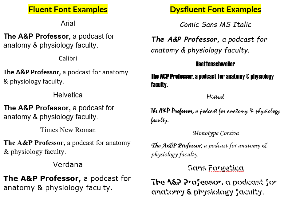
samples of fluent and dysfluent fonts


People

Contributors: David Curole, Terry Thompson

Mentions: Wendy Riggs, Natalie Wade, Jaime Chapman, Robert Bringhurst, Carla Delgado

Production: Aileen Park (announcer), Andrés Rodriguez (theme composer,  recording artist), Rev.com team (transcription), Kevin Patton (writer, editor, producer, host)


If the hyperlinks here are not active, go to TAPPradio.org to find the episode page.

★ More details at the episode page: theAPprofessor.org/podcast-episode-123.html

★ Transcript available in the transcript box: theAPprofessor.org/podcast-episode-123.html

★ Need help accessing resources locked behind a paywall? Check out this advice from Episode 32 to get what you need! my-ap.us/paywall

Take The A&P Professor experience to the next level!

theAPprofessor.org/community

Earn cash by referring other A&P faculty to this podcast:

theAPprofessor.org/refer

Tools & Resources

★ TAPP Science & Education Updates: theAPprofessor.org/updates

★ Amazon: amzn.to/2r6Qa3J

★ Text Expander: theapprofessor.org/textexpander

★ Rev.com: try.rev.com/Cw2nZ

★ Snagit & Camtasia: techsmith.pxf.io/9MkPW

★ Krisp Free Noise-Cancelling App: theAPprofessor.org/krisp

★ JotForm (build forms for free): theAPprofessor.org/jotform

★ QuillBot (writing tools): theAPprofessor.org/quillbot

★ The A&P Professor Logo Items: https://www.teepublic.com/stores/the-a-p-professor

Sponsors

★ Transcript and captions for this episode are supported by the American Association for Anatomy | anatomy.org

★ The Human Anatomy & Physiology Society provides marketing support for this podcast | theAPprofessor.org/haps

★ Distribution of this episode is supported by the Northeast College of Health Sciences online graduate program in Human Anatomy & Physiology Instruction (HAPI) | northeast.edu/hapi

Clicking on sponsor links helps let them know you appreciate their support of this podcast!

Follow The A&P Professor on  Twitter, Facebook, Blogger, Revue, Tumblr, or Instagram @theAPprofessor

The A&P Professor® and Lion Den® are registered trademarks of Lion Den Inc. (Kevin Patton)

As an Amazon Associate I earn from qualifying purchases. I may be compensated for links to sponsors and certain other links.


Click here to listen to this episode—or access the detailed notes and transcript.

Monday, February 11, 2019

The Last Best Story in Teaching Anatomy & Physiology | Episode 37


01:17 | Feedback in Online Tests
08:17 | The Anatomical Compass
14:47 | Sponsored by AAA
15:12 | Reserve hematopoiesis
18:09 | Sponsored by HAPS
18:54 | Featured: Last Best Story in Adult Neurogenesis & ANS Pathways
If you cannot see or activate the audio player click here.

Questions & Feedback: 1-833-LION-DEN (1-833-546-6336)
Follow The A&P Professor on Twitter, Facebook, Blogger, Nuzzel, Tumblr, or Instagram!

Scientific theories are tested every time someone makes an observation or conducts an experiment, so it is misleading to think of science as an edifice, built on foundations. Rather, scientific knowledge is more like a web. The difference couldn’t be more crucial. A tall edifice can collapse – if the foundations upon which it was built turn out to be shaky. But a web can be torn in several parts without causing the collapse of the whole. The damaged threads can be patiently replaced and re-connected with the rest – and the whole web can become stronger, and more intricate. (Massimo Pigliucci)

1 | Feedback in Online tests

7 minutes
In Episode 36, Adam Rich called in regarding how we can provide feedback to students taking online tests. I responded that I encourage students to get the correct response from their study buddies—or from me. After the episode aired, Krista Rompolski pointed out that this could be a challenge in very large courses. What do y'all think? Tell us. Really.


2 | The Anatomical Compass

6.5 minutes
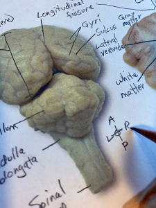
Although you and I are comfortable in orienting ourselves to anatomical directions when looking at diagrams, photographs, and specimens in anatomy, our beginning student often are not. The simple process of adding an "anatomical rosette" reflecting the anatomical directions in each encountered diagram can  help students develop the skill of understanding anatomical perspective.

 anatomical rosette


3 | Sponsored by AAA

0.5 minutes
The searchable transcript for this episode, as well as the captioned audiogram of this episode, are sponsored by The American Association of Anatomists (AAA) at anatomy.org. Their big meeting is in April at the Experimental Biology (EB) meeting in Orlando FL. Check it out!
 American Association of Anatomists

4 | Reserve Hematopoiesis

3 minutes
Hematopoietic stem cells  (HSCs) may have a "back-up system" that helps out after damage to the working population. These "reserve" HSCs (rHSCs) may step up when the primed HSCs (pHSCs) cannot keep up with the demand for hematopoiesis.
  • Scientists have identified a bone marrow backup system (summary article) my-ap.us/2BmcoE0
  • N-Cadherin-Expressing Bone and Marrow Stromal Progenitor Cells Maintain Reserve Hematopoietic Stem Cells
    (report by Zhao, et al. in Cell Reports) my-ap.us/2Bk7vLN

 hematopoietic stem cell


5 | Sponsored by HAPS

0.5 minutes
The Human Anatomy & Physiology Society (HAPS) is a sponsor of this podcast. Did you know there's a one-day regional HAPS conference in March? Check it out. You can help appreciate their support by clicking the link below and checking out the many resources and benefits found there.
 HAPS logo

6 | Featured: Last Best Story in Adult Neurogenesis & ANS Pathways

12 minutes
The "last best story" is what I tell my students I'm providing to them. That approach emphasizes the evolving nature of scientific understanding. In this episode, I mention two stories that are evolving right now.
reading a book

If the hyperlinks here are not active, go to TAPPradio.org to find the episode page.

Amazon and TextExpander referrals help defray podcasting expenses.

Transcript and captions for this episode
are supported by theAmerican Association of Anatomists.anatomy.org


The Human Anatomy & Physiology Societyalso provides support for this podcast.
theAPprofessor.org/haps


(Clicking on sponsor links 
helps let them know you appreciatetheir support of this podcast!)

Click here to listen to this episode—or access the detailed notes and transcript.


Friday, January 5, 2018

Platelets vs. Bacteria

Platelets as potent scavengers of bacteria? Really?

Something like 750 billion tiny cell fragments called platelets circulate in the human blood stream. When an injury to a blood vessel occurs, they stick to the exposed collagen in groups—forming platelet plug. And trigger additional reactions that eventually result in a blood clot.

But did you know that they have other helpful jobs, too? Like rounding up bacteria and feeding them up to immune cells, which devour them to make us safe.

This innate immune function of platelets has recently been outlined by researchers, as the information below summarizes.

Read through the quick points below to get an overview of some immune functions of platelets. Then read the full articles if you want to know more about these discoveries—including some great diagrams, micrographs, and videos.

[A short item on this topic also appears in today's edition of my daily Nuzzel newsletter of curated headlines for A&P professors.]

Quick points about platelets as bacterial scavengers


  • At sites of vessel injury/inflammation, platelets that contact intact collagen stick together—but platelets that do not contact collagen are motile.

  • Motile platelets change shape from a "fried egg" to a polarized "half moon" to better navigate the shearing forces of blood flow.

    • They can even navigate "upstream" against the flow of blood.

  • Platelets can use mechanical force to pull particles—including bacteria—from surrounding substrates.

  • Platelets collect and bundle bacteria, which facilitates neutrophil activation and subsequent phagocytosis. 

Migration pattern of motile platelet (left). Platelets collecting bacteria into bundles.

What can we use from this in teaching undergraduate A&P?


  • Yeah, okay we don't have time to go into all the ins and outs of platelets in a typical A&P course, but we can mention that platelets are now known to have immune functions.

    • Consider circling back to this mention later, when (if) you cover innate immune mechanisms a bit later in the course.

  • Consider calling attention to the sensory functions needed for platelets to analyze their microenvironment within the bloodstream.

  • Consider pointing out the specialized structure and function of the platelet's plasma membrane.

    • Integrins (integral membrane proteins) have a role in detecting particles for adhesion, binding to them, and sorting them.

    • Invaginations of the plasma membrane facilitate bundling of bacteria.

  • The shape changes needed for migration and handling of bacteria require actin-myosin reactions to power them. As in muscle fibers, these contractions are triggered by influx of extracellular calcium. In case you want to circle back to that.

  • Perhaps we should make a stronger point in reminding students that although they are "cell fragments" without a nucleus, they're more than just bags of hemostatic chemicals.

  • All these opportunities to "circle back" to previously studied concepts helps students make connections in their developing conceptual framework. And help them form a better understanding of the "big picture."

Want to know more?


Platelets, On Your Marks, Get Set, Migrate!

  • Bambach S, Lämmermann T. Cell. 2017 vol: 171 (6) pp: 1256-1258
  • Introduction to the Gaertner, et. al., paper below—giving background and overview to enhance understanding of the new discoveries. Great diagram, too! Click "Supplemental information" in the article to access video clips. 
  • my-ap.us/2EbU6Eu


Migrating Platelets Are Mechano-scavengers that Collect and Bundle Bacteria.

  • Gaertner F et. al. Cell. 2017 vol: 171 (6) pp: 1368-1382.e23
  • Journal article describing the scavenger role of platelets. Includes a few very nice, simple diagrams—and some cool micrographs and data graphs. These can also be downloaded as PowerPoint slides. Click "Supplemental information" in the article to access video clips. 
  • my-ap.us/2F5iM2U


Platelets Mediate Host Defense against Staphylococcus aureus through Direct Bactericidal Activity and by Enhancing Macrophage Activities.

  • Ali R et. al. Journal of immunology. 2017 vol: 198 (1) pp: 344-351
  • Journal article that supports the concept that platelets can kill MRSA bacteria and enhance their phagocytosis by macrophages.
  • my-ap.us/2EaO5be


[NOTE: If you can't access the full text of any resource, ask your school's reference librarian for help. If they can't provide direct access, they'll probably know how to get a copy of the resource for you. Quickly.]


Sketch: パタゴニア
Photos: LMU

Thursday, February 9, 2017

How Dietary Fiber Prevents Disease

So why, exactly, is it that we should consume a lot of fiber in our diet to remain healthy? Are refined fiber supplements just as good as, say, an "apple a day?"

Recently, an article in the journal Cell answer seems to verify some of the answers for us.

As the paper cited below indicates, research seems to confirm that dietary fiber provides nutrients for the inhabitants of our intestinal microbiome.  When dietary fiber is missing, then the microbes undergo a shift in populations and start consuming our GI mucus as an alternate source of nutrition.  That, as you might guess, reduces the thickness of the protective mucus—hus increasing the likelihood that pathogens can more easily attack the intestinal lining. Ouch.

Apparently, refined prebiotic fibers don't fix the problem.

Here are some highlights of the research article (quoted from their online preview):

  • Characterized synthetic bacterial communities enable functional insights in vivo
  • Low-fiber diet promotes expansion and activity of colonic mucus-degrading bacteria
  • Purified prebiotic fibers do not alleviate degradation of the mucus layer
  • Fiber-deprived gut microbiota promotes aggressive colitis by an enteric pathogen

Image for unlabelled figure


What can we use from this in teaching undergraduate A&P?


  • When asked by students about dietary fiber, you have more information from which to draw an answer.
  • When discussing any of these topics, you'll now have a bit more to add to your story:
    • nutrition
    • function of mucus
    • the human microbial system (or specifically, the GI microbiome)
    • how pathogens cause disease (or specifically, GI disorders)

Want to know more?


Veggies and Intact Grains a Day Keep the Pathogens Away

  • Francesca S. Gazzaniga. Dennis L. Kasper. Cell. Available online 17 November 2016
  • Brief preview of the M. Desai article cited below.
  • my-ap.us/2lijkbE


A Dietary Fiber-Deprived Gut Microbiota Degrades the Colonic Mucus Barrier and Enhances Pathogen Susceptibility

  • Mahesh S. Desai et al. Cell, Volume 167, Issue 5, 17 November 2016, Pages 1339-1353.e21
  • The detailed research article.
  • my-ap.us/2lijz6w

Microbiome articles

  • Kevin Patton. The A&P Professor. Various dates.
  • Collection of previous posts on this topic from this blog.
  • my-ap.us/2liCrSP


Photos: Youssef KH (top) Cell (bottom)

Monday, November 28, 2016

Sacral Efferent Pathways are Sympathetic, Not Parasympathetic

A recent report in the journal Science proposed a big change in how we understand the sympathetic and parasympathetic pathways of the autonomic nervous system (ANS).

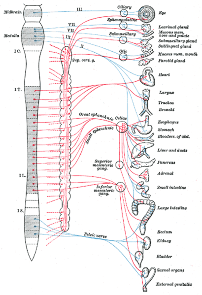
In a nutshell, the new model stipulates that the outflow (efferent pathways) are divided into a cranial division and spinal division—not the craniosacral and thoracolumbar divisions that we learned (and that exist in all A&P textbooks):

Current model:
  • Craniosacral division (parasympathetic outflow)
  • Thoracolumbar division (sympathetic outflow)
New model:
  • Cranial division (parasympathetic outflow)
  • Spinal division (sympathetic outflow)
The authors lay out embryological and genetic phenotype evidence to show that the sacral components of the ANS outflow pathways are similar to sympathetic thoracic pathways—not to cranial parasympathetic pathways as we have long supposed. 

But wait, you say, what about the parasympathetic control of the genitals, rectum, bladder? What about, well, all kinds of things that now seem to unravel? I suggest reading the rather brief and plainly written article in Science for the full answer. 

However, a few quick points may reduce your blood pressure a bit—and perhaps pique your interest.

Quick points about the new ANS model

  • Thoracic and sacral pathways share common embryologic development by location and when looking at transcriptional markers associated with neurotransmitters that differ from the developmental pattern of cranial pathways.

  • Thoracic and sacral pathways have a ventral exit point from the spinal cord; cranial pathways have a dorsal exit point.

  • The pelvic ganglion has been considered a "mixed" sympathetic/parasympathetic ganglion because it receives fibers from both the upper lumbar and sacral segments. But if the sacral pathways are sympathetic, the pelvic ganglion is clearly a sympathetic ganglion (not mixed). 

  • Analyses of transcription factors show that cells of the pelvic ganglia resemble those sympathetic ganglia and do not resemble cells in cranial ganglia.

  • The supposed lumbar vs. sacral antagonism in the urinary bladder's detrusor muscle does not seem to hold up, with the lumbar inhibitory effects either not demonstrable in experiments or of questionable functional relevance.

  • The effects on vessel dilation in genitals can be explained as a "continuity of action—rather than antagonism"

  • The sacral pathway to the rectum seems to resemble sympathetic structure, not cranial (parasympathetic) structure.

What can we use from this in teaching undergraduate A&P?

  • When covering the craniosacral/thoracolumbar scheme, consider mentioning this newly proposed model.

  • Consider using this scenario to illustrate the dynamic nature of science. Perhaps discuss that long-held dogma is occasionally challenged using newer methods and ways of thinking.

  • Consider discussing pros and cons of adopting the new model. For example, can evidence from mice extend to all vertebrates? Which is stronger, evidence for the current model or the new model? Which model is most useful in understanding principles of ANS regulation? A little critical thinking never hurt anyone (at least not much).

Want to know more?


The sacral autonomic outflow is sympathetic
  • I. Espinosa-Medina, O. Saha, F. Boismoreau, Z. Chettouh, F. Rossi, W. D. Richardson, J.-F. Brunet. Science  18 Nov 2016: Vol. 354, Issue 6314, pp. 893-897 DOI: 10.1126/science.aah5454
  • Peer-reviewed research report describing this discovery, Includes an updated version of the classic diagram of sympathetic and parasympathetic pathways.
  • my-ap.us/2fNdcF3

Neural circuitry gets rewired
  • Adameyko, I. Science 18 Nov 2016: Vol. 354, Issue 6314, pp. 833-834 DOI: 10.1126/science.aal2810
  • Companion article to the report cited above, stating that "This finding provokes a serious shift in textbook knowledge, and, as with any fundamental discovery, it brings important practical implications..." and goes on to mention of a few of the implications (e.g., how to treat bladder dysfunction).
  • my-ap.us/2gg9O8P

The Autonomic Nervous System. Part I.
  • John Newport Langley. W. Heffer & Sons Ltd., Cambridge, 1921.80pp.
  • Classic "primary source" that codified the modern concept of the ANS. 
  • my-ap.us/2fYHt3M

Gray's Anatomy ANS diagram
  • Henry Gray. 1918 (online edition at Bartleby)
  • Classic diagram by Henry Vandyke Carter of ANS pathways from an early edition of Gray's Anatomy.
  • my-ap.us/2fYGMaT or my-ap.us/2gcAmaW

Tuesday, July 14, 2015

Dissolving Microneedle Vaccinations

Researchers recently demonstrated that a flu vaccine delivered using microneedles that dissolve in the skin can protect people against infection even better than the standard needle-delivered vaccine.


The new microneedle patch is made of dissolvable material, eliminating needle-related risks. Not to mention the sea change it may mean for patients with severe needle anxiety!  I suspect this approach may also be more tolerable for many patients than oral and nasal vaccination methods. It is also easy to use without the need for trained medical personnel—making it ideal for use where healthcare resources are limited.

“Our novel transcutaneous vaccination using a dissolving microneedle patch is the only application vaccination system that is readily adaptable for widespread practical use,” said Professor Shinsaku Nakagawa, one of the authors of the study from Osaka University. “Because the new patch is so easy to use, we believe it will be particularly effective in supporting vaccination in developing countries.”

The new microneedle patch – MicroHyala – is dissolvable in water. The tiny needles are made of hyaluronic acid, a naturally occurring substance in tissue matrix and the synovial fluid that cushions the joints. When the patch is applied sort of like a Band-Aid, the needles pierce the epidermis of skin and dissolve into the body, taking the vaccine with them.

The researchers compared the new system to traditional needle delivery by vaccinating two groups of people against three strains of influenza: A/H1N1, A/H3N2 and B. None of the subjects had a bad reaction to the vaccine, showing that it is safe to use in humans. The patch was also effective: people given the vaccine using the microneedles had an immune reaction that was equal to or stronger than those given the vaccine by injection.

“We were excited to see that our new microneedle patch is just as effective as the needle-delivered flu vaccines, and in some cases even more effective,” said Professor Nakagawa.

Previous research has evaluated the use of microneedles made of silicon or metal, but they were not shown to be safe. Microneedles made from these materials also run the risk of breaking off in the skin, leaving tiny fragments behind. The new dissolvable patch eliminates this risk because the microneedles are designed to dissolve in the skin.

What can we use from this in teaching undergraduate A&P?


  • Consider mentioning this advance when discussing the layers of the skin, this giving a clinical application to pique student interest.

  • When discussing immunity and vaccination, consider mentioning this discovery.

  • If you discuss hyaluronic acid when covering histology, this information may help students realize the importance of knowing such details because of clinical applications of materials science.

Want to know more?


  • Clinical study and stability assessment of a novel transcutaneous influenza vaccination using a dissolving microneedle patch.
    • Sachiko Hirobe, et al. Biomaterials. Vol 57 (July 2015), Elsevier. doi: 10.1016/j.biomaterials.2015.04.007
    • The original research article.
    • my-ap.us/1eXzAud


Microneedle image courtesy of S. Nakagawa
Some content adapted from an Elsevier newsroom release

Wednesday, June 3, 2015

A Brain-Lymphatic Connection

Existing dogma in neuroscience states that the brain does not possess the classical lymphatic drainage system found in other parts of body. However, a recent letter in the journal Nature reports the discovery of lymphatic vessels lining the dural sinuses in mice. These were shown to drain immune cells and cerebrospinal fluid (CSF) into the deep cervical lymph nodes.

Although more work is yet to be done in humans, this discovery will cause neuroscientists to revisit a number of concepts related to CSF and lymphatic drainage, as well as immune functions in the brain.

For example, do these new data truly challenge the notion of immune privilege in the nervous tissue of the central nervous system—or do they apply to the brain as an organ and allow for lymphatic drainage of tissues outside the nervous tissue of the brain?

What can we use from this in teaching undergraduate A&P?

  • When discussing CSF drainage, consider mentioning the possibility that filtration of CSF directly into dural sinuses may be augmented by the newly discovered lymphatic drainage.

  • If you discuss the dogma of the "immune privilege" of the brain, consider mentioning this possible challenge to the concept. This may trigger a great discussion of whether these newly discovered lymphatic vessels are truly "in the brain."

  • If you discuss disorders involving altered immunity, such as multiple sclerosis, consider mentioning this discovery.

  • Bringing up this new information may be useful in discussions related to the process of science—how existing concepts are sometimes challenged by new information, for example. Perhaps a discussion of the need for more investigation would stimulate students to think about what future steps can be taken to map out a possible lymphatic network in or around the brain.
Current concept of lymphatic drainage (left) compared to updated version to reflect new data (right).

Want to know more?


Brain Drain | The brain contains lymphatic vessels similar to those found elsewhere in the body, a mouse study shows.
  • Ashley P. Taylor, The Scientist, June 1, 2015 (online)
  • Plain-English article summarizing the study and its significance.
  • my-ap.us/1IezLLF
Structural and functional features of central nervous system lymphatic vessels,
  • A. Louveau et al., Nature, June 1, 2015.doi:10.1038/nature14432, 
  • The original research article. Includes images and video.
  • my-ap.us/1KNFPy6
Missing link found between brain, immune system; major disease implications
  • University of Virginia Health System, Science Daily, June 1, 2015
  • Illustrated press release describing the research.
  • my-ap.us/1HQys2J

Images: DBCLS (top)
Univ Va Health System (bottom)

Friday, January 2, 2015

Fat Cells in Skin Kill Bacteria


Scientists reported today that adipocytes in mouse and human skin produce an antimicrobial peptide (AMP) called cathelicidin is response to Staph aureus infections, including MRSA. Experimental animals that were deficient in the AMP were more susceptible to skin infections.

Adipocytes may recognize S. aureus by detecting bacterial peptides with toll-like receptors (TLRs), but more work is needed to fully understand the mechanisms.

This finding adds more to our understanding of human skin as a vital part of our body's defenses against infection. It also opens the door to understanding how diabetes, metabolic syndrome, and other conditions can reduce resistance to skin infections by altering the availability of AMPs in the fat associated with skin.

All of this may eventually lead to additional—perhaps more effective—strategies in preventing or curing serious skin infections such as MRSA.

I realize that we generally think of fat cells as belonging to the hypodermis, not the dermis, as described in the research. However, recent evidence shows the presence of adipocytes in the dermis that are distinct from those in the hypodermis. These adipocytes derive from a common precursor cell that produces both dermal fibroblasts and intradermal adipocytes. These dermal adipocytes have been shown to have a role in wound healing and the regeneration of hair follicles. And the research summarized here suggests that they also have a role in immunity.


What can we use from this in teaching undergraduate A&P?

  • Mention this discovery when discussing the roles of adipose tissue and adipocytes in your coverage of tissues of the body.

    • Consider clarifying that dermal adipocytes are distinct from fat cells in the hypdermis. And perhaps mention that it's a detail often left out of introductory discussions of skin.

  • This is a good point to mention when discussing the protective functions of the skin when covering the integumentary system.

  • When discussing the immune system, this concept helps illustrate several important principles:

    • The role of the skin as the first line of defense against infection

      • The variety of mechanisms available in the skin to act defensively

    • The role of TLRs and pattern recognition in immunity

    • The fact that immunity is a role for many tissues—not just lymphocytes and other WBCs

  • Take a moment NOW to add this to your course notes!

Want to know more?


Killer Fat
  • J. Alcorn and J. Kolls. Science 2 January 2015: Science Vol. 347 no. 6217 pp. 26-27 DOI: 10.1126/science.aaa4567
  • Editorial summary of the research in plain English. Includes a really nice, simple illustration of the concept (includes FREE teaching slide)
  • my-ap.us/1vBWbNP

Dermal adipocytes protect against invasive Staphylococcus aureus skin infection
  • L. Zhang1, et al. Science 2 January 2015: Vol. 347 no. 6217 pp. 67-71 DOI: 10.1126/science.126097
  • Original research article. Additional images available here, including some nice micrographs showing increase in adipocytes in response to S. aureus infection
  • my-ap.us/1xePNS4

Defining dermal adipose tissue.
  • Driskell RR, et al. Exp Dermatol. 2014. Exp Dermatol. 2014 Sep;23(9):629-31. doi: 10.1111/exd.12450.
  • Review article describing dermal adipocytes.
  • my-ap.us/1GuH3bL

FREE teaching slide
Click the image
to download a


Adipose image credit: my-ap.us/13MYGWO
This post was updated 6 OCT 2015

Wednesday, October 8, 2014

Nobel Prize 2014: Super-resolved fluorescence microscopy


The Royal Swedish Academy of Sciences has decided to award the Nobel Prize in Chemistry for 2014 to

Eric Betzig
Janelia Farm Research Campus, Howard Hughes Medical Institute, Ashburn, VA, USA,

Stefan W. Hell
Max Planck Institute for Biophysical Chemistry, Göttingen, and German Cancer Research Center, Heidelberg, Germany

and

William E. Moerner
Stanford University, Stanford, CA, USA

“for the development of
super-resolved fluorescence microscopy”


Surpassing the limitations of the light microscope


For a long time optical microscopy was held back by a presumed limitation: that it would never obtain a better resolution than half the wavelength of light. Helped by fluorescent molecules the Nobel Laureates in Chemistry 2014 ingeniously circumvented this limitation. Their ground-breaking work has brought optical microscopy into the nanodimension.

In what has become known as nanoscopy, scientists visualize the pathways of individual molecules inside living cells. They can see how molecules create synapses between nerve cells in the brain; they can track proteins involved in Parkinson’s, Alzheimer’s and Huntington’s diseases as they aggregate; they follow individual proteins in fertilized eggs as these divide into embryos.

It was all but obvious that scientists should ever be able to study living cells in the tiniest molecular detail. In 1873, the microscopist Ernst Abbe stipulated a physical limit for the maximum resolution of traditional optical microscopy: it could never become better than 0.2 micrometres. Eric Betzig, Stefan W. Hell and William E. Moerner are awarded the Nobel Prize in Chemistry 2014 for having bypassed this limit. Due to their achievements the optical microscope can now peer into the nanoworld.

Two separate principles are rewarded. 


One enables the method stimulated emission depletion (STED) microscopy, developed by Stefan Hell in 2000. Two laser beams are utilized; one stimulates fluorescent molecules to glow, another cancels out all fluorescence except for that in a nanometre-sized volume. Scanning over the sample, nanometre for nanometre, yields an image with a resolution better than Abbe’s stipulated limit.

Eric Betzig and William Moerner, working separately, laid the foundation for the second method, single-molecule microscopy. The method relies upon the possibility to turn the fluorescence of individual molecules on and off. Scientists image the same area multiple times, letting just a few interspersed molecules glow each time. Superimposing these images yields a dense super-image resolved at the nanolevel. In 2006 Eric Betzig utilized this method for the first time.

Today, nanoscopy is used world-wide and new knowledge of greatest benefit to mankind is produced on a daily basis.


This video is a brief animation of how STED works and how it improves resolution of individual particles.


This video is a longer, more detailed presentation by one of the Nobel laureates (Hell).


What can we use from this in teaching undergraduate A&P?

  • Discuss how this technology has enabled us to better visualize the chemicals and structures within our cells, enabling scientists to better understand the structure and function of cell, organelles, microbiome constituents, and other structures of the human body.

  • If you do a brief run-through of the theory of microscopy—perhaps in your A&P lab—you can add a mention of this technology.  

  • Your textbook or other learning resource may already have an example of this type of microscopy.

  • A discussion of this  Nobel Prize could evolve into a meaningful example of how science works, including how incremental improvements in classical tools for observation expand the number of questions that can be answered.

  • Use the links below (and images above) to use for a handout and/or teaching slides.


Want to know more?


Resources from Nobelprize.org

  • Popular Information 
  • Scientific Background
    • Handout: More detailed information includes references to original research articles
    • my-ap.us/ZdLJ69
  • Advanced Information
  • Images
  • Biographies
    • Eric Betzig, 
      • U.S. citizen. Born 1960 in Ann Arbor, MI, USA. Ph.D. 1988 from Cornell University, Ithaca, NY, USA. Group Leader at Janelia Farm Research Campus, Howard Hughes Medical Institute, Ashburn, VA, USA.
      • http://janelia.org/lab/betzig-lab
    • Stefan W. Hell, German citizen. 
      • Born 1962 in Arad, Romania. Ph.D. 1990 from the University of Heidelberg, Germany. Director at the Max Planck Institute for Biophysical Chemistry, Göttingen, and Division head at the German Cancer Research Center, Heidelberg, Germany.
      • http://www3.mpibpc.mpg.de/groups/hell
    • William E. Moerner, U.S. citizen. 
      • Born 1953 in Pleasanton, CA, USA. Ph.D. 1982 from Cornell University, Ithaca, NY, USA. Harry S. Mosher Professor in Chemistry and Professor, by courtesy, of Applied Physics at Stanford University, Stanford, CA, USA.
      • http://web.stanford.edu/group/moerner


Diagrram cretit: Ganbaatar
Micrograph credit: Tesselkaffee
Text adapted from press release from Nobel Media

Monday, July 21, 2014

Biological Pacemaker Using Gene Therapy


Researchers recently induced ordinary cardiac muscle fibers into becoming functioning pacemaker cells by injecting a therapeutic gene.

Working with pigs, a common model for human cardiovascular research, researchers first destroyed the natural pacemaker cells in each subject's heart and installed an electronic pacemaker. They then inserted a gene for transcription factor TBX18 into cardiac muscle tissue using an adenovirus.  Using adenovirous vectors for inserting genes is a common strategy in gene therapy.

Within a couple of days, ordinary myocardial fibers had developed the structure and function of pacemaker cells.  In about 5 days, the electronic pacemakers were no longer needed.

However, this biological pacemaking peaked at about 8 days, then eventually disappeared.  This may occur because the virus-infected cells are probably destroyed by the body's immune defenses.  So researchers are thinking that perhaps, at the very least, this could eventually lead to a temporary treatment for certain arrythmias in humans.


What can we use from this in teaching undergraduate A&P?  


  • This is an interesting bit of news that helps illustrate the frontiers of human biomedical sciences.
  • This story provides a good case to provoke a discussion of the nature of gene therapy.  
    • Why did the effect last only 8 or so day?  
    • What does this tell us about transcription factor TBX18?  
    • What benefit might this treatment have if developed for humans?
  • This may add interest to an discussion of the function of the electrical system of the heart in general, and artificial pacemakers in particular.
  • The case also provides a scenario in which the body attacks and destroys virus-infected cells.
FREE image you can use in your course

Want to know more?


Next Generation: Biological Pacemakers

  • R Williams, The Scientist (the-scientist.com) July 16, 2014
  • Plain-English article summarizing the discovery.  Includes quotes from the researchers.
  • my-ap.us/1ruocYu


Biological pacemaker created by minimally invasive somatic reprogramming in pigs with complete heart block
  • Y-F. Hu et al., Science Translational Medicine, 6:245ra94, 2014. DOI:10.1126/scitranslmed.3008681
  • Original research report.
  • my-ap.us/1nfErcA
FREE image you can use in your course

Photo credit: vfdbsn

Monday, October 7, 2013

Nobel Prize: Vesicle Transport

The Nobel Assembly at Karolinska Institutet has today decided to award the 2013 Nobel Prize in Physiology or Medicine jointly to James E. Rothman, Randy W. Schekman and Thomas C. Südhof for their discoveries of machinery regulating vesicle traffic, a major transport system in our cells.

Those of you now teaching A&P 1 have probably recently covered the essential concepts vesicle transport and have perhaps already had the opportunity to apply them to specific functions of the body such as the release of acetylcholine at the neuromuscular junction.  Bringing up today's news gives us a great chance to underscore the importance of a topic that many students wonder, "why do we have to know this?"

Near the bottom of  this post, you'll find a link to a nice handout that you can distribute to your class (or link to from an email or webpage).  I've often added a question on my midterm exam covering that year's relevant Nobel Prize concept.

Summary

The 2013 Nobel Prize honors three scientists who have solved the mystery of how the cell organizes its transport system. Each cell is a factory that produces and exports molecules. For instance, insulin is manufactured and released into the blood and chemical signals called neurotransmitters are sent from one nerve cell to another. These molecules are transported around the cell in small packages called vesicles. The three Nobel Laureates have discovered the molecular principles that govern how this cargo is delivered to the right place at the right time in the cell.

Randy Schekman discovered a set of genes that were required for vesicle traffic. James Rothman  unravelled protein machinery that allows vesicles to fuse with their targets to permit transfer of cargo. Thomas Südhof revealed how signals instruct vesicles to release their cargo with precision.

Through their discoveries, Rothman, Schekman and Südhof have revealed the exquisitely precise control system for the transport and delivery of cellular cargo. Disturbances in this system have deleterious effects and contribute to conditions such as neurological diseases, diabetes, and immunological disorders.

How cargo is transported in the cell

In a large and busy port, systems are required to ensure that the correct cargo is shipped to the correct destination at the right time. The cell, with its different compartments called organelles, faces a similar problem: cells produce molecules such as hormones, neurotransmitters, cytokines and enzymes that have to be delivered to other places inside the cell, or exported out of the cell, at exactly the right moment. Timing and location are everything. Miniature bubble-like vesicles, surrounded by membranes, shuttle the cargo between organelles or fuse with the outer membrane of the cell and release their cargo to the outside. This is of major importance, as it triggers nerve activation in the case of transmitter substances, or controls metabolism in the case of hormones. How do these vesicles know where and when to deliver their cargo?

Traffic congestion reveals genetic controllers

Randy Schekman was fascinated by how the cell organizes its transport system and in the 1970s decided to study its genetic basis by using yeast as a model system. In a genetic screen, he identified yeast cells with defective transport machinery, giving rise to a situation resembling a poorly planned public transport system. Vesicles piled up in certain parts of the cell. He found that the cause of this congestion was genetic and went on to identify the mutated genes. Schekman identified three classes of genes that control different facets of the cell´s transport system, thereby providing new insights into the tightly regulated machinery that mediates vesicle transport in the cell.

Docking with precision

James Rothman was also intrigued by the nature of the cell´s transport system. When studying vesicle transport in mammalian cells in the 1980s and 1990s, Rothman discovered that a protein complex enables vesicles to dock and fuse with their target membranes. In the fusion process, proteins on the vesicles and target membranes bind to each other like the two sides of a zipper. The fact that there are many such proteins and that they bind only in specific combinations ensures that cargo is delivered to a precise location. The same principle operates inside the cell and when a vesicle binds to the cell´s outer membrane to release its contents.

It turned out that some of the genes Schekman had discovered in yeast coded for proteins corresponding to those Rothman identified in mammals, revealing an ancient evolutionary origin of the transport system. Collectively, they mapped critical components of the cell´s transport machinery.

Timing is everything

Thomas Südhof was interested in how nerve cells communicate with one another in the brain. The signalling molecules, neurotransmitters, are released from vesicles that fuse with the outer membrane of nerve cells by using the machinery discovered by Rothman and Schekman. But these vesicles are only allowed to release their contents when the nerve cell signals to its neighbours. How is this release controlled in such a precise manner? Calcium ions were known to be involved in this process and in the 1990s, Südhof searched for calcium sensitive proteins in nerve cells. He identified molecular machinery that responds to an influx of calcium ions and directs neighbour proteins rapidly to bind vesicles to the outer membrane of the nerve cell. The zipper opens up and signal substances are released. Südhof´s discovery explained how temporal precision is achieved and how vesicles´ contents can be released on command.

Vesicle transport gives insight into disease processes

The three Nobel Laureates have discovered a fundamental process in cell physiology. These discoveries have had a major impact on our understanding of how cargo is delivered with timing and precision within and outside the cell.  Vesicle transport and fusion operate, with the same general principles, in organisms as different as yeast and man. The system is critical for a variety of physiological processes in which vesicle fusion must be controlled, ranging from signalling in the brain to release of hormones and immune cytokines. Defective vesicle transport occurs in a variety of diseases including a number of neurological and immunological disorders, as well as in diabetes. Without this wonderfully precise organization, the cell would lapse into chaos.

About this year's Nobel laureates

James E. Rothman was born 1950 in Haverhill, Massachusetts, USA. He received his PhD from Harvard Medical School in 1976, was a postdoctoral fellow at Massachusetts Institute of Technology, and moved in 1978 to Stanford University in California, where he started his research on the vesicles of the cell. Rothman has also worked at Princeton University, Memorial Sloan-Kettering Cancer Institute and Columbia University. In 2008, he joined the faculty of Yale University in New Haven, Connecticut, USA, where he is currently Professor and Chairman in the Department of Cell Biology.

Randy W. Schekman was born 1948 in St Paul, Minnesota, USA, studied at the University of California in Los Angeles and at Stanford University, where he obtained his PhD in 1974 under the supervision of Arthur Kornberg (Nobel Prize 1959) and in the same department that Rothman joined a few years later. In 1976, Schekman joined the faculty of the University of California at Berkeley, where he is currently Professor in the Department of Molecular and Cell biology. Schekman is also an investigator of Howard Hughes Medical Institute.

Thomas C. Südhof was born in 1955 in Göttingen, Germany. He studied at the Georg-August-Universität in Göttingen, where he received an MD in 1982 and a Doctorate in neurochemistry the same year. In 1983, he moved to the University of Texas Southwestern Medical Center in Dallas, Texas, USA, as a postdoctoral fellow with Michael Brown and Joseph Goldstein (who shared the 1985 Nobel Prize in Physiology or Medicine). Südhof became an investigator of Howard Hughes Medical Institute in 1991 and was appointed Professor of Molecular and Cellular Physiology at Stanford University in 2008.


Want to know more?


Handout showing mechanisms for which this prize was awarded

  • Diagrams and brief description of the contributions of each Nobel laureate
  • my-ap.us/1cnPDhF


Machinery Regulating Vesical Traffic, A Major Transport System in our Cells

  • Nobelprize.org accessed 7 October 2013
  • [Nice summary of the scientific concepts involved.  An expanded version of the handout, with additional diagrams and explanations.]
  • my-ap.us/GDLiZR


Original Journal Articles

  • Seminal papers describing the original work of this year's Nobel Laureates
  • Novick P, Schekman R: Secretion and cell-surface growth are blocked in a temperature-sensitive mutant of Saccharomyces cerevisiae. Proc Natl Acad Sci USA 1979; 76:1858-1862.
  • Balch WE, Dunphy WG, Braell WA, Rothman JE: Reconstitution of the transport of protein between successive compartments of the Golgi measured by the coupled incorporation of N-acetylglucosamine. Cell 1984; 39:405-416.
  • Kaiser CA, Schekman R: Distinct sets of SEC genes govern transport vesicle formation and fusion early in the secretory pathway. Cell 1990; 61:723-733.
  • Perin MS, Fried VA, Mignery GA, Jahn R, Südhof TC: Phospholipid binding by a synaptic vesicle protein homologous to the regulatory region of protein kinase C. Nature 1990; 345:260-263.
  • Sollner T, Whiteheart W, Brunner M, Erdjument-Bromage H, Geromanos S, Tempst P, Rothman JE: SNAP receptor implicated in vesicle targeting and fusion. Nature 1993; 362:318-324.
  • Hata Y, Slaughter CA, Südhof TC: Synaptic vesicle fusion complex contains unc-18 homologue bound to syntaxin. Nature 1993; 366:347-351.

Related textbook content

  • Anatomy & Physiology 8th ed.  Chapter 3 (see p. 75, 82), Chapter 4 (see p. 99-100), and throughout book my-ap.us/QZTbK1
  • Essentials of Anatomy & Physiology Chapter 3 (see p. 52), Chapter 4 (see p. 69), and throughout book my-ap.us/SCfNlj  
  • The Human Body in Health and Disease 6th ed. Chapter 3 (see p. 49, 56) and throughout book my-ap.us/X71LJO 
  • Structure & Function of the Body 14th ed. Chapter 3 (see p. 48, 53) and throughout book http://my-ap.us/10s50MH


Portions of this post adapted from Nobel Prize news release

Thursday, May 23, 2013

Acupuncture and connective tissue

A recent article in TheScientist proposes a novel theory about how acupuncture may produce some of its therapeutic effects.

It turns out that when an acupuncture needle is inserted into the connective tissue under the skin and twisted, the needle becomes wound with connective fibers in much the same way as noodles enwrap a fork as it twirls.  This pulls on fibroblasts and causes local changes that may be transmitted throughout a large area of connective tissue and possibly produce therapeutic effects.

ATP released from stretched fibroblasts may also contribute to a pain-relieving effect.

An interesting new direction for further study.  And a good little story to weave into a discussion of fibrous connective tissues in our A&P course to illustrate that "it's not just glue."

Want to know more?

The Science of Stretch
  • H.M. Langevin
  • TheScientist published online 1 May 2013
  • Article summarizing the context and results of the research.  Includes nice graphic showing stretched fibers wrapped around a needle.
  • http://my-ap.us/16VQK5X


Biomechanical response to acupuncture needling in humans
  • Helene M. Langevin
  • Journal of Applied Physiology December 1, 2001 vol. 91 no. 6 2471-2478
  • The original research article.
  • http://my-ap.us/121Rxkp

Monday, December 31, 2012

Rita Levi-Montalcini, growth factor pioneer

Yesterday, the scientific community lost another of its great people, Rita Levi-Montalcini.

In The Human Body in Health & Disease and Structure & Function of the Body, I wrote this about Levi-Montalcini:
Rita Levi-Montalcini had just finished a medical degree in her native Italy when in 1938 the Fascist government under Mussolini barred all “non-Aryans” from working in academic and professional careers. Being Jewish, Levi-Montalcini was forced to move to Belgium to work. But when Belgium was about to be invaded by the Nazis, she decided to return home to Italy and work in secret. Her home laboratory was very crude, but in it she made some important discoveries about how the nervous system develops during embryonic development. After World War II, she was invited to Washington University in St. Louis to work. There, she discovered the existence of nerve growth factor (NGF), for which she later won the 1986 Nobel Prize. Her discovery of a chemical that regulates the growth of new nerves during early brain development has led to many different paths of investigation. For example, by learning more about growth regulators we now know more about how the nervous system develops, as well as other tissues, organs, and systems of the body.
Note that I put in a little plug for my hometown of St. Louis, where we continue to be proud of this remarkable woman and her pioneering work.

As I said in a recent post about the passing of transplant pioneer Joseph Murray, I think the occasional story of a pioneer in the history of human science adds a lot to the A&P course.  Such stories give a human dimension to the pursuit of science and provide the context needed for students to understand how we know what we know.  Levi-Montalcini's story gives us the further opportunities to weave into our courses the themes of global collaboration among the scientific community as the role of women in science.

Want to know more?

  • Nobel Scientist Rita Levi-Montalcini Dies in Rome
  • Oldest Nobel winner Rita Levi-Montalcini dies at 103
  • Nobel Lecture by Rita Levi-Montalcini 
    • Media Player at Nobelprize.org
    • [Full video (in English) of Nobel lecture by Rita Levi-Montalcini in which she fully credits "good luck"; 57 minutes]
    • http://my-ap.us/Vf6sPz

  • Rita Levi-Montalcini Interview
    • Adam Smith, Editor-in-Chief of Nobelprize.org.
    • Nobel Interview, November 2008
    • [Video interview with Rita Levi-Montalcini, who talks about her daily work, why she had to make a laboratory in her bedroom to conduct research during World War II (3:06), the benefits of working in isolation (5:03), her post-war move to the United States (6:25), her work with Stanley Cohen and the discovery of nerve growth factor (7:15), the roles of intuition and chance in biological research (15:14), her current research (16:58), her advice to young scientists (17:41), and why this period of her life has been the best so far (28:10).]
    • http://my-ap.us/Ug6gA5

  • The Nobel Prize in Physiology or Medicine 1986 Press Release
    • Nobelprize.org
    • [Detailed news release that includes some simple diagrams that help illustrate the concepts involved.]
    • http://my-ap.us/ZPyacv
  • In Praise of Imperfection: My Life and Work
    • Rita Levi-Montalcini
    • Sloan Foundation Science Series, October 1989
    • [Her autobiography]
    • http://amzn.to/Wgjk7b


Related textbook content
  • Anatomy & Physiology 8th ed.  p. 409, 1111-1113, A&P Connect: The Nobel Legacy my-ap.us/QZTbK1
  • Essentials of Anatomy & Physiology p. 231-232, 241, 610-612 my-ap.us/SCfNlj  
  • The Human Body in Health and Disease 5th ed. p. 236-237, 644-645, 658 my-ap.us/fNN00N 
  • Structure & Function of the Body 14th ed. p. 168-169, 472-473 my-ap.us/X6QxqE


Photo: Presidenza della Repubblica Italiana