Showing posts with label free stuff. Show all posts

Saturday, November 18, 2017

Got High Blood Pressure Covered? The 2017 Hypertension Guidelines.

With the new guidelines for high blood pressure popping up all over the news recently, we may wonder what we need to know when this comes up in our A&P classrooms. And we know it will—students love, love, love to connect what they are learning in A&P with what they are experiencing in their lives. 

It turns out that although the new 2017 Guideline For the Prevention, Detection, Evaluation, and Management of High Blood Pressure in Adults is focused on how physicians should make diagnoses and manage patient care, the definitions of exactly what constitutes high blood pressure (hypertension or HTN) are important learning points in the undergrad A&P course.

I'll outline the main things for us A&P professors to know here, but do check out the resources I've linked below to deepen your understanding of current thinking regarding approaches to blood pressure (BP) health.

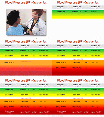
First, there are revised guidelines as to what constitutes high blood pressure or HTN:

  • Normal BP: Less than 120/80 mm Hg;
  • Elevated BP: Systolic between 120-129 and diastolic less than 80;
  • Stage 1 HTN: Systolic between 130-139 or diastolic between 80-89;
  • Stage 2 HTN: Systolic at least 140 or diastolic at least 90 mm Hg;
  • Hypertensive crisis: Systolic over 180 and/or diastolic over 120, with patients needing prompt changes in medication if there are no other indications of problems, or immediate hospitalization if there are signs of organ damage.


Regardless of the precise cutoffs listed above, in an interview discussing the new guidelines, the main author states that, "120/80 is normal, the same as we had before" the new guidelines. So I think we're safe in using 120/80 as an example of BP when discussing the normal science, even though technically it could be designated as "elevated." Not that we can't use an elevated variable measurement as an example when discussing the physiology of anything. The fact that even the main author of the guidelines uses 120/80 as the starting point of discussion makes me feel more confident in using it as the starting point of my course discussions, too.

The main thing to note in the categories above is that the cutoffs for HTN categories have been lowered. This puts many more people in an HTN category that were not there before. The main goal is for those folks to have conversations with their physicians to evaluate their risk for complications and develop a personalized prevention and care plan.

Note also that the category of prehypertension has been eliminated.

The new guidelines also recommend prescribing medication for Stage 1 HTN if the patient already had a cardiovascular event—or is at a high risk for such an event. They also recognize that many patients will need more than one medication to manage BP and that combining meds into one pill is likely to help folks take them consistently.

There are a lot of other recommendations, so reviewing the Executive Summary or similar resource (see below) may be a good idea.


What can we use from this in teaching undergraduate A&P?

  • If you discuss hypertension, or use case studies in teaching, you need to update the cutoff BPs you are using.
  • A BP of 120/80 is still considered the starting point for discussing blood pressure.
  • Consider discussing the impact of the changes in the new guidelines for ordinary people.
  • Discuss why such diagnosis, prevention, and treatment recommendations often change over time. Consider discussion other recent clinical updates.
  • Consider discussing specific changes suggested in the new guidelines.
  • Consider having students explore the Executive Summary and/or other documents and write their own summary or interpretation of key points. Perhaps they can create their own chart or concept map.


Need some free teaching materials?


SLIDE SET: High Blood Pressure
  • Kevin Patton. Lion Den Slide Collection. 18 Nov 2017
  • Small slide deck that includes an animated version of the BP Category chart pictured above. Part of the Lion Den Slide Collection (requires free registration to download). You can also download a static PNG image file of the chart in the slide collection set.
  • my-ap.us/2ivoql5


VIDEO: AHA 2017 | New High Blood Pressure Guidelines
  • America Heart Association. 13 Nov 2017.
  • Free video (viewable in the player above) features a chat with the main author of the new guidelines and summarizes the main points. Very practical and easy to understand.
  • youtu.be/rvYL-7ergDs


SLIDE SET: 2017 Guideline For the Prevention, Detection, Evaluation and Management of High Blood Pressure in Adults
  • American College of Cardiology. 13 Nov 2017
  • Free set of almost 100 PowerPoint slides to use in teaching. And it has a decided focus on clinical applications, rather than the basic science. These are way, way beyond the coverage desirable in an undergrad A&P course. But some slides may be useful to you.
  • my-ap.us/2itMBjY


Want to know more?


New blood pressure guidelines put half of U.S. adults in unhealthy range
  • A. Cunningham Science News. 13 Nov 2017 
  • Plain-English article summarizing the first major update since 2003 aims to spur heart-healthy lifestyle changes. Has a useful graph and links to other articles and resources.
  • my-ap.us/2itVnOR

New Multisociety Hypertension Guideline Is Released
  • Allan S. Brett, MD reviewing Whelton PK et al. J Am Coll Cardiol 2017 Nov 13. NEJM Journal Watch Nov 2017.
  • Brief review of the larger report (listed below), summarize key take-away points.
  • my-ap.us/2iumCc1

2017 ACC/AHA/AAPA/ABC/ACPM/AGS/APhA/ASH/ASPC/NMA/PCNA Guideline for the Prevention, Detection, Evaluation, and Management of High Blood Pressure in Adults: Executive Summary
A Report of the American College of Cardiology/American Heart Association Task Force on Clinical Practice Guidelines
  • PK Whelton et al. Hypertension, Dec 2017, Volume 70, Issue 6.  DOI: 10.1161/HYP.0000000000000066
  • Free PDF of the Executive Summary of the larger report. I recommend reading this first, then decide if you need to read the whole report.
  • my-ap.us/2iuzvCT

2017 ACC/AHA/AAPA/ABC/ACPM/AGS/APhA/ASH/ASPC/NMA/PCNA Guideline for the Prevention, Detection, Evaluation, and Management of High Blood Pressure in Adults
A Report of the American College of Cardiology/American Heart Association Task Force on Clinical Practice Guidelines
  • PK Whelton et al. Hypertension. Dec 2017, Volume 70, Issue 6.  DOI: 10.1161/HYP.0000000000000065
  • Free PDF of the entire report. It's huge, so make a whole pot of tea before starting it.
  • my-ap.us/2irTUZj

Potential U.S. Population Impact of the 2017 American College of Cardiology/American Heart Association High Blood Pressure Guideline
  • Paul Muntner et al. Journal of the American College of Cardiology. November 2017. DOI: 10.1016/j.jacc.2017.10.073
  • Free abstract briefly outlines the impact of the new HTN guidelines.
  • my-ap.us/2iu8BLi


Monday, August 8, 2016

Student Success Increases When They Debrief After Tests

A new study published in Advances in Physiology Education adds additional evidence of the effectiveness that students do better when they take the time to analyze their tests immediately after taking them.

In my blog The A&P Student, I published an article in October of 2009 that outlines an easy and effective way for A&P students to "debrief" after each test and exam so that they can both clarify misconceptions and gain insights into possible weakness in test preparation.

The new research confirms that
By having students focus on missed questions coupled with addressing deficiencies in their test preparation strategies and behaviors, they likely engage in more self-regulated learning to better prepare for exams and avoid repeating past mistakes. (Favero & Hendricks 2016).

What can we use from this in teaching undergraduate A&P?


  • Encouraging students to debrief after every test and exam—individually or in study groups—can be more effective than whole-class reviews of exams or tests.

  • Test self-analysis can help students learn (or re-learn) concepts they are unable to retrieve on a test.

  • Test self-analysis can help students identify patterns of misconceptions, poor test preparation, and poor test-taking skills that make them better aware of their own thinking (metacognition) and thus more likely to succeed in later testing.

  • Consider encouraging your students to debrief after every test by providing them with the tools needed (see the links below).

  • In my many years of using this technique, I've found that the process described in the links below works equally well for either/both online tests (including adaptive quizzing) and traditional paper tests and exams


Want to know more?


Student exam analysis (debriefing) promotes positive changes in exam preparation and learning
  • Terence G. Favero, Nora Hendricks. Advances in Physiology Education Published 1 September 2016 Vol. 40 no. 3, 323-328 DOI: 10.1152/advan.00060.2016
  • The new research confirming the value of self-analysis of recent tests in an A&P course.
  • my-ap.us/2aGEoYM


Learn from your mistakes!
  • Kevin Patton The A&P Student. October 21, 2009
  • Brief blog post directed at A&P students giving them resources to perform this on their own, including an instructional video (see below), and access to a sample analysis form (see below). Consider linking to this post in your syllabus (and/or the other embedded resources).
  • bit.ly/7aK7YZ


Test Item Analysis
  • Kevin Patton Lion Den retrieved August 8, 2016
  • Entry from Kevin's library of Study Tips & Tools for A&P students, briefly runs through the advantages of debriefing after a test and provides an instructional video (see below) and sample analysis form that students can download and use (or adapt). Consider adding a link from your syllabus or course website/LMS.
  • lionden.com/testreview.htm


Learn from Your Mistakes: TEST ANALYSIS
  • Kevin Patton YouTube retrieved August 8, 2016
  • Brief instructional video that teaches students how to perform a test analysis (and why it's important). Link to this video in your course, or embed the video.
  • youtu.be/nIiZCov_fDI


Survival Guide for Anatomy & Physiology (2 ed)
  • Kevin Patton. 2014. Mosby, Inc., an affiliate of Elsevier Inc. St. Louis MO ISBN: 978-0-323-11280-2 
  • Brief paperback book that you can make available in your school or classroom library or require/suggest for purchase in your college bookstore. Covers the process of test debriefing and gives tips on how to resolves inefficient patterns of test preparation and test taking. Also contains A&P specific content tips and analogies.
  • lionden.com/tips-survival-guide.htm

Photo credit (top): relaenin

Monday, June 15, 2015

Virtual Cardiology Lab


Looking for a supplemental hands-on activity with cardiology in your course?

Try the FREE online interactive Cardiology Virtual Lab from the Howard Hughes Medical Institute.

It covers these concepts:
  • Symptoms of a selection of heart diseases, to serve as examples of what kinds of things can go wrong with the heart. 
    • How are symptoms detected and why?
  • Tools and techniques used for diagnosis. 
    • What can the different techniques detect and how do they work?
  • Principles of pedigree analysis.


What can we use from this in teaching undergraduate A&P?


  • Link to this virtual lab activity from your online syllabus, course web page, or LMS (or in an email  or tweet to students)
  • If you want to give course points for the lab, consider an online quiz or lab report submitted through your learning management system (LMS) or emailed to you.
  • Gives students a "real life" clinical lab application for the concepts they are learning in A&P.
  • Provides a cardiology lab option for online/hybrid courses or wet labs that don't have funding for cardiology experiments.
  • Helps integrate principles of genetics with cardiology, so it can be used in your genetics unit.


Want to know more?


Cardiology Virtual Lab

  • BioInteractive. Howard Hughes Medical Institute. Accessed 16 Sep 2014.
  • This virtual lab will familiarize you with heritable diseases of the heart. Learn about the diagnostic tools used to examine and diagnose patients.
  • my-ap.us/1qKWiK7

BioInteractive Virtual Labs

  • BioInteractive. Howard Hughes Medical Institute. Accessed 16 Sep 2014.
  • List of all the FREE virtual labs offered by HHMI's BioInteractive project.
  • my-ap.us/1wAX92q

Cardiovascular Topics

  • Kevin Patton. The A&P Professor. Various dates.
  • List of previous blog posts on cardiovascular topics
  • my-ap.us/1uEGngu

Monday, February 9, 2015

Cytotoxic T Cell Horror Flick


Liven up your A&P class with a great video showing a gruesome attack by a killer T cell on a cancer cell. It's a fantastic bit of video microscopy produced by Cambridge University.

Okay, with the oddly soothing music score instead of a more appropriate score for the graphic violence shown in this video, it's not much of a horror flick.  Especially when you consider that it's the "bad guy" cell getting whacked.  But it is graphic and dramatic and impressive.

Just the thing to liven up a discussion of adaptive immunity, which (let's face it) can often cause a catatonic state in many students.

It's a free resource available on YouTube.

Monday, February 2, 2015

Virtual Immunology Lab


Looking for a supplemental hands-on activity with the immune system in your course?

Try the FREE online interactive Immunology Virtual Lab from the Howard Hughes Medical Institute.

It covers these concepts:

  • The basis of humoral immunity 
  • The foundation for ELISA (enzyme-linked immunosorbent assay)
  • Potential errors in conducting an ELISA
  • Sensitivity and specificity of a diagnostic test


What can we use from this in teaching undergraduate A&P?


  • Link to this virtual lab activity from your online syllabus, course web page, or LMS (or in an email  or tweet to students)
  • If you want to give course points for the lab, consider an online quiz or lab report submitted through your learning management system (LMS) or emailed to you.
  • Gives students a "real life" clinical lab application for the concepts they are learning in A&P.
  • Provides an immunology lab option for online/hybrid courses or wet labs that don't have funding for immunology experiments.


Want to know more?


Immunology Virtual Lab

  • BioInteractive. Howard Hughes Medical Institute. Accessed 16 Sep 2014.
  • This virtual lab teaches the procedures of performing an ELISA test to determine whether a particular antibody is present in a patient's blood sample.
  • my-ap.us/YPWv3f
ELISA
  • MedlinePlus. National Library of Medicine. Accessed 16 Sep 2014
  • Brief overview of what ELISA is.  You can link your students to this as a brief intro to the virtual lab.
  • my-ap.us/1o1dVjg

BioInteractive Virtual Labs

  • BioInteractive. Howard Hughes Medical Institute. Accessed 16 Sep 2014.
  • List of all the FREE virtual labs offered by HHMI's BioInteractive project.
  • my-ap.us/1wAX92q

Immunity Topics

  • Kevin Patton. The A&P Professor. Various dates.
  • List of previous blog posts on the topic of immunity.
  • my-ap.us/XdWVyO


Friday, January 2, 2015

Fat Cells in Skin Kill Bacteria


Scientists reported today that adipocytes in mouse and human skin produce an antimicrobial peptide (AMP) called cathelicidin is response to Staph aureus infections, including MRSA. Experimental animals that were deficient in the AMP were more susceptible to skin infections.

Adipocytes may recognize S. aureus by detecting bacterial peptides with toll-like receptors (TLRs), but more work is needed to fully understand the mechanisms.

This finding adds more to our understanding of human skin as a vital part of our body's defenses against infection. It also opens the door to understanding how diabetes, metabolic syndrome, and other conditions can reduce resistance to skin infections by altering the availability of AMPs in the fat associated with skin.

All of this may eventually lead to additional—perhaps more effective—strategies in preventing or curing serious skin infections such as MRSA.

I realize that we generally think of fat cells as belonging to the hypodermis, not the dermis, as described in the research. However, recent evidence shows the presence of adipocytes in the dermis that are distinct from those in the hypodermis. These adipocytes derive from a common precursor cell that produces both dermal fibroblasts and intradermal adipocytes. These dermal adipocytes have been shown to have a role in wound healing and the regeneration of hair follicles. And the research summarized here suggests that they also have a role in immunity.


What can we use from this in teaching undergraduate A&P?

  • Mention this discovery when discussing the roles of adipose tissue and adipocytes in your coverage of tissues of the body.

    • Consider clarifying that dermal adipocytes are distinct from fat cells in the hypdermis. And perhaps mention that it's a detail often left out of introductory discussions of skin.

  • This is a good point to mention when discussing the protective functions of the skin when covering the integumentary system.

  • When discussing the immune system, this concept helps illustrate several important principles:

    • The role of the skin as the first line of defense against infection

      • The variety of mechanisms available in the skin to act defensively

    • The role of TLRs and pattern recognition in immunity

    • The fact that immunity is a role for many tissues—not just lymphocytes and other WBCs

  • Take a moment NOW to add this to your course notes!

Want to know more?


Killer Fat
  • J. Alcorn and J. Kolls. Science 2 January 2015: Science Vol. 347 no. 6217 pp. 26-27 DOI: 10.1126/science.aaa4567
  • Editorial summary of the research in plain English. Includes a really nice, simple illustration of the concept (includes FREE teaching slide)
  • my-ap.us/1vBWbNP

Dermal adipocytes protect against invasive Staphylococcus aureus skin infection
  • L. Zhang1, et al. Science 2 January 2015: Vol. 347 no. 6217 pp. 67-71 DOI: 10.1126/science.126097
  • Original research article. Additional images available here, including some nice micrographs showing increase in adipocytes in response to S. aureus infection
  • my-ap.us/1xePNS4

Defining dermal adipose tissue.
  • Driskell RR, et al. Exp Dermatol. 2014. Exp Dermatol. 2014 Sep;23(9):629-31. doi: 10.1111/exd.12450.
  • Review article describing dermal adipocytes.
  • my-ap.us/1GuH3bL

FREE teaching slide
Click the image
to download a


Adipose image credit: my-ap.us/13MYGWO
This post was updated 6 OCT 2015

Monday, December 8, 2014

5 Useful Features of The A&P Professor Blog That You May Have Missed


Okay, the title is a bit misleading because a some of the five features you may have missed are new, so it may just be too early to have noticed them yet.  But there are some that have been around a while and you may not have given them much thought until now.

  1. Top menu bar.  

    I recently added a menu bar to the top of the blog page, just under the blog description.   You can link to the Home Page, Site Map, Subscription and various off-blog resources.  Explore each one to see if it's useful to you.

  2. Site Map.

    This new feature is a table of contents that lists blog posts in alphabetical lists by topic.  It's pretty big because I've been doing this blog for many years, and some posts are listed under more than one topic.  But if you are "into" the teaching of A&P, this might be a fun treasure hunt to find things you may have missed—or have simply forgotten about.

  3. Email Newsletter.

    The email newsletter for this blog, powered by Feedblitz, is a way to subscribe to this blog and be notified by email each time a new article is posted in blog.  It's free and you can safely unsubscribe at any time.  Then re-subscribe later when you find that you missed getting updates!  You may not realize that by subscribing, you also get additional content.  For example, once a month, I send out a Throwback Thursday newsletter reprising a popular article from the past that remain relevant.  I occasionally send out other "extra issues" that do not appear in the blog itself.

  4. Social Media.

    As more and more of you join the rest of the world on Facebook, Twitter, and other social media networks, you may want to follow The A&P Professor there, too.  As with the email newsletters, my Twitter and Facebook followers get additional content not found on the blog or other channels.  You can even follow my YouTube channel, where I post videos with study tips for A&P that you can share with your students.

  5. Family of Blogs and Websites.

    I have other blogs that are focused on particular topics that overlap your interest in teaching A&P.  For example, The A&P Student blog is directed at, er, A&P students.  Teachers who follow that blog pick up ideas for sharing with students who are having difficulty in your class, or link to blog posts in their course pages or syllabus.  My o-log-y blog relates specifically to the terminology of human science and medicine.  There, you may find tips for helping your students learn the language of A&P or you may find information about some of the tricky aspects of terminology that you may not already know. Check out each one to see if any of them interest you.

    Oh, and don't forget the separate website theAPprofessor.org, where you can find my free image library, sign up to get free bookmarks for your students, participate in teaching seminars, and gain access to my free library of teaching slides.

Play around the edges of this blog and see what other helpful little tools you may have missed.

Tuesday, November 11, 2014

Shepherd's Apps for Teaching Physiology


Here are some computer-based activities that help students learn specific concepts of physiology produced by veteran professor Pete Shepherd.  Dr. Shepherd has been developing over the course of many years based on his extensive teaching experience.

A while back, I told you about the Life Science Teaching Resource Community and its archive of free teaching and learning resources.  Here's an example of a collection of resources from that archive that your students can use to "play around with" physiological conditions to see what changes occur in the body as a result.  All of them can either be used in the classroom/lab setting or can be used individually by students for self-learning.

Included in the collection are these apps, most of which are available on multiple platforms:

Capillary Pressure, which illustrates the vascular control of capillary hydrostatic pressure.

Alveolar Gas, which lets you study some of the physiological factors that affect the composition of alveolar and expired gases. Such factors include dead space, tidal volume, the frequency of breathing, and the rates of oxygen consumption and carbon dioxide production. A worksheet is included.

Blood Oxygen, which enables the user to change variables like the PO2, hemoglobin concentration, and hemoglobin's affinity for oxygen and calculate the concentrations of oxygen in the form of oxyhemoglobin and dissolved oxygen. Simulations include anemia, polycythemia, comparing the effects of oxygen inhalation in a pulmonary "patient" with a normal person, carbon monoxide poisoning, hyperbaria, etc., as well as the concepts of the Fick Principle and the arteriovenous oxygen difference.

Pulse Pressure, which simulates the arterial pressure pulse. The user explores factors like heart rate, stroke volume, arterial compliance, and arterial resistance and see how they affect the arterial pulse pressure.

Sat Curves, which allows the user to demonstrate the effects of pH, PCO2, DPG, and temperature on the oxyhemoglobin dissociation curve. Two graphs are displayed so that one can serve as a control and compared with the other. Cursors on both graphs can be manipulated to obtain exact readings of oxyhemoglobin saturation as specified PO2s.


If you want to check out these amazing resources, please access them in the Life Science Teaching Resource Community at


Image credit: Shepherd

Monday, October 13, 2014

RNA Interference. Again.


Five years ago, I extolled the virtues of teaching a little bit about RNA interference (RNAi) in undergraduate A&P courses.  But for a while it looked like the promise of RNAi in basic and clinical research might be sputtering.  However, a recent article by Eric Bender called The Second Coming of RNAi shows that RNAi "the gene-silencing technique [now] begins to fulfill some of its promises."

I recommend reading the entire article at my-ap.us/1BbxvB9  Before you read it, allow me to reprise my reasons of five years ago supporting my proposal to include RNAi in your course.

What can we use from this in teaching undergraduate A&P?


  • RNAi plays a role in defending our cells against viruses by stopping viral genetic code from being translated in host cells

  • RNAi likely plays a role in regulating gene activity in a cell by preventing translation of the gene product(s)

  • RNAi is increasingly used as method for "knocking out" a particular gene's effects in research animals in order to study the gene's functions

  • RNAi is being used to treat genetic disease. . . an application that will likely expand greatly over the next few decades
I'll add two more items to my previous list:
  • RNA interference is a mechanism of human disease, as has been demonstrated in some cases of inherited progressive hearing loss (for example).

  • Learning about RNAi helps clarify a general understanding of the many roles played by RNA in our lives—some perhaps still undiscovered.

I'm not sure that it's useful to expect beginning undergraduate students to learn the nitty-gritty details of RNAi mechanisms.  But I do think it's valuable to be exposed to the general concept of RNA interference and gene silencing.  A&P students are going to run up against these eventually as they learn about and then administer RNAi-based therapies, after all.  And perhaps we should prepare them.

Want to know more?


The Second Coming of RNAi
  • Eric Bender. The Scientist. September 1, 2014
  • Article mentioned above. In plain English, it shows that clinical progress in RNAi therapy against liver diseases, the gene-silencing technique begins to fulfill some of its promises. Includes useful illustrations and links to other resources.
  • my-ap.us/1BbxvB9

Why do we need to know about RNA interference?
  • Kevin Patton. The A&P Professor. 14 April 2009
  • My first article promoting the idea of teaching RNAi in the A&P course.  It links to an expanded article with additional teaching resources.
  • my-ap.us/1xbq4v6

RNA interference revisited
  • Kevin Patton. The A&P Professor. 9 June 2009
  • Brief follow-up article that references the role of RNA interference as a mechanism of human disease.  Links to other resources.
  • my-ap.us/1oDflzw

RNA Interference Animation and Slideshow
  • Nature Reviews Genetics. Accessed 3 September 2014
  • FREE animation, slideshow, and poster on RNAi, as well as a link to more details.
  • my-ap.us/1roNGYm

RNA Interference BioInteractive
  • Howard Hughes Medical Institute. Accessed 3 September 2014
  • FREE slideshow with worksheet that students fill out as they view the slideshow.  Links to FREE DVD from HMMI called The Double Life of RNA.
  • my-ap.us/1pJ9r5V


Wednesday, October 8, 2014

Nobel Prize 2014: Super-resolved fluorescence microscopy


The Royal Swedish Academy of Sciences has decided to award the Nobel Prize in Chemistry for 2014 to

Eric Betzig
Janelia Farm Research Campus, Howard Hughes Medical Institute, Ashburn, VA, USA,

Stefan W. Hell
Max Planck Institute for Biophysical Chemistry, Göttingen, and German Cancer Research Center, Heidelberg, Germany

and

William E. Moerner
Stanford University, Stanford, CA, USA

“for the development of
super-resolved fluorescence microscopy”


Surpassing the limitations of the light microscope


For a long time optical microscopy was held back by a presumed limitation: that it would never obtain a better resolution than half the wavelength of light. Helped by fluorescent molecules the Nobel Laureates in Chemistry 2014 ingeniously circumvented this limitation. Their ground-breaking work has brought optical microscopy into the nanodimension.

In what has become known as nanoscopy, scientists visualize the pathways of individual molecules inside living cells. They can see how molecules create synapses between nerve cells in the brain; they can track proteins involved in Parkinson’s, Alzheimer’s and Huntington’s diseases as they aggregate; they follow individual proteins in fertilized eggs as these divide into embryos.

It was all but obvious that scientists should ever be able to study living cells in the tiniest molecular detail. In 1873, the microscopist Ernst Abbe stipulated a physical limit for the maximum resolution of traditional optical microscopy: it could never become better than 0.2 micrometres. Eric Betzig, Stefan W. Hell and William E. Moerner are awarded the Nobel Prize in Chemistry 2014 for having bypassed this limit. Due to their achievements the optical microscope can now peer into the nanoworld.

Two separate principles are rewarded. 


One enables the method stimulated emission depletion (STED) microscopy, developed by Stefan Hell in 2000. Two laser beams are utilized; one stimulates fluorescent molecules to glow, another cancels out all fluorescence except for that in a nanometre-sized volume. Scanning over the sample, nanometre for nanometre, yields an image with a resolution better than Abbe’s stipulated limit.

Eric Betzig and William Moerner, working separately, laid the foundation for the second method, single-molecule microscopy. The method relies upon the possibility to turn the fluorescence of individual molecules on and off. Scientists image the same area multiple times, letting just a few interspersed molecules glow each time. Superimposing these images yields a dense super-image resolved at the nanolevel. In 2006 Eric Betzig utilized this method for the first time.

Today, nanoscopy is used world-wide and new knowledge of greatest benefit to mankind is produced on a daily basis.


This video is a brief animation of how STED works and how it improves resolution of individual particles.


This video is a longer, more detailed presentation by one of the Nobel laureates (Hell).


What can we use from this in teaching undergraduate A&P?

  • Discuss how this technology has enabled us to better visualize the chemicals and structures within our cells, enabling scientists to better understand the structure and function of cell, organelles, microbiome constituents, and other structures of the human body.

  • If you do a brief run-through of the theory of microscopy—perhaps in your A&P lab—you can add a mention of this technology.  

  • Your textbook or other learning resource may already have an example of this type of microscopy.

  • A discussion of this  Nobel Prize could evolve into a meaningful example of how science works, including how incremental improvements in classical tools for observation expand the number of questions that can be answered.

  • Use the links below (and images above) to use for a handout and/or teaching slides.


Want to know more?


Resources from Nobelprize.org

  • Popular Information 
  • Scientific Background
    • Handout: More detailed information includes references to original research articles
    • my-ap.us/ZdLJ69
  • Advanced Information
  • Images
  • Biographies
    • Eric Betzig, 
      • U.S. citizen. Born 1960 in Ann Arbor, MI, USA. Ph.D. 1988 from Cornell University, Ithaca, NY, USA. Group Leader at Janelia Farm Research Campus, Howard Hughes Medical Institute, Ashburn, VA, USA.
      • http://janelia.org/lab/betzig-lab
    • Stefan W. Hell, German citizen. 
      • Born 1962 in Arad, Romania. Ph.D. 1990 from the University of Heidelberg, Germany. Director at the Max Planck Institute for Biophysical Chemistry, Göttingen, and Division head at the German Cancer Research Center, Heidelberg, Germany.
      • http://www3.mpibpc.mpg.de/groups/hell
    • William E. Moerner, U.S. citizen. 
      • Born 1953 in Pleasanton, CA, USA. Ph.D. 1982 from Cornell University, Ithaca, NY, USA. Harry S. Mosher Professor in Chemistry and Professor, by courtesy, of Applied Physics at Stanford University, Stanford, CA, USA.
      • http://web.stanford.edu/group/moerner


Diagrram cretit: Ganbaatar
Micrograph credit: Tesselkaffee
Text adapted from press release from Nobel Media

Monday, September 15, 2014

Updated Cell Transport Slides


Many longtime readers of this blog know that I have a set of animated PowerPoint-compatible slides available for you to use FREE in your A&P classes.  These slides—the Lion Den Slide Collection— supplement the publisher-supplied slides or homegrown slides that you are already using.

I recently updated, improved, and expanded the set of slides that animate several key cell transport processes such as diffusion, osmosis, endocytosis, etc.

If you've accessed the Lion Den Slide Collection in the last several months, you've already registered in the system and probably have already received an email notification of the update.

If you're new to the collection (or accessed it before June 2014) then you need to go to the Lion Den Slide Collection page and click on the link to the form.  It takes a few steps, but by filling out the brief forms you register for a service that will notify you (if you want it) of any updates or additions to the collection.

Here is a preview the newly updated Membrane Transport Animations
Click image for download

When you download the linked file, you'll be able to play it in PowerPoint or any other program that plays "show" or PPTSX files.  The slides can only be viewed, not edited or added to your own slide deck.  To do that, you must download the fully editable PPTX files from the Lion Den Slide Collection

Once you download any slide deck from the Lion Den Slide Collection, you can mix and match them to blend them with your existing presentations.  You can also alter the content or timings to suit your needs.  

I'm hoping you'll also get some ideas from them to create new additional slides!  If you modify, create, or already have any slides (for which you own the content) that you want to place in the collection for sharing, please contact me directly. 

Want to know more?


Presentation Zen
  • Kevin Patton The Electronic Professor 6 August 2014
  • The zen approach to presentations.  Includes a video and links to resources.
  • my-ap.us/1mZjY7e

Are your students dodging bullets?
  • Kevin Patton. The Electronic Professor. 28 July 2011
  • My blog article on improving slide presentations.  With illustrated examples.
  • my-ap.us/1ni8c72

Handling bullets safely
  • Kevin Patton. The Electronic Professor.  3 August 2012
  • Another of my blog articles on improving slide presentations. Includes my own video on how to trim down those wordy slides to something that actually works in a slide.
  • my-ap.us/1ooEced

Monday, September 1, 2014

Help Your A&P Students Get Off to a Good Start

Your Brain on A&P


I recently posted an article in The A&P Student called Getting a Good Start in Your Anatomy & Physiology Course. In it, I run down a brief list of practical strategies students can employ from the start of their course to get a solid start in a rigorous course—a course that intimidates many beginners who are not fully prepared.

My suggested strategies are organized under three subheadings:

  • Learn to read and raid your textbook
  • Brush up on your study skills
  • Take A&P seriously

For each of these broad categories, I list several practical and proven tips for A&P students to get a handle on things early in the course.  All have links to more detailed and specific advice from various resources.


What can we use from this in teaching undergraduate A&P?


  • Early in your course, mention the importance of getting off to a good start—not waiting until the first test looms to get organized and begin working.
  • Link to the article in your syllabus and/or your course web page or LMS course shell.
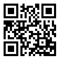
  • QR code for the Getting a Good Start article
    • Consider putting an unlabeled QR code (shown) in your syllabus or on your classroom wall.  Many students will scan the code and find the article simply out of curiosity.  Just like a snare trap!
  • Have a link to the article ready to give to students who contact you about being overwhelmed with the rigor of your course.
  • Consider suggesting to your students that they subscribe to The A&P Student newsletter (delivered by email, FB, LinkedIn or Twitter).
    • Consider subscribing yourself—a great way to keep up with various tips and strategies you can pass on to your students.
  • Even if your course has already started it's not too late to share these strategies with your students!


Want to know more?


Getting a Good Start in Your Anatomy & Physiology Course

  • Kevin Patton The A&P Student 26 August 2014
  • Outlines several practical strategies to begin the anatomy and/or physiology course on solid footing.  Link to this article from your syllabus and/or course web page.
  • my-ap.us/1onII8I

Subscription for The A&P Student

  • Choice of FREE delivery by email, Facebook, Twitter, or LinkedIn
  • my-ap.us/1oxkJIM

A&P is the Foundation

  • Kevin Patton Lion Den Slide Collection
  • FREE animated PowerPoint slide you can use in your introductory course presentation.  The link takes you to information on accessing the entire collection.  Find this one in v2, Study Tips, AP-is-foundation
  • lionden.com/slides-form.htm
Download free slides, including this one, to use in your A&P course
Free animated slide

Monday, August 18, 2014

Human Microbial System


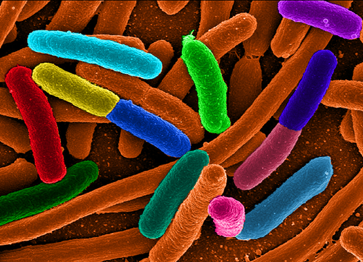
A recent article in The Scientist once again reminds us of the ongoing explosion in the scientific understanding of the human microbial system.  In a few short years, this area of exploration has moved to the forefront of medical and basic science research in human biology.

I think it's becoming clear that the most useful way to think of human body function is to recognize that an "organism" is really a sort of "habitat."  And like any habitat, it functions best when all the inhabitants are within a limited range of balanced relationships.

Who are the inhabitants?  Besides our own cells?  Well, one could think of mitochondria and cilia and other organelles as symbiotic internal inhabitants of our cells.  They're not that literally, of course, but I think its a useful metaphor for understanding the human body.  Then there are the many microbes and animals that cover our internal and external surfaces, burrow into some of our pores and glands, and inhabit our body fluids.

I call the balanced functional relationship among the various microbomes of the body and our own tissues the human microbial system.  And I am certain that it won't be long before we will be discussing this system alongside the major organ systems of the body.  That is if we truly want to understand how the body really works.

The article in The Scientist I mention is a great summary of some of the major roles that the human microbial system plays in the human body—and a good survey of some of the areas of the body where the human-microbial functional relationships play out.  See the link to the article below.


What can we use from this in teaching undergraduate A&P?


  • Why not introduce the concept of the human microbial system at the beginning of our A&P course, when we set the stage by explain how scientists understand the body and its functions as an integrated system of different parts?
  • We can mention the different microbiomes of the body when we explore each organ system where they play an important role—which is pretty much all of them!
  • Consider discussing what happens to normal human function when microbiomes get out of balance.  For example, in the gut a microbial imbalance can lead to ulcers, diarrhea, and other dysfunctions.  On the skin a pathogenic microbe may become dominant and cause a rash.
  • Promote a discussion of what kinds of wellness strategies might be employed to prevent microbial imbalances.
  • Our students can leave our A&P course with an up-to-date understanding of human biology that will help them understand new clinical concepts and treatment strategies.

Want to Know More?

The Body’s Ecosystem

  • By The Scientist Staff.  The Scientist. August 1, 2014
  • Plain-English article (cited above) on how research on the human microbiome is booming, and scientists have moved from simply taking stock of gut flora to understanding the influence of microbes throughout the body.
  • my-ap.us/1vgOu5y

Articles from The A&P Professor


Moving pictures of the human microbiome

  • J Gregory Caporaso et al. Genome Biology 2011, 12:R50  doi:10.1186/gb-2011-12-5-r50
  • Open-access journal article that includes FREE videos that show how dynamic the human microbial system is
  • my-ap.us/V7St3Q

Human Microbiome Project


The Microbiome and Disease

  • List of diseases associated with microbiome imbalances from Genetic Science Learning Center
  • my-ap.us/1nDpxr1

Audio

  • Radio stories from National Public Radio on human microbiomes and their role in health and disease.  The growing number of these stories tells us something as A&P teachers: maybe we better be covering this!
  • my-ap.us/1uA7Qyg

Thursday, July 24, 2014

Number of Human Genes Revised Downward. Again.

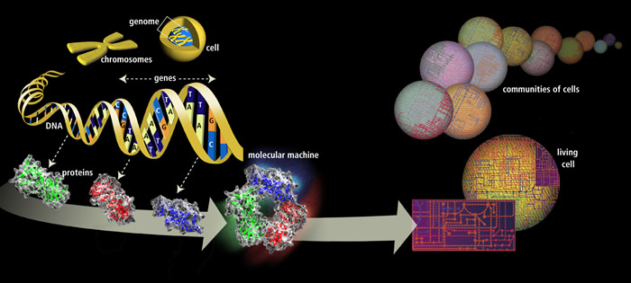
Genomic researchers in Spain have recently proposed a new, lower number of protein-coding genes in the human genome.  Previously, the number of coding genes was estimated by some at nearly 22,000 in the human genome.  The new estimate is approximately 19,000 protein-coding genes.

How can we use this new information in teaching undergraduate A&P?  Well, first we can update the numbers we use when discussing the role of genes in protein synthesis.  If it fits with our course objectives, we can use this as a way to transition to a discussion of coding vs. noncoding genes.

We also have an opportunity to discuss how science works—we are constantly checking our facts and revising our conclusions to improve the accuracy of our knowledge.  And that the story of genomics is far from complete.

I often tell students that I'm trying to tell them "the last, best story" of the human body's structure and function.  So if my story changes over time, that's a good thing!

FREE image you can use in your course


Want to know more?

Size of the human genome reduced to 19,000 genes

  • Science Daily. July 3, 2014
  • Press release in plain English based on information provided by researchers.
  • my-ap.us/1qrtHWf


Multiple evidence strands suggest that there may be as few as 19 000 human protein-coding genes. 

  • I. Ezkurdia, et al. Human Molecular Genetics, 2014; DOI: 10.1093/hmg/ddu309
  • Research article proposing the newly revised number.  Open access to full text of article.
  • my-ap.us/1oL7aTh

Want a FREE digital image of the nuclear genome that you can use in your presentation, handout, or other course material?  The image above is in the public domain and can be used in your course materials.


Monday, July 21, 2014

Biological Pacemaker Using Gene Therapy


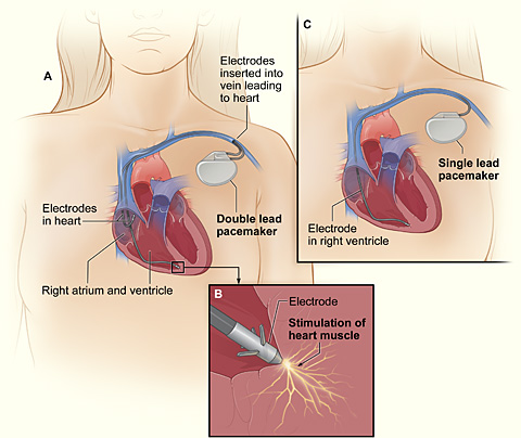
Researchers recently induced ordinary cardiac muscle fibers into becoming functioning pacemaker cells by injecting a therapeutic gene.

Working with pigs, a common model for human cardiovascular research, researchers first destroyed the natural pacemaker cells in each subject's heart and installed an electronic pacemaker. They then inserted a gene for transcription factor TBX18 into cardiac muscle tissue using an adenovirus.  Using adenovirous vectors for inserting genes is a common strategy in gene therapy.

Within a couple of days, ordinary myocardial fibers had developed the structure and function of pacemaker cells.  In about 5 days, the electronic pacemakers were no longer needed.

However, this biological pacemaking peaked at about 8 days, then eventually disappeared.  This may occur because the virus-infected cells are probably destroyed by the body's immune defenses.  So researchers are thinking that perhaps, at the very least, this could eventually lead to a temporary treatment for certain arrythmias in humans.


What can we use from this in teaching undergraduate A&P?  


  • This is an interesting bit of news that helps illustrate the frontiers of human biomedical sciences.
  • This story provides a good case to provoke a discussion of the nature of gene therapy.  
    • Why did the effect last only 8 or so day?  
    • What does this tell us about transcription factor TBX18?  
    • What benefit might this treatment have if developed for humans?
  • This may add interest to an discussion of the function of the electrical system of the heart in general, and artificial pacemakers in particular.
  • The case also provides a scenario in which the body attacks and destroys virus-infected cells.
FREE image you can use in your course

Want to know more?


Next Generation: Biological Pacemakers

  • R Williams, The Scientist (the-scientist.com) July 16, 2014
  • Plain-English article summarizing the discovery.  Includes quotes from the researchers.
  • my-ap.us/1ruocYu


Biological pacemaker created by minimally invasive somatic reprogramming in pigs with complete heart block
  • Y-F. Hu et al., Science Translational Medicine, 6:245ra94, 2014. DOI:10.1126/scitranslmed.3008681
  • Original research report.
  • my-ap.us/1nfErcA
FREE image you can use in your course

Photo credit: vfdbsn

Wednesday, July 16, 2014

The Life Science Teaching Resource Community

Have you heard that the American Physiological Society (APS) has expanded their Archive of Teaching Resources into The Life Science Teaching Resource Community?

This new online community—LifeSciTRC for short—offers thousands of FREE resources that you can use in your A&P course!

This transition marks a culmination of efforts of APS and several other scientific societies to advance the Archive of Teaching Resources beyond an online library into a community of practice for life science educators at the K-12, undergraduate, graduate, and professional levels.

The LifeSciTRC offers more than 6,000 free, scientifically-accurate teaching resources along with many new tools that allow educators to share their ideas and teaching expertise including:
  • Community pages with news and recommended teaching resources
  • Blogs focusing on classroom and science topics relevant to educators
  • Forums for educator-led discussions
  • Resource rating and commenting areas where educators can share their experiences of using resources
  • Monthly newsletters highlighting community members, news, and resources
I can't speak highly enough of the quality and diversity of teaching resources available in the archive.  These are peer-reviewed submissions from all levels of teaching and learning.  Case studies, experiments, demonstrations, slide presentations—all kinds of great stuff!

The new system is really easy (and fun) to use.  It has a starred rating system, so you can see how others have rated each resource.  And you can earn badges by submitting and reviewing items.

In addition to the new name, the LifeSciTRC will feature three new scientific society partners:
  • The Physiological Society
  • Genetics Society of America
  • American Society of Plant Biologists
These societies will join the current partners:
  • APS
  • Human Anatomy and Physiology Society
  • Society for Developmental Biology
  • American Association of Anatomists
  • Massachusetts Society for Medical Research
  • Northwest Association for Biomedical Research
Educators looking for new teaching ideas or interaction with other educators are invited to visit LifeSciTRC.org and discover what they have to offer.

For more information, contact our colleague at APS, Miranda Byse, PhD, Program Manager at mbyse@the-aps.org or 240-743-8045.  Be sure to tell her where you heard the message!

Some content in this post came from APS

Thursday, July 10, 2014

Mutations in Mitochondrial DNA

A new study suggests that DNA mutations in some of the mitochondria of healthy people may be a lot more common than scientists thought.

The term heteroplasmy describes a situation in which some mitochondria of a cell have mutant mitochondrial DNA (mtDNA) and other mitochondria have normal mtDNA.  Cell function can become disordered, perhaps producing disease, when the balance of mutant vs. normal mtDNA crosses a certain threshold.

The recent study shows that about 90% of healthy people studied in the 1000 Genomes Project had at least one heteroplasmy.  Some of these (about 20%) have been shown to correlate to disease.  That's a lot more than we were thinking prior to the study (25%-65% heteroplasmy rate).

We don't  know the significance of this finding yet, but it could influence how likely it is for mitochondrial diseases to develop over time—or to be inherited.  Could the mutant/normal mtDNA balance get skewed as oocytes form, thus giving different offspring different probabilities of inheriting mitochondrial disorders?  Or change the probabilities from one generation to the next?  Mitochondrial dysfunction is thought to be a mechanism of agingcould the rate of heteroplasmy be part of the aging mechanism?

What can we use from this in teaching undergraduate A&P?  The fact that we now know that mtDNA mutations are common in healthy people will be interesting and useful to students.


Want to know more?

Mutations Pervade Mitochondrial DNA

  • Jyoti Madhusoodanan. The Scientist (the-scientist.com) July 7, 2014
  • This is a plain-English article summarizing the new findings; includes quotes from the researchers.
  • my-ap.us/1oo18X8 


Extensive pathogenicity of mitochondrial heteroplasmy in healthy human individuals

  • K. Ye et al., Proceedings of the National Academy of Science (PNAS), doi:10.1073/pnas.1403521111, 2014. 
  • This is the original research report.
  • my-ap.us/1sDPV8S


Want a FREE labeled image of mtDNA that you can use in your presentation, handout, or other course material?
my-ap.us/1ncmAxl


Wednesday, November 6, 2013

New knee ligament confirmed

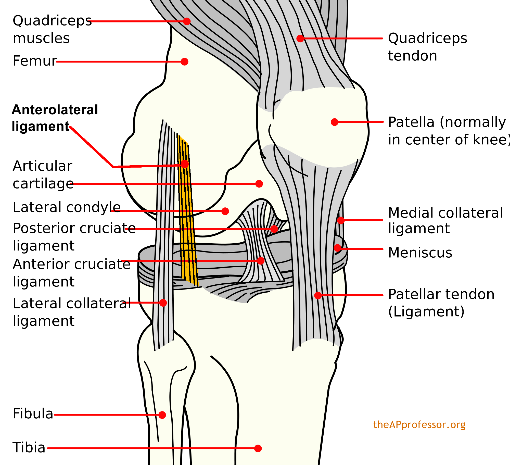
It's been there all along.  In most of us, at least.  Back in 1879, French surgeon Paul Ferdinand Segond first described it as it related to a particular type of avulsion fracture of the knee—the Segond fracture. But it was never really confirmed as separate from the joint capsule and named as a normal ligament of the human knee.  Until now.

Dubbed the anterolateral ligament (ALL), it originates at the prominence of the lateral femoral epicondyle (just anterior to the lateral collateral ligament) and running obliquely to the anterolateral part of the tibia (attached at the lateral meniscus).

Segond
So maybe we should pencil the ALL into our anatomic atlases, eh?  And wait for some research to confirm its biomechanical function—probably related to controlling internal rotation of the tibia.

This might also provide a good opportunity to talk about the dynamic nature of anatomical science—and the fact that human anatomy is not "finished."


Want to know more?

Anatomy of the anterolateral ligament of the knee
  • Steven Claes et al. Journal of Anatomy. Volume 223, Issue 4, pages 321–328, October 2013 (First published online: 1 AUG 2013) DOI: 10.1111/joa.12087
  • This the original journal article (free abstract).
  • my-ap.us/1aFUVV3

The Anterolateral Ligament of the Knee: Anatomy, Radiology, Biomechanics and Clinical Implications
  • Steven Claes, et al. American Academy of Orthopedic Surgeons (AAOS) Annual Meeting, SE73, 20 March 2013
  • This is an abstract (with image) of a preliminary presentation giving prior to journal publication.
  • my-ap.us/1b7bU0Q

Photo of the ALL
  • Steven Claes, et al. American Academy of Orthopedic Surgeons (AAOS) Annual Meeting, SE73, 20 March 2013
  • my-ap.us/1cF4sgC

Diagram of ALL


Monday, October 7, 2013

Nobel Prize: Vesicle Transport

The Nobel Assembly at Karolinska Institutet has today decided to award the 2013 Nobel Prize in Physiology or Medicine jointly to James E. Rothman, Randy W. Schekman and Thomas C. Südhof for their discoveries of machinery regulating vesicle traffic, a major transport system in our cells.

Those of you now teaching A&P 1 have probably recently covered the essential concepts vesicle transport and have perhaps already had the opportunity to apply them to specific functions of the body such as the release of acetylcholine at the neuromuscular junction.  Bringing up today's news gives us a great chance to underscore the importance of a topic that many students wonder, "why do we have to know this?"

Near the bottom of  this post, you'll find a link to a nice handout that you can distribute to your class (or link to from an email or webpage).  I've often added a question on my midterm exam covering that year's relevant Nobel Prize concept.

Summary

The 2013 Nobel Prize honors three scientists who have solved the mystery of how the cell organizes its transport system. Each cell is a factory that produces and exports molecules. For instance, insulin is manufactured and released into the blood and chemical signals called neurotransmitters are sent from one nerve cell to another. These molecules are transported around the cell in small packages called vesicles. The three Nobel Laureates have discovered the molecular principles that govern how this cargo is delivered to the right place at the right time in the cell.

Randy Schekman discovered a set of genes that were required for vesicle traffic. James Rothman  unravelled protein machinery that allows vesicles to fuse with their targets to permit transfer of cargo. Thomas Südhof revealed how signals instruct vesicles to release their cargo with precision.

Through their discoveries, Rothman, Schekman and Südhof have revealed the exquisitely precise control system for the transport and delivery of cellular cargo. Disturbances in this system have deleterious effects and contribute to conditions such as neurological diseases, diabetes, and immunological disorders.

How cargo is transported in the cell

In a large and busy port, systems are required to ensure that the correct cargo is shipped to the correct destination at the right time. The cell, with its different compartments called organelles, faces a similar problem: cells produce molecules such as hormones, neurotransmitters, cytokines and enzymes that have to be delivered to other places inside the cell, or exported out of the cell, at exactly the right moment. Timing and location are everything. Miniature bubble-like vesicles, surrounded by membranes, shuttle the cargo between organelles or fuse with the outer membrane of the cell and release their cargo to the outside. This is of major importance, as it triggers nerve activation in the case of transmitter substances, or controls metabolism in the case of hormones. How do these vesicles know where and when to deliver their cargo?

Traffic congestion reveals genetic controllers

Randy Schekman was fascinated by how the cell organizes its transport system and in the 1970s decided to study its genetic basis by using yeast as a model system. In a genetic screen, he identified yeast cells with defective transport machinery, giving rise to a situation resembling a poorly planned public transport system. Vesicles piled up in certain parts of the cell. He found that the cause of this congestion was genetic and went on to identify the mutated genes. Schekman identified three classes of genes that control different facets of the cell´s transport system, thereby providing new insights into the tightly regulated machinery that mediates vesicle transport in the cell.

Docking with precision

James Rothman was also intrigued by the nature of the cell´s transport system. When studying vesicle transport in mammalian cells in the 1980s and 1990s, Rothman discovered that a protein complex enables vesicles to dock and fuse with their target membranes. In the fusion process, proteins on the vesicles and target membranes bind to each other like the two sides of a zipper. The fact that there are many such proteins and that they bind only in specific combinations ensures that cargo is delivered to a precise location. The same principle operates inside the cell and when a vesicle binds to the cell´s outer membrane to release its contents.

It turned out that some of the genes Schekman had discovered in yeast coded for proteins corresponding to those Rothman identified in mammals, revealing an ancient evolutionary origin of the transport system. Collectively, they mapped critical components of the cell´s transport machinery.

Timing is everything

Thomas Südhof was interested in how nerve cells communicate with one another in the brain. The signalling molecules, neurotransmitters, are released from vesicles that fuse with the outer membrane of nerve cells by using the machinery discovered by Rothman and Schekman. But these vesicles are only allowed to release their contents when the nerve cell signals to its neighbours. How is this release controlled in such a precise manner? Calcium ions were known to be involved in this process and in the 1990s, Südhof searched for calcium sensitive proteins in nerve cells. He identified molecular machinery that responds to an influx of calcium ions and directs neighbour proteins rapidly to bind vesicles to the outer membrane of the nerve cell. The zipper opens up and signal substances are released. Südhof´s discovery explained how temporal precision is achieved and how vesicles´ contents can be released on command.

Vesicle transport gives insight into disease processes

The three Nobel Laureates have discovered a fundamental process in cell physiology. These discoveries have had a major impact on our understanding of how cargo is delivered with timing and precision within and outside the cell.  Vesicle transport and fusion operate, with the same general principles, in organisms as different as yeast and man. The system is critical for a variety of physiological processes in which vesicle fusion must be controlled, ranging from signalling in the brain to release of hormones and immune cytokines. Defective vesicle transport occurs in a variety of diseases including a number of neurological and immunological disorders, as well as in diabetes. Without this wonderfully precise organization, the cell would lapse into chaos.

About this year's Nobel laureates

James E. Rothman was born 1950 in Haverhill, Massachusetts, USA. He received his PhD from Harvard Medical School in 1976, was a postdoctoral fellow at Massachusetts Institute of Technology, and moved in 1978 to Stanford University in California, where he started his research on the vesicles of the cell. Rothman has also worked at Princeton University, Memorial Sloan-Kettering Cancer Institute and Columbia University. In 2008, he joined the faculty of Yale University in New Haven, Connecticut, USA, where he is currently Professor and Chairman in the Department of Cell Biology.

Randy W. Schekman was born 1948 in St Paul, Minnesota, USA, studied at the University of California in Los Angeles and at Stanford University, where he obtained his PhD in 1974 under the supervision of Arthur Kornberg (Nobel Prize 1959) and in the same department that Rothman joined a few years later. In 1976, Schekman joined the faculty of the University of California at Berkeley, where he is currently Professor in the Department of Molecular and Cell biology. Schekman is also an investigator of Howard Hughes Medical Institute.

Thomas C. Südhof was born in 1955 in Göttingen, Germany. He studied at the Georg-August-Universität in Göttingen, where he received an MD in 1982 and a Doctorate in neurochemistry the same year. In 1983, he moved to the University of Texas Southwestern Medical Center in Dallas, Texas, USA, as a postdoctoral fellow with Michael Brown and Joseph Goldstein (who shared the 1985 Nobel Prize in Physiology or Medicine). Südhof became an investigator of Howard Hughes Medical Institute in 1991 and was appointed Professor of Molecular and Cellular Physiology at Stanford University in 2008.


Want to know more?


Handout showing mechanisms for which this prize was awarded

  • Diagrams and brief description of the contributions of each Nobel laureate
  • my-ap.us/1cnPDhF


Machinery Regulating Vesical Traffic, A Major Transport System in our Cells

  • Nobelprize.org accessed 7 October 2013
  • [Nice summary of the scientific concepts involved.  An expanded version of the handout, with additional diagrams and explanations.]
  • my-ap.us/GDLiZR


Original Journal Articles

  • Seminal papers describing the original work of this year's Nobel Laureates
  • Novick P, Schekman R: Secretion and cell-surface growth are blocked in a temperature-sensitive mutant of Saccharomyces cerevisiae. Proc Natl Acad Sci USA 1979; 76:1858-1862.
  • Balch WE, Dunphy WG, Braell WA, Rothman JE: Reconstitution of the transport of protein between successive compartments of the Golgi measured by the coupled incorporation of N-acetylglucosamine. Cell 1984; 39:405-416.
  • Kaiser CA, Schekman R: Distinct sets of SEC genes govern transport vesicle formation and fusion early in the secretory pathway. Cell 1990; 61:723-733.
  • Perin MS, Fried VA, Mignery GA, Jahn R, Südhof TC: Phospholipid binding by a synaptic vesicle protein homologous to the regulatory region of protein kinase C. Nature 1990; 345:260-263.
  • Sollner T, Whiteheart W, Brunner M, Erdjument-Bromage H, Geromanos S, Tempst P, Rothman JE: SNAP receptor implicated in vesicle targeting and fusion. Nature 1993; 362:318-324.
  • Hata Y, Slaughter CA, Südhof TC: Synaptic vesicle fusion complex contains unc-18 homologue bound to syntaxin. Nature 1993; 366:347-351.

Related textbook content

  • Anatomy & Physiology 8th ed.  Chapter 3 (see p. 75, 82), Chapter 4 (see p. 99-100), and throughout book my-ap.us/QZTbK1
  • Essentials of Anatomy & Physiology Chapter 3 (see p. 52), Chapter 4 (see p. 69), and throughout book my-ap.us/SCfNlj  
  • The Human Body in Health and Disease 6th ed. Chapter 3 (see p. 49, 56) and throughout book my-ap.us/X71LJO 
  • Structure & Function of the Body 14th ed. Chapter 3 (see p. 48, 53) and throughout book http://my-ap.us/10s50MH


Portions of this post adapted from Nobel Prize news release

Monday, September 16, 2013

Teaching bones & bone features

I've found that one of the things that stand in the way of student success in learning the anatomy of the skeleton is that they don't know the underlying language.

If they already knew that a foramen is a hole and a condyle is a bump, then they could make short work of identifying and learning a long list of bones and bone features. And they'd have a better understanding of the concepts, plus an easier time remembering each structure for the long term.

We can't do anything about the fact that they just don't teach Latin as a required subject  in grade schools and high schools anymore.  But that doesn't mean there's nothing we can do to give them a head start on learning the skull!

For a long time, I've started out my students learning the general types of bone features before starting on the specific skeletal structures.  I find that they have a much easier time identifying structures because they have a better understanding of what the bone names mean.  They know they're looking for a hole when they get to the foramen ovale.  That oblong dark spot in the skull diagram or photo could be a bump, for all they know.

I use the list of general types of bone features found in my textbooks, lab manuals, and other publications (see list below).  I've also provided you with a downloadable version of this list you can link to in your course or distribute as a handout (see links below).

I've just uploaded two videos to my YouTube channel that help students get started this way.  You can link to the videos in an email, syllabus, or LMS; embed them in your course website or LMS; or embed them in a PowerPoint slide.  Or you could use the slides I used in the video to make your own video or live presentationperhaps even customize it for your course.  This links to all of these resources are below.

I'll be posting these videos on my study-tip blog for students, The A&P Student, soon.  If your students are not yet using this blog, you can distribute my FREE bookmarks to get them started.  See below for details.


Want to know more?

Bone Names Have Meaning - Part 1 - Get Started (video)
  • by Kevin Patton, Kevin's YouTube Channel,  uploaded September 2013
  • Video briefly explains the importance of knowing the meaning of bone names and how to get started doing that.  Includes link to list of bone features.
  • Link URL: youtu.be/heBjZIZP328
  • Embed code is available once you link to the above URL and click "Share"
Bone Names Have Meaning - Part 2 - Types of Bone Markings (video)
  • by Kevin Patton, Kevin's YouTube Channel, uploaded September 2013
  • Brief video runs through each of the major bone feature types, showing one or more examples. Includes link to list of bone features.
  • Link URL: youtu.be/NRR-t93jyFw
  • Embed code is available once you link to the above URL and click "Share"
Bone-Names-1-2 (PowerPoint slides)
  • by Kevin Patton, Lion Den Slide Collection,  September 2013
  • Download these animated slides I used to make the videos and use them in your recorded or live presentation.  The slide notes include the narration, which you could customize (or ignore).  You can use some or all of the slides.
  • To get the download password, fill out the form at lionden.com/slides-form.htm
Bone Names (web page)
  • by Kevin Patton, Lion Den Study Tips & Tools
  • Web page for students briefly outlining how to get started learning bone structures. The videos above are embedded in the page, so you could link to this page in your course instead of linking to the individual videos.  Links to related, downloadable lists. Part of the Field Guide to the Body.
  • lionden.com/tips-lab-anatomy-bone-names.htm
List of Bone Markings
Lion Den Slide Collection
  • by Kevin Patton, Lion Den Slide Collection
  • Download all kinds of  animated slides you can use in your own course to supplement your own slides and those provided with your textbook or school. Most slides are animated.
  • To get access to the whole collection, fill out the form at lionden.com/slides-form.htm
Free Bookmarks
  • Get packs of 50 bookmarks to distribute to students.  The bookmarks tell students how to access The A&P Student blog, which has a collection of study tips for A&P.
  • theapprofessor.org/free-bookmarks.html