You know that online graduate degree in Human Anatomy & Physiology Instruction—the HAPI degree—that I'm always talking about? I just found out that they're offering a virtual Open House this coming Monday, July 15, at 8 pm ET.
This event is completely online, like the MS-HAPI degree itself. And it'll give you a better idea of what it's all about. And you can ask all those questions that have been occurring to you.
Whether it's because you realize the benefit of additional training for yourself in "how to teach A&P" or you want to know more so you can pass it along to colleagues or prospective hires in your department, it'll be worth the small investment of time.
Yeah, I know. It's late notice. I just found out about it recently, myself. If you miss the Open House, that's okay—you can go to nycc.edu/hapi anytime to find out more about the program.
Note: I'm a founding faculty member in HAPI and the program currently sponsors the free distribution of my podcast The A&P Professor.
Please forward this notice to all those who you think might be interested!
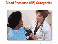
With the new guidelines for high blood pressure popping up all over the news recently, we may wonder what we need to know when this comes up in our A&P classrooms. And we know it will—students love, love, love to connect what they are learning in A&P with what they are experiencing in their lives.
It turns out that although the new 2017 Guideline For the Prevention, Detection, Evaluation, and Management of High Blood Pressure in Adults is focused on how physicians should make diagnoses and manage patient care, the definitions of exactly what constitutes high blood pressure (hypertension or HTN) are important learning points in the undergrad A&P course.
I'll outline the main things for us A&P professors to know here, but do check out the resources I've linked below to deepen your understanding of current thinking regarding approaches to blood pressure (BP) health.
First, there are revised guidelines as to what constitutes high blood pressure or HTN:
Normal BP: Less than 120/80 mm Hg;
Elevated BP: Systolic between 120-129 and diastolic less than 80;
Stage 1 HTN: Systolic between 130-139 or diastolic between 80-89;
Stage 2 HTN: Systolic at least 140 or diastolic at least 90 mm Hg;
Hypertensive crisis: Systolic over 180 and/or diastolic over 120, with patients needing prompt changes in medication if there are no other indications of problems, or immediate hospitalization if there are signs of organ damage.
Regardless of the precise cutoffs listed above, in an interview discussing the new guidelines, the main author states that, "120/80 is normal, the same as we had before" the new guidelines. So I think we're safe in using 120/80 as an example of BP when discussing the normal science, even though technically it could be designated as "elevated." Not that we can't use an elevated variable measurement as an example when discussing the physiology of anything. The fact that even the main author of the guidelines uses 120/80 as the starting point of discussion makes me feel more confident in using it as the starting point of my course discussions, too.
The main thing to note in the categories above is that the cutoffs for HTN categories have been lowered. This puts many more people in an HTN category that were not there before. The main goal is for those folks to have conversations with their physicians to evaluate their risk for complications and develop a personalized prevention and care plan.
Note also that the category of prehypertension has been eliminated.
The new guidelines also recommend prescribing medication for Stage 1 HTN if the patient already had a cardiovascular event—or is at a high risk for such an event. They also recognize that many patients will need more than one medication to manage BP and that combining meds into one pill is likely to help folks take them consistently.
There are a lot of other recommendations, so reviewing the Executive Summary or similar resource (see below) may be a good idea.
What can we use from this in teaching undergraduate A&P?
If you discuss hypertension, or use case studies in teaching, you need to update the cutoff BPs you are using.
A BP of 120/80 is still considered the starting point for discussing blood pressure.
Consider discussing the impact of the changes in the new guidelines for ordinary people.
Discuss why such diagnosis, prevention, and treatment recommendations often change over time. Consider discussion other recent clinical updates.
Consider discussing specific changes suggested in the new guidelines.
Consider having students explore the Executive Summary and/or other documents and write their own summary or interpretation of key points. Perhaps they can create their own chart or concept map.
Need some free teaching materials?
SLIDE SET: High Blood Pressure
Kevin Patton. Lion Den Slide Collection. 18 Nov 2017
Small slide deck that includes an animated version of the BP Category chart pictured above. Part of the Lion Den Slide Collection (requires free registration to download). You can also download a static PNG image file of the chart in the slide collection set.
VIDEO: AHA 2017 | New High Blood Pressure Guidelines
America Heart Association. 13 Nov 2017.
Free video (viewable in the player above) features a chat with the main author of the new guidelines and summarizes the main points. Very practical and easy to understand.
SLIDE SET: 2017 Guideline For the Prevention, Detection, Evaluation and Management of High Blood Pressure in Adults
American College of Cardiology. 13 Nov 2017
Free set of almost 100 PowerPoint slides to use in teaching. And it has a decided focus on clinical applications, rather than the basic science. These are way, way beyond the coverage desirable in an undergrad A&P course. But some slides may be useful to you.
New blood pressure guidelines put half of U.S. adults in unhealthy range
A. Cunningham Science News. 13 Nov 2017
Plain-English article summarizing the first major update since 2003 aims to spur heart-healthy lifestyle changes. Has a useful graph and links to other articles and resources.
2017 ACC/AHA/AAPA/ABC/ACPM/AGS/APhA/ASH/ASPC/NMA/PCNA Guideline for the Prevention, Detection, Evaluation, and Management of High Blood Pressure in Adults: Executive Summary A Report of the American College of Cardiology/American Heart Association Task Force on Clinical Practice Guidelines
PK Whelton et al. Hypertension, Dec 2017, Volume 70, Issue 6. DOI: 10.1161/HYP.0000000000000066
Free PDF of the Executive Summary of the larger report. I recommend reading this first, then decide if you need to read the whole report.
2017 ACC/AHA/AAPA/ABC/ACPM/AGS/APhA/ASH/ASPC/NMA/PCNA Guideline for the Prevention, Detection, Evaluation, and Management of High Blood Pressure in Adults A Report of the American College of Cardiology/American Heart Association Task Force on Clinical Practice Guidelines
PK Whelton et al. Hypertension. Dec 2017, Volume 70, Issue 6. DOI: 10.1161/HYP.0000000000000065
Free PDF of the entire report. It's huge, so make a whole pot of tea before starting it.
The Nobel Assembly at Karolinska Institutet has today decided to award the 2017 Nobel Prize in Physiology or Medicine jointly to Jeffrey C. Hall,Michael Rosbash, and Michael W. Young for their discoveries of molecular mechanisms controlling the circadian rhythm.
Summary
Life on Earth is adapted to the rotation of our planet. For many years we have known that living organisms, including humans, have an internal, biological clock that helps them anticipate and adapt to the regular rhythm of the day. But how does this clock actually work? Jeffrey C. Hall, Michael Rosbash and Michael W. Young were able to peek inside our biological clock and elucidate its inner workings. Their discoveries explain how plants, animals and humans adapt their biological rhythm so that it is synchronized with the Earth's revolutions.
Using fruit flies as a model organism, this year's Nobel laureates isolated a gene that controls the normal daily biological rhythm. They showed that this gene encodes a protein that accumulates in the cell during the night, and is then degraded during the day. Subsequently, they identified additional protein components of this machinery, exposing the mechanism governing the self-sustaining clockwork inside the cell. We now recognize that biological clocks function by the same principles in cells of other multicellular organisms, including humans.
With exquisite precision, our inner clock adapts our physiology to the dramatically different phases of the day. The clock regulates critical functions such as behavior, hormone levels, sleep, body temperature and metabolism. Our wellbeing is affected when there is a temporary mismatch between our external environment and this internal biological clock, for example when we travel across several time zones and experience "jet lag". There are also indications that chronic misalignment between our lifestyle and the rhythm dictated by our inner timekeeper is associated with increased risk for various diseases.
Our inner clock
Most living organisms anticipate and adapt to daily changes in the environment. During the 18th century, the astronomer Jean Jacques d'Ortous de Mairan studied mimosa plants, and found that the leaves opened towards the sun during daytime and closed at dusk. He wondered what would happen if the plant was placed in constant darkness. He found that independent of daily sunlight the leaves continued to follow their normal daily oscillation (Figure 1). Plants seemed to have their own biological clock.
Other researchers found that not only plants, but also animals and humans, have a biological clock that helps to prepare our physiology for the fluctuations of the day. This regular adaptation is referred to as the circadian rhythm, originating from the Latin words circa meaning "around" and dies meaning "day". But just how our internal circadian biological clock worked remained a mystery.
Figure 1. An internal biological clock. The leaves of the mimosa plant open towards the sun during day but close at dusk (upper part). Jean Jacques d'Ortous de Mairan placed the plant in constant darkness (lower part) and found that the leaves continue to follow their normal daily rhythm, even without any fluctuations in daily light.
Identification of a clock gene
During the 1970's, Seymour Benzer and his student Ronald Konopka asked whether it would be possible to identify genes that control the circadian rhythm in fruit flies. They demonstrated that mutations in an unknown gene disrupted the circadian clock of flies. They named this gene period. But how could this gene influence the circadian rhythm?
This year's Nobel Laureates, who were also studying fruit flies, aimed to discover how the clock actually works. In 1984, Jeffrey Hall and Michael Rosbash, working in close collaboration at Brandeis University in Boston, and Michael Young at the Rockefeller University in New York, succeeded in isolating the period gene. Jeffrey Hall and Michael Rosbash then went on to discover that PER, the protein encoded by period, accumulated during the night and was degraded during the day. Thus, PER protein levels oscillate over a 24-hour cycle, in synchrony with the circadian rhythm.
A self-regulating clockwork mechanism
The next key goal was to understand how such circadian oscillations could be generated and sustained. Jeffrey Hall and Michael Rosbash hypothesized that the PER protein blocked the activity of the period gene. They reasoned that by an inhibitory feedback loop, PER protein could prevent its own synthesis and thereby regulate its own level in a continuous, cyclic rhythm (Figure 2A).
Figure 2B. A simplified illustration of the molecular components of the circadian clock.
Such a regulatory feedback mechanism explained how this oscillation of cellular protein levels emerged, but questions lingered. What controlled the frequency of the oscillations? Michael Young identified yet another gene, doubletime, encoding the DBT protein that delayed the accumulation of the PER protein. This provided insight into how an oscillation is adjusted to more closely match a 24-hour cycle.
The paradigm-shifting discoveries by the laureates established key mechanistic principles for the biological clock. During the following years other molecular components of the clockwork mechanism were elucidated, explaining its stability and function. For example, this year's laureates identified additional proteins required for the activation of the period gene, as well as for the mechanism by which light can synchronize the clock.
Keeping time on our human physiology
The biological clock is involved in many aspects of our complex physiology. We now know that all multicellular organisms, including humans, utilize a similar mechanism to control circadian rhythms. A large proportion of our genes are regulated by the biological clock and, consequently, a carefully calibrated circadian rhythm adapts our physiology to the different phases of the day (Figure 3). Since the seminal discoveries by the three laureates, circadian biology has developed into a vast and highly dynamic research field, with implications for our health and wellbeing.
Figure 3. The circadian clock anticipates and adapts our physiology to the different phases of the day. Our biological clock helps to regulate sleep patterns, feeding behavior, hormone release, blood pressure, and body temperature.
About the Nobel Laureates
Jeffrey C. Hall was born 1945 in New York, USA. He received his doctoral degree in 1971 at the University of Washington in Seattle and was a postdoctoral fellow at the California Institute of Technology in Pasadena from 1971 to 1973. He joined the faculty at Brandeis University in Waltham in 1974. In 2002, he became associated with University of Maine.
Michael Rosbash was born in 1944 in Kansas City, USA. He received his doctoral degree in 1970 at the Massachusetts Institute of Technology in Cambridge. During the following three years, he was a postdoctoral fellow at the University of Edinburgh in Scotland. Since 1974, he has been on faculty at Brandeis University in Waltham, USA.
Michael W. Young was born in 1949 in Miami, USA. He received his doctoral degree at the University of Texas in Austin in 1975. Between 1975 and 1977, he was a postdoctoral fellow at Stanford University in Palo Alto. From 1978, he has been on faculty at the Rockefeller University in New York.
What can we use from this in teaching undergraduate A&P?
When you discuss biological clocks and rhythms in your course, this could be a way to garner student interest—considering that this is a current and ongoing effort in science. I begin discussing this at the beginning of the course—when covering homeostasis.
Consider using the annual Nobel Prize announcements as a springboard to discuss the process of scientific discovery.
Consider mentioning the other major awards for scientific achievement and discuss what the judges seem to value most about scientific discoveries. The Nobel Prize is the one everyone has heard of, so it's a great place to start.
Use the Nobel Prizes (and other awards) over time as a way to keep students aware of the history of, and progress, of human biology. One could also address the global diversity of laureates. Or the lack of other kinds of diversity among laureates.
The sources below are great places to find media for teaching and for great, pithy explanations of complex topics for a "beginner" audience like our A&P students.
This trend in misleading "click bait" headlines among science news outlets continues to spiral into infinity. Okay, "infinity" is an exaggeration, but apparently that's what it takes these days to get us reading the actual content of science articles. And a growing phenomenon is that the articles themselves include exaggerations within their content. That's the topic of my rant, er, post today.
I've been thinking about this for a long while. I often discuss it in class with my students. Yesterday, I ran across a recent (January 2017) example of the perennial "scientists discover that the appendix has a function" headline: Your Appendix Might Serve an Important Biological Function After All
That example actually has a pretty good article about a study analyzing the evolutionary appearances and reappearances of the appendix in mammals and what that may tell us about this organ's function. But we already know enough about the functions of the vermiform appendix in humans that it's hardly true that its functions are completely unknown. The article clearly acknowledges that fact within the content, despite that attention-grabbing headline.
Another recent example was the round of excited shares on social media regarding the "discovery" that hematopoiesis (blood development) occurs in lung tissue. There were a lot of "wow, who knew?" tweets that week. Even from highly trained experts in A&P. But my Anatomy & Physiology textbook (p. 624) already has this information—and it surely cannot be the only textbook to do so.
The journal article that prompted this wave of tweets and posts described some research in mice that expands our knowledge about this phenomenon—turns out that more is going in the lungs than we thought. The lungs may be the primary site for thrombopoiesis (platelet development), if human lungs work like mice lungs. But the fact that the lungs are sites of hematopoiesis—specifically platelet formation—is not new.
I've shared these and other posts with exaggerated headlines myself—mostly on Twitter,Facebook, or my new daily newsletter from Nuzzel.
However, I think it's way to easy to succumb to the excitement of a potential "new discovery" that turns out to be not new, or even a discovery, at all. As a blogger I know full well that exaggerated headlines get more "engagement", which leads to more "followers," which leads to better "brand recognition" and thus, more future "engagement." Who wants to spend time researching and writing when nobody is reading?
But in science, maybe the public perception of how science works is better served by a more toned-down approach that recognizes what we already think we know, why we think we know it, and what any new studies can do to clarify, correct, or extend what we know.
I know that none of us individuals can stop the tide of exaggerated science news headlines. I'm just using a platform I have to express my concern that we may be making a mistake by doing so. If everything is a "breakthrough" or even a "huge breakthrough," then maybe casual observers will miss those truly game-changing ideas when they come along.
At least it's something to keep in the back our minds and we do our daily scan of science new headlines.
What can we use from this in teaching undergraduate A&P?
Consider challenging your student to find the first new "science discovers the function of the appendix" article or post of the semester. (or spleen or gallbladder or any organ).
Find some posts or articles that have exciting "new discovery" headlines and analyze them as a class. The may help us all learn better the critical analysis needed when reading science content.
Have a class discussion regarding the balance between the excitement of discovery that drives science and the exaggerations of discovery that may mislead.
Consider making sure that your students know that the appendix has functions (and that the lungs make platelets). Just in case they become science journalists.
Consider throwing out science journalism or science writing as career options. They already have an interest in human biology—and they may soon discover they don't like the career path they first chose, after all.
Want to know more?
Your Appendix Might Serve an Important Biological Function After All
BEC CREW Science Alert 10 JAN 2017
Article about an evolution study of the appendix in many organisms and how that may relate to the organ's function.
An Unexpected New Lung Function Has Been Found - They Make Blood
This article, with the subtitle Things just got complicated, outlines the recent work done in mice to show that most platelets (not just some platelets) may form in lungs.
The Nobel Assembly at Karolinska Institutet has today decided to award the 2016 Nobel Prize in Physiology or Medicine to Yoshinori Ohsumi for his discoveries of mechanisms for autophagy.
Overview
This year's Nobel Laureate discovered and elucidated mechanisms underlying autophagy, a fundamental process for degrading and recycling cellular components.
The word autophagy (aw-toh-FAY-jee) originates from the Greek words auto-, meaning "self", and phagein, meaning "to eat". Thus, autophagy denotes "self eating".
This concept emerged during the 1960's, when researchers first observed that the cell could destroy its own contents by enclosing it in membranes, forming sack-like vesicles that were transported to a recycling compartment, called the lysosome, for degradation.
Difficulties in studying the phenomenon meant that little was known until, in a series of brilliant experiments in the early 1990's, Yoshinori Ohsumi used baker's yeast to identify genes essential for autophagy. He then went on to elucidate the underlying mechanisms for autophagy in yeast and showed that similar sophisticated machinery is used in our cells.
Ohsumi's discoveries led to a new paradigm in our understanding of how the cell recycles its content. His discoveries opened the path to understanding the fundamental importance of autophagy in many physiological processes, such as in the adaptation to starvation or response to infection. Mutations in autophagy genes can cause disease, and the autophagic process is involved in several conditions including cancer and neurological disease.
Degradation – a central function in all living cells
In the mid 1950's scientists observed a new specialized cellular compartment, called an organelle, containing enzymes that digest proteins, carbohydrates and lipids. This specialized compartment is referred to as a "lysosome" and functions as a workstation for degradation of cellular constituents. The Belgian scientist Christian de Duve was awarded the Nobel Prize in Physiology or Medicine in 1974 for the discovery of the lysosome.
New observations during the 1960's showed that large amounts of cellular content, and even whole organelles, could sometimes be found inside lysosomes. The cell therefore appeared to have a strategy for delivering large cargo to the lysosome. Further biochemical and microscopic analysis revealed a new type of vesicle transporting cellular cargo to the lysosome for degradation (Figure 1).
Christian de Duve, the scientist behind the discovery of the lysosome, coined the term autophagy, "self-eating", to describe this process. The new vesicles were named autophagosomes.
Figure 1: Autophagosome. Our cells have different specialized compartments. Lysosomes constitute one such compartment and contain enzymes for digestion of cellular contents. A new type of vesicle called autophagosome was observed within the cell. As the autophagosome forms, it engulfs cellular contents, such as damaged proteins and organelles. Finally, it fuses with the lysosome, where the contents are degraded into smaller constituents. This process provides the cell with nutrients and building blocks for renewal.
During the 1970's and 1980's researchers focused on elucidating another system used to degrade proteins, namely the "proteasome". Within this research field Aaron Ciechanover, Avram Hershko and Irwin Rose were awarded the 2004 Nobel Prize in Chemistry for "the discovery of ubiquitin-mediated protein degradation". The proteasome efficiently degrades proteins one-by-one, but this mechanism did not explain how the cell got rid of larger protein complexes and worn-out organelles. Could the process of autophagy be the answer and, if so, what were the mechanisms?
A groundbreaking experiment
Yoshinori Ohsumi had been active in various research areas, but upon starting his own lab in 1988, he focused his efforts on protein degradation in the vacuole, an organelle that corresponds to the lysosome in human cells.
Yeast cells are relatively easy to study and consequently they are often used as a model for human cells. They are particularly useful for the identification of genes that are important in complex cellular pathways. But Ohsumi faced a major challenge; yeast cells are small and their inner structures are not easily distinguished under the microscope and thus he was uncertain whether autophagy even existed in this organism.
Ohsumi reasoned that if he could disrupt the degradation process in the vacuole while the process of autophagy was active, then autophagosomes should accumulate within the vacuole and become visible under the microscope. He therefore cultured mutated yeast lacking vacuolar degradation enzymes and simultaneously stimulated autophagy by starving the cells.
The results were striking! Within hours, the vacuoles were filled with small vesicles that had not been degraded (Figure 2). The vesicles were autophagosomes and Ohsumi's experiment proved that authophagy exists in yeast cells. But even more importantly, he now had a method to identify and characterize key genes involved this process. This was a major break-through and Ohsumi published the results in 1992.
Figure 2: Yeast. In yeast (left panel) a large compartment called the vacuole corresponds to the lysosome in mammalian cells. Ohsumi generated yeast lacking vacuolar degradation enzymes. When these yeast cells were starved, autophagosomes rapidly accumulated in the vacuole (middle panel). His experiment demonstrated that autophagy exists in yeast. As a next step, Ohsumi studied thousands of yeast mutants (right panel) and identified 15 genes that are essential for autophagy.
Autophagy genes are discovered
Ohsumi now took advantage of his engineered yeast strains in which autophagosomes accumulated during starvation. This accumulation should not occur if genes important for autophagy were inactivated. Ohsumi exposed the yeast cells to a chemical that randomly introduced mutations in many genes, and then he induced autophagy.
His strategy worked! Within a year of his discovery of autophagy in yeast, Ohsumi had identified the first genes essential for autophagy. In his subsequent series of elegant studies, the proteins encoded by these genes were functionally characterized. The results showed that autophagy is controlled by a cascade of proteins and protein complexes, each regulating a distinct stage of autophagosome initiation and formation (Figure 3).
Figure 3: Stages of autophagosome formation. Ohsumi studied the function of the proteins encoded by key autophagy genes. He delineated how stress signals initiate autophagy and the mechanism by which proteins and protein complexes promote distinct stages of autophagosome formation.
Autophagy – an essential mechanism in our cells
After the identification of the machinery for autophagy in yeast, a key question remained. Was there a corresponding mechanism to control this process in other organisms? Soon it became clear that virtually identical mechanisms operate in our own cells. The research tools required to investigate the importance of autophagy in humans were now available.
Thanks to Ohsumi and others following in his footsteps, we now know that autophagy controls important physiological functions where cellular components need to be degraded and recycled.
Autophagy can rapidly provide fuel for energy and building blocks for renewal of cellular components, and is therefore essential for the cellular response to starvation and other types of stress.
After infection, autophagy can eliminate invading intracellular bacteria and viruses. Autophagy contributes to embryo development and cell differentiation. Cells also use autophagy to eliminate damaged proteins and organelles, a quality control mechanism that is critical for counteracting the negative consequences of aging.
Disrupted autophagy has been linked to Parkinson's disease, type 2 diabetes and other disorders that appear in the elderly. Mutations in autophagy genes can cause genetic disease. Disturbances in the autophagic machinery have also been linked to cancer. Intense research is now ongoing to develop drugs that can target autophagy in various diseases.
Autophagy has been known for over 50 years but its fundamental importance in physiology and medicine was only recognized after Yoshinori Ohsumi's paradigm-shifting research in the 1990's. For
Yoshinori Ohsumi was born 1945 in Fukuoka, Japan. He received a Ph.D. from University of Tokyo in 1974. After spending three years at Rockefeller University, New York, USA, he returned to the University of Tokyo where he established his research group in 1988. He is since 2009 a professor at the Tokyo Institute of Technology.
More background on the winner and the prize
Yoshinori Ohsumi was born in Fukuoka, Japan, in 1945. He is affiliated with the Tokyo Institute of Technology in Tokyo, Japan. His monetary award will be nearly one million dollars.
The Nobel Assembly, consisting of 50 professors at Karolinska Institutet, awards the Nobel Prize in Physiology or Medicine. Its Nobel Committee evaluates the nominations. Since 1901 the Nobel Prize has been awarded to scientists who have made the most important discoveries for the benefit of mankind.his discoveries, he is awarded this year's Nobel Prize in physiology or medicine.
Nobel Prize® is the registered trademark of the Nobel Foundation
What can we use from this in teaching undergraduate A&P?
Consider using the Nobel Prizes as a discussion-starter in your class about
How science influences society
How society influences science
How science progresses
Rewarding of science discoveries
What makes a discovery "important"
Relate this discovery to prior (or upcoming) discussions of
Cell function
Organelle specialization
How cells handle protein
How autophagosomes work with lysosomes
Compare/contrast with phagocytosis
Compare/contrast with proteasome function and protein "quality control
Relate this discovery to the general idea of cellular mechanisms of disease
Consider taking this opportunity to emphasize "why we need to know all this" detail about cellular structure and function.
Want to know more?
Scientific Background Discoveries of Mechanisms for Autophagy
Larsson, N-G, Msucci, M. G. Nobelprize.org accessed 8 October 2016
A more advanced summary of the prizewinning discovery, including a handy glossary of terms.
A ubiquitin-like system mediates protein lipidation.
Ichimura, Y., Kirisako T., Takao, T., Satomi, Y., Shimonishi, Y., Ishihara, N., Mizushima, N., Tanida, I., Kominami, E., Ohsumi, M., Noda, T. and Ohsumi, Y. (2000). Nature, 408, 488-492
Honoring the 2016 Nobel laureates with free access to selections of their research
Elisa Nelissen Elsevier Connect. October 3, 2016
This blog post provides links to download the most cited papers the laureates published with Elsevier, a major publisher of scientific journals and references.
The World Health Organization says it is likely that the virus will spread, as the mosquitoes that carry the virus are found in almost every country in the Americas.
Zika virus was discovered almost 70 years ago, but wasn’t associated with outbreaks until 2007. So how did this formerly obscure virus wind up causing so much trouble in Brazil and other nations in South America?
Where did Zika come from?
Zika virus was first detected in Zika Forest in Uganda in 1947 in a rhesus monkey, and again in 1948 in the mosquito Aedes africanus, which is the forest relative of Aedes aegypti. Aedes aegypti and Aedes albopictus can both spread Zika. Sexual transmission between people has also been reported.
Zika has a lot in common with dengue and chikungunya, another emergent virus. All three originated from West and central Africa and Southeast Asia, but have recently expanded their range to include much of the tropics and subtropics globally. And they are all spread by the same species of mosquitoes.
Until 2007 very few cases of Zika in humans were reported. Then an outbreak occurred on Yap Island of Micronesia, infecting approximately 75 percent of the population. Six years later, the virus appeared in French Polynesia, along with outbreaks of dengue and chikungunya viruses.
How did Zika get to the Americas?
Genetic analysis of the virus revealed that the strain in Brazil was most similar to one that had been circulating in the Pacific.
Brazil had been on alert for an introduction of a new virus following the 2014 FIFA World Cup, because the event concentrated people from all over the world. However, no Pacific island nation with Zika transmission had competed at this event, making it less likely to be the source.
There is another theory that Zika virus may have been introduced following an international canoe event held in Rio de Janeiro in August of 2014, which hosted competitors from various Pacific islands.
Another possible route of introduction was overland from Chile, since that country had detected a case of Zika disease in a returning traveler from Easter Island.
Most people with Zika don’t know they have it
According to research after the Yap Island outbreak, the vast majority of people (80 percent) infected with Zika virus will never know it – they do not develop any symptoms at all. A minority who do become ill tend to have fever, rash, joint pains, red eyes, headache and muscle pain lasting up to a week. And no deaths had been reported.
In early 2015, Brazilian public health officials sounded the alert that Zika virus had been detected in patients with fevers in northeast Brazil. Then there was a similar uptick in the number of cases of Guillain-Barré in Brazil and El Salvador. And in late 2015 in Brazil, cases of microcephaly started to emerge.
At present, the link between Zika virus infection and microcephaly isn’t confirmed, but the virus has been found in amniotic fluid and brain tissue of a handful of cases.
How Zika might affect the brain is unclear, but a study from the 1970s revealed that the virus could replicate in neurons of young mice, causing neuronal destruction. Recent genetic analyses suggest that strains of Zika virus may be undergoing mutations, possibly accounting for changes in virulence and its ability to infect mosquitoes or hosts.
The Swiss cheese model for system failure
The Swiss cheese model of accident causation. Davidmack via Wikimedia Commons, CC BY-SA
One way to understand how Zika spread is to use something called the Swiss cheese model. Imagine a stack of Swiss cheese slices. The holes in each slice are a weakness, and throughout the stack, these holes aren’t the same size or the same shape. Problems arise when the holes align.
With any disease outbreak, multiple factors are at play, and each may be necessary but not sufficient on its own to cause it. Applying this model to our mosquito-borne mystery makes it easier to see how many different factors, or layers, coincided to create the current Zika outbreak.
A hole through the layers
The first layer is a fertile environment for mosquitoes. That’s something my colleagues and I have studied in the Amazon rain forest. We found that deforestation followed by agriculture and regrowth of low-lying vegetation provided a much more suitable environment for the malaria mosquito carrier than pristine forest.
Increasing urbanization and poverty create a fertile environment for the mosquitoes that spread dengue by creating ample breeding sites. In addition, climate change may raise the temperature and/or humidity in areas that previously have been below the threshold required for the mosquitoes to thrive.
The second layer is the introduction of the mosquito vector. Aedes aegypti and Aedes albopictus have expanded their geographic range in the past few decades. Urbanization, changing climate, air travel and transportation, and waxing and waning control efforts that are at the mercy of economic and political factors have led to these mosquitoes spreading to new areas and coming back in areas where they had previously been eradicated.
A woman walks away from her apartment as health workers fumigate the Altos del Cerro neighborhood as part of preventive measures against the Zika virus and other mosquito-borne diseases in Soyapango, El Salvador January 21, 2016.
Jose Cabezas/Reuters
For instance, in Latin America, continental mosquito eradication campaigns in the 1950s and 1960s led by the Pan American Health Organization conducted to battle yellow fever dramatically shrunk the range of Aedes aegypti. Following this success, however, interest in maintaining these mosquito control programs waned, and between 1980 and the 2000s the mosquito had made a full comeback.
The third layer, susceptible hosts, is critical as well. For instance, chikungunya virus has a tendency to infect very large portions of a population when it first invades an area. But once it blows through a small island, the virus may vanish because there are very few susceptible hosts remaining.
Since Zika is new to the Americas, there is a large population of susceptible hosts who haven’t previously been exposed. In a large country, Brazil for instance, the virus can continue circulating without running out of susceptible hosts for a long time.
The fourth layer is the introduction of the virus. It can be very difficult to pinpoint exactly when a virus is introduced in a particular setting. However, studies have associated increasing air travel with the spread of certain viruses such as dengue.
When these multiple factors are in alignment, it creates the conditions needed for an outbreak to start.
Putting the layers together
My colleagues and I are studying the role of these “layers” as they relate to the outbreak of yet another mosquito-borne virus, Madariaga virus (formerly known as Central/South American eastern equine encephalitis virus), which has caused numerous cases of encephalitis in the Darien jungle region of Panama.
There, we are examining the association between deforestation, mosquito vector factors, and the susceptibility of migrants compared to indigenous people in the affected area.
In our highly interconnected world which is being subjected to massive ecological change, we can expect ongoing outbreaks of viruses originating in far-flung regions with names we can barely pronounce – yet.
Researchers recently demonstrated that a flu vaccine delivered using microneedles that dissolve in the skin can protect people against infection even better than the standard needle-delivered vaccine.
The new microneedle patch is made of dissolvable material, eliminating needle-related risks. Not to mention the sea change it may mean for patients with severe needle anxiety! I suspect this approach may also be more tolerable for many patients than oral and nasal vaccination methods. It is also easy to use without the need for trained medical personnel—making it ideal for use where healthcare resources are limited.
“Our novel transcutaneous vaccination using a dissolving microneedle patch is the only application vaccination system that is readily adaptable for widespread practical use,” said Professor Shinsaku Nakagawa, one of the authors of the study from Osaka University. “Because the new patch is so easy to use, we believe it will be particularly effective in supporting vaccination in developing countries.”
The new microneedle patch – MicroHyala – is dissolvable in water. The tiny needles are made of hyaluronic acid, a naturally occurring substance in tissue matrix and the synovial fluid that cushions the joints. When the patch is applied sort of like a Band-Aid, the needles pierce the epidermis of skin and dissolve into the body, taking the vaccine with them.
The researchers compared the new system to traditional needle delivery by vaccinating two groups of people against three strains of influenza: A/H1N1, A/H3N2 and B. None of the subjects had a bad reaction to the vaccine, showing that it is safe to use in humans. The patch was also effective: people given the vaccine using the microneedles had an immune reaction that was equal to or stronger than those given the vaccine by injection.
“We were excited to see that our new microneedle patch is just as effective as the needle-delivered flu vaccines, and in some cases even more effective,” said Professor Nakagawa.
Previous research has evaluated the use of microneedles made of silicon or metal, but they were not shown to be safe. Microneedles made from these materials also run the risk of breaking off in the skin, leaving tiny fragments behind. The new dissolvable patch eliminates this risk because the microneedles are designed to dissolve in the skin.
What can we use from this in teaching undergraduate A&P?
Consider mentioning this advance when discussing the layers of the skin, this giving a clinical application to pique student interest.
When discussing immunity and vaccination, consider mentioning this discovery.
If you discuss hyaluronic acid when covering histology, this information may help students realize the importance of knowing such details because of clinical applications of materials science.
Want to know more?
Clinical study and stability assessment of a novel transcutaneous influenza vaccination using a dissolving microneedle patch.
The Royal Swedish Academy of Sciences has decided to award the Nobel Prize in Chemistry for 2014 to
Eric Betzig
Janelia Farm Research Campus, Howard Hughes Medical Institute, Ashburn, VA, USA,
Stefan W. Hell
Max Planck Institute for Biophysical Chemistry, Göttingen, and German Cancer Research Center, Heidelberg, Germany
and
William E. Moerner
Stanford University, Stanford, CA, USA
“for the development of super-resolved fluorescence microscopy”
Surpassing the limitations of the light microscope
For a long time optical microscopy was held back by a presumed limitation: that it would never obtain a better resolution than half the wavelength of light. Helped by fluorescent molecules the Nobel Laureates in Chemistry 2014 ingeniously circumvented this limitation. Their ground-breaking work has brought optical microscopy into the nanodimension.
In what has become known as nanoscopy, scientists visualize the pathways of individual molecules inside living cells. They can see how molecules create synapses between nerve cells in the brain; they can track proteins involved in Parkinson’s, Alzheimer’s and Huntington’s diseases as they aggregate; they follow individual proteins in fertilized eggs as these divide into embryos.
It was all but obvious that scientists should ever be able to study living cells in the tiniest molecular detail. In 1873, the microscopist Ernst Abbe stipulated a physical limit for the maximum resolution of traditional optical microscopy: it could never become better than 0.2 micrometres. Eric Betzig, Stefan W. Hell and William E. Moerner are awarded the Nobel Prize in Chemistry 2014 for having bypassed this limit. Due to their achievements the optical microscope can now peer into the nanoworld.
Two separate principles are rewarded.
One enables the method stimulated emission depletion (STED) microscopy, developed by Stefan Hell in 2000. Two laser beams are utilized; one stimulates fluorescent molecules to glow, another cancels out all fluorescence except for that in a nanometre-sized volume. Scanning over the sample, nanometre for nanometre, yields an image with a resolution better than Abbe’s stipulated limit.
Eric Betzig and William Moerner, working separately, laid the foundation for the second method, single-molecule microscopy. The method relies upon the possibility to turn the fluorescence of individual molecules on and off. Scientists image the same area multiple times, letting just a few interspersed molecules glow each time. Superimposing these images yields a dense super-image resolved at the nanolevel. In 2006 Eric Betzig utilized this method for the first time.
Today, nanoscopy is used world-wide and new knowledge of greatest benefit to mankind is produced on a daily basis.
This video is a brief animation of how STED works and how it improves resolution of individual particles.
This video is a longer, more detailed presentation by one of the Nobel laureates (Hell).
What can we use from this in teaching undergraduate A&P?
Discuss how this technology has enabled us to better visualize the chemicals and structures within our cells, enabling scientists to better understand the structure and function of cell, organelles, microbiome constituents, and other structures of the human body.
If you do a brief run-through of the theory of microscopy—perhaps in your A&P lab—you can add a mention of this technology.
Your textbook or other learning resource may already have an example of this type of microscopy.
A discussion of this Nobel Prize could evolve into a meaningful example of how science works, including how incremental improvements in classical tools for observation expand the number of questions that can be answered.
Use the links below (and images above) to use for a handout and/or teaching slides.
U.S. citizen. Born 1960 in Ann Arbor, MI, USA. Ph.D. 1988 from Cornell University, Ithaca, NY, USA. Group Leader at Janelia Farm Research Campus, Howard Hughes Medical Institute, Ashburn, VA, USA.
Born 1962 in Arad, Romania. Ph.D. 1990 from the University of Heidelberg, Germany. Director at the Max Planck Institute for Biophysical Chemistry, Göttingen, and Division head at the German Cancer Research Center, Heidelberg, Germany.
Born 1953 in Pleasanton, CA, USA. Ph.D. 1982 from Cornell University, Ithaca, NY, USA. Harry S. Mosher Professor in Chemistry and Professor, by courtesy, of Applied Physics at Stanford University, Stanford, CA, USA.
The Nobel Assembly at Karolinska Institutet has today decided to award
The 2014 Nobel Prize in Physiology or Medicine
with one half to
John O´Keefe
and the other half jointly to
May-Britt Moser and Edvard I. Moser
for their discoveries of cells that constitute a positioning
system in the brain.
How do we know where we are? How can we find the way from one place to another? And how can we store this information in such a way that we can immediately find the way the next time we trace the same path? This year´s Nobel Laureates have discovered a positioning system, an “inner GPS” in the brain that makes it possible to orient ourselves in space, demonstrating a cellular basis for higher cognitive function.
In 1971, John O´Keefe discovered the first component of this positioning system. He found that a type of nerve cell in an area of the brain called the hippocampus that was always activated when a rat was at a certain place in a room. Other nerve cells were activated when the rat was at other places. O´Keefe concluded that these “place cells” formed a map of the room.
More than three decades later, in 2005, May-Britt and Edvard Moser discovered another key component of the brain’s positioning system. They identified another type of nerve cell, which they called “grid cells”, that generate a coordinate system and allow for precise positioning and pathfinding.
Their subsequent research showed how place and grid cells make it possible to determine position and to navigate.
The discoveries of John O´Keefe, May-Britt Moser and Edvard Moser have solved a problem that has occupied philosophers and scientists for centuries – how does the brain create a map of the space surrounding us and how can we navigate our way through a complex environment?
How do we experience our environment?
The sense of place and the ability to navigate are fundamental to our existence. The sense of place gives a perception of position in the environment. During navigation, it is interlinked with a sense of distance that is based on motion and knowledge of previous positions.
Questions about place and navigation have engaged philosophers and scientists for a long time. More than 200 years ago, the German philosopher Immanuel Kant argued that some mental abilities exist as a priori knowledge, independent of experience. He considered the concept of space as an inbuilt principle of the mind, one through which the world is and must be perceived. With the advent of behavioural psychology in the mid-20th century, these questions could be addressed experimentally. When Edward Tolman examined rats moving through labyrinths, he found that they could learn how to navigate, and proposed that a “cognitive map” formed in the brain allowed them to find their way. But questions still lingered - how would such a map be represented in the brain?
John O´Keefe and the place in space
John O´Keefe was fascinated by the problem of how the brain controls behaviour and decided, in the late 1960s, to attack this question with neurophysiological methods. When recording signals from individual nerve cells in a part of the brain called the hippocampus, in rats moving freely in a room, O’Keefe discovered that certain nerve cells were activated when the animal assumed a particular place in the environment (Figure 1). He could demonstrate that these “place cells” were not merely registering visual input, but were building up an inner map of the environment. O’Keefe concluded that the hippocampus generates numerous maps, represented by the collective activity of place cells that are activated in different environments. Therefore, the memory of an environment can be stored as a specific combination of place cell activities in the hippocampus.
May-Britt and Edvard Moser find the coordinates
May-Britt and Edvard Moser were mapping the connections to the hippocampus in rats moving in a room when they discovered an astonishing pattern of activity in a nearby part of the brain called the entorhinal cortex. Here, certain cells were activated when the rat passed multiple locations arranged in a hexagonal grid (Figure 2). Each of these cells was activated in a unique spatial pattern and collectively these “grid cells” constitute a coordinate system that allows for spatial navigation. Together with other cells of the entorhinal cortex that recognize the direction of the head and the border of the room, they form circuits with the place cells in the hippocampus. This circuitry constitutes a comprehensive positioning system, an inner GPS, in the brain (Figure 3).
A place for maps in the human brain
Recent investigations with brain imaging techniques, as well as studies of patients undergoing neurosurgery, have provided evidence that place and grid cells exist also in humans. In patients with Alzheimer´s disease, the hippocampus and entorhinal cortex are frequently affected at an early stage, and these individuals often lose their way and cannot recognize the environment. Knowledge about the brain´s positioning system may, therefore, help us understand the mechanism underpinning the devastating spatial memory loss that affects people with this disease.
The discovery of the brain’s positioning system represents a paradigm shift in our understanding of how ensembles of specialized cells work together to execute higher cognitive functions. It has opened new avenues for understanding other cognitive processes, such as memory, thinking and planning.
What can we use from this in teaching undergraduate A&P?
The role of the hippocampus and it's place cells could be briefly discussed as you explore the cognitive functions of the brain.
A discussion of this year's Nobel Prize could evolve into a meaningful example of how science works, including the use of discoveries in animals that can later be applied to learning more about human structure and function.
Information revealed by this discovery could be discussed when discussing human disorders, such as Alzheimer disease, that involve impairments of spatial orientation and/or place memory.
Want to know more?
Here's a video in which May-Britt Moser and Edvard I. Moser explain their research, which you can use in your course discussion.
2014 Nobel Prize announcement
Official announcement, which includes photos of the laureates and links to related information.
In A&P, correct spelling could be a life-or-death issue. Really.
The topic of correct spelling—and the consequences of incorrectly spelled terms—was brought to mind recently with the news story about a student on the TV game show Jeopardy! whose answer was disqualified because it was misspelled. A lot of folks were angry, as though the boy was cheated, but the producers calmly pointed out that it’s not an acceptable answer if it’s not spelled correctly. Like Scrabble or Words with Friends, Jeopardy! is a game with rules, after all.
But the A&P course is not “just a game.” It is the foundation for many health professions. Professions where misspellings can be the basis for life-threatening medical errors.
A few years ago, I called our attention to Doing our part to reduce medical errors by enforcing accuracy in our courses—including correct spelling of scientific and medical terms.
Here’s what I tell my own students:
“That's part of learning how to communicate accurately and professionally. For those of you going into patient care or managing patient records, accuracy can affect a person's life . . . so it's best to learn that lesson here and now—where no one's life is in danger.”
There really IS a difference between perineum and peritoneum. Just two letters, and the whole meaning of a sentence or paragraph—or medical record—is changed. It may still make sense, even in context, but is now wrong.
Some of my students counter that current software platforms used in hospitals and clinics have safety features that autocorrect or call attention to potential errors. That’s true—to some extent. But just like the autocorrect features found in word processing software, they cannot be relied upon entirely. We really must know which term is which by its correct spelling.
Now’s a good time to think about how we are preparing our students for their profession. I want my healthcare providers to get it right. So let’s make that happen!
On Wednesday of this week, just a few miles from my home in Missouri, Virginia Johnson Masters passed away at age 88.
Most of you are aware of the pioneering work in human sexual physiology she and her late ex-husband, William Masters, undertook at Washington University in St. Louis during the mid-20th century. I briefly underscored that work in several of my textbooks:
"The study of human reproduction, and especially sexual function, has many cultural implications. So it is no wonder that American researchers William Masters and Virginia Johnson encountered a great deal of controversy during their decades of pioneering work in the field of human sex and reproduction. They were the first to study human sexual physiology in the laboratory. William Masters was a gynecologist (physician specializing in women's health) and Virginia Johnson was a psychologist. In 1966, their book Human Sexual Response clearly explained the physiology of sex for the first time. Besides making discoveries in the physiology of human sex and reproduction, they also developed therapies for treating sex-related conditions, and they trained therapists from around the world. In addition to the broad fields of biology, medicine, psychology, and the behavioral sciences, the pioneering work of Masters and Johnson paved the way for advances in such diverse and specialized areas of knowledge as comparative neuroscience and social dynamics. Today, there are many opportunities to apply knowledge of reproductive science in a variety of professions."
You may also recall my previous article Masters of Sex, in which I related some of my experiences with Johnson's late ex-husband and collaborator, Bill Masters.
With the Showtime network about to debut their new miniseries Masters of Sex, in which the character of Virginia Johnson plays a pivotal role, students will likely be bringing their curiosity about Masters and Johnson's work to their A&P courses.
Want to know more?
Virginia Johnson, Widely Published Collaborator in Sex Research, Dies at 88
Brief article about Masters, Johnson, the recent book about them (on which the Showtime series is based), and Masters's unforgettable presentation at the HAPS Conference in 1995

 You know that online graduate degree in Human Anatomy & Physiology Instruction—the HAPI degree—that I'm always talking about? I just found out that they're offering a virtual Open House this coming Monday, July 15, at 8 pm ET.


