Host Kevin Patton previews the content of the upcoming full episode, which focuses on the big ideas (essential concepts) of the A&P course.
There's more... a few content updates... plus some word dissections, a toast to Elaine Marieb, and a recommendation from The A&P Professor Book Club.
If you cannot see or activate the audio player click here and scroll down to the Preview section. Questions & Feedback: 1-833-LION-DEN (1-833-546-6336) Follow The A&P Professor on Twitter, Facebook, Blogger, Nuzzel, Tumblr, or Instagram!
Topics
.5 minute
News about how smell relates to stress
News about how oxytocin works
Cholesterol testing for cardiac risk - are changes coming?
also provides support for this podcast.theAPprofessor.org/haps (Clicking on sponsor links helps let them know you appreciate their support of this podcast!)
Medical mitochondria (4 min) Syllabuses: I need your help (1 min) Review of first two parts of this series (3 min) Featured: EVEN MORE Tricks for Retention & Success in Online Courses(18.5 min)
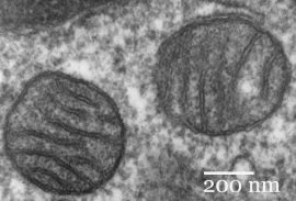
(0:42) New research proposes using mitochondria isolated from healthy tissue in a patient's body to treat ischemic heart muscle and perhaps other dysfunctional tissues or organs.
(4:44) Syllabuses, syllabi. Whatever. It's almost time to think about tweaking our course documents for the fall semester. I'll cover that in an upcoming episode, so I need you to send your contributions now!
Please share your syllabus ideas, questions, or comments at:
1-833-LION-DEN or 1-833-546-6336
podcast@theAPprofessor.org
(5:55) It's too long for one episode, so it's a series of three episodes: 21, 22 (previous episodes), and 23 (this episode).
If you're not teaching online now, you will be someday! Most of these tips apply to face-to-face courses, anyway.
In the previous two episodes, Kevin suggested:
It's all about connections.
Cultivate a friendly, informal, and supportive "online teaching persona"
Express empathy, don't just have empathy.
Use customer-service skills when communicating with students
Use our own pain points and frustrations to tap into how our students might feel
How we can literally make our online course a face to face course
How to use faces, voices, and scheduled course announcements to enhance the connections necessary to retain students and promote student success.
(8:47) Online courses are notorious for high dropout rates and high failure rates, compared to traditional face-to-face classes. Kevin continues to share even more strategies he has found to work in creating and nurturing the kinds of connections that help retain students and support their success in the course.
This episode focuses on:
Why reaching out to individual students who may be at risk is important--and how to do that.
Why feedback to students is important in nurturing connections.
Some final thoughts.
If you experience a repeated section starting about about timestamp 21:36, it's not your imagination. Probably. A pre-release version had such a hiccup and it may have been downloaded into your app. If so, you can simply re-download in the app. Or enjoy twice the fun by leaving the repeat in there!
If the hyperlinks here are not active, go to TAPPradio.org to find the episode page.
Pre-testing is not just for assessment—it helps learning, too. A weird sneeze injury. The Anatomical Society's list of online resources. How many proteins are there in a cell?
A recent analysis suggests that a reasonable average number of proteins in a cell is 42 million. How might we incorporate that bit of trivia in our A&P courses? (0:41)
Pre-Testing isn't just for measuring prior competence before new learning starts. By itself, regardless of its use in course assessment, it's a powerful learning tool. Listen to Kevin's experience with pre-testing in his A&P courses. (10:28)
Platelets as potent scavengers of bacteria? Really?
Something like 750 billion tiny cell fragments called platelets circulate in the human blood stream. When an injury to a blood vessel occurs, they stick to the exposed collagen in groups—forming platelet plug. And trigger additional reactions that eventually result in a blood clot.
But did you know that they have other helpful jobs, too? Like rounding up bacteria and feeding them up to immune cells, which devour them to make us safe.
This innate immune function of platelets has recently been outlined by researchers, as the information below summarizes.
Read through the quick points below to get an overview of some immune functions of platelets. Then read the full articles if you want to know more about these discoveries—including some great diagrams, micrographs, and videos.
Quick points about platelets as bacterial scavengers
At sites of vessel injury/inflammation, platelets that contact intact collagen stick together—but platelets that do not contact collagen are motile.
Motile platelets change shape from a "fried egg" to a polarized "half moon" to better navigate the shearing forces of blood flow.
They can even navigate "upstream" against the flow of blood.
Platelets can use mechanical force to pull particles—including bacteria—from surrounding substrates.
Platelets collect and bundle bacteria, which facilitates neutrophil activation and subsequent phagocytosis.
Migration pattern of motile platelet (left). Platelets collecting bacteria into bundles.
What can we use from this in teaching undergraduate A&P?
Yeah, okay we don't have time to go into all the ins and outs of platelets in a typical A&P course, but we canmention that platelets are now known to have immune functions.
Consider circling back to this mention later, when (if) you cover innate immune mechanisms a bit later in the course.
Consider calling attention to the sensory functions needed for platelets to analyze their microenvironment within the bloodstream.
Consider pointing out the specialized structure and function of the platelet's plasma membrane.
Integrins (integral membrane proteins) have a role in detecting particles for adhesion, binding to them, and sorting them.
Invaginations of the plasma membrane facilitate bundling of bacteria.
The shape changes needed for migration and handling of bacteria require actin-myosin reactions to power them. As in muscle fibers, these contractions are triggered by influx of extracellular calcium. In case you want to circle back to that.
Perhaps we should make a stronger point in reminding students that although they are "cell fragments" without a nucleus, they're more than just bags of hemostatic chemicals.
All these opportunities to "circle back" to previously studied concepts helps students make connections in their developing conceptual framework. And help them form a better understanding of the "big picture."
Introduction to the Gaertner, et. al., paper below—giving background and overview to enhance understanding of the new discoveries. Great diagram, too! Click "Supplemental information" in the article to access video clips.
Journal article describing the scavenger role of platelets. Includes a few very nice, simple diagrams—and some cool micrographs and data graphs. These can also be downloaded as PowerPoint slides. Click "Supplemental information" in the article to access video clips.
[NOTE: If you can't access the full text of any resource, ask your school's reference librarian for help. If they can't provide direct access, they'll probably know how to get a copy of the resource for you. Quickly.]
Today, the Royal Swedish Academy of Sciences has decided to award the Nobel Prize in Chemistry 2017 to Jacques Dubochet (University of Lausanne, Switzerland) and Joachim Frank (Columbia University, New York, USA), and Richard Henderson (MRC Laboratory of Molecular Biology, Cambridge, UK). The award is given "for developing cryo-electron microscopy for the high-resolution structure determination of biomolecules in solution"
We may soon have detailed images of life’s complex machineries in atomic resolution. The Nobel Prize in Chemistry 2017 is awarded to Jacques Dubochet, Joachim Frank and Richard Henderson for the development of cryo-electron microscopy, which both simplifies and improves the imaging of biomolecules. This method has moved biochemistry into a new era.
A picture is a key to understanding. Scientific breakthroughs often build upon the successful visualization of objects invisible to the human eye. However, biochemical maps have long been filled with blank spaces because the available technology has had difficulty generating images of much of life’s molecular machinery. Cryo-electron microscopy changes all of this. Researchers can now freeze biomolecules mid-movement and visualize processes they have never previously seen, which is decisive for both the basic understanding of life’s chemistry and for the development of pharmaceuticals.
Electron microscopes were long believed to only be suitable for imaging dead matter, because the powerful electron beam destroys biological material. But in 1990, Richard Henderson succeeded in using an electron microscope to generate a three-dimensional image of a protein at atomic resolution. This breakthrough proved the technology’s potential.
Joachim Frank made the technology generally applicable. Between 1975 and 1986 he developed an image processing method in which the electron microscope’s fuzzy two-dimensional images are analysed and merged to reveal a sharp three-dimensional structure.
Jacques Dubochet added water to electron microscopy. Liquid water evaporates in the electron microscope’s vacuum, which makes the biomolecules collapse. In the early 1980s, Dubochet succeeded in vitrifying water – he cooled water so rapidly that it solidified in its liquid form around a biological sample, allowing the biomolecules to retain their natural shape even in a vacuum.
Following these discoveries, the electron microscope’s every nut and bolt have been optimised. The desired atomic resolution was reached in 2013, and researchers can now routinely produce three-dimensional structures of biomolecules. In the past few years, scientific literature has been filled with images of everything from proteins that cause antibiotic resistance, to the surface of the Zika virus. Biochemistry is now facing an explosive development and is all set for an exciting future.
Joachim Frank, born 1940 in Siegen, Germany. Ph.D. 1970, Technical University of Munich, Germany. Professor of Biochemistry and Molecular Biophysics and of Biological Sciences, Columbia University, New York, USA. http://franklab.cpmc.columbia.edu/franklab/ Richard Henderson, born 1945 in Edinburgh, Scotland. Ph.D. 1969, Cambridge University, UK. Programme Leader, MRC Laboratory of Molecular Biology, Cambridge, UK. www2.mrc-lmb.cam.ac.uk/groups/rh15/
What can we use from this in teaching undergraduate A&P?
If you talk about imaging molecules in your course, this could be a way to garner student interest—considering that this is a current and ongoing effort in science. I always have a brief "shape is important in biological chemistry and here's what we can see with current tools" because they're going to see all those little odd-shaped rutabaga blobs in illustrations in their textbooks.
If you bring up microscopy in your course, perhaps describing the types of microscopy, adding a bit of info on this could help show students that microscopy is still evolving—in exciting ways.
Consider using the annual Nobel Prize announcements as a springboard to discuss the process of scientific discovery.
Consider mentioning the other major awards for scientific achievement and discuss what the judges seem to value most about scientific discoveries. The Nobel Prize is the one everyone has heard of, so it's a great place to start.
Use the Nobel Prizes (and other awards) over time as a way to keep students aware of the history of, and progress, of human biology. One could also address the global diversity of laureates. Or the lack of other kinds of diversity among laureates.
The Nobel Assembly at Karolinska Institutet has today decided to award the 2017 Nobel Prize in Physiology or Medicine jointly to Jeffrey C. Hall,Michael Rosbash, and Michael W. Young for their discoveries of molecular mechanisms controlling the circadian rhythm.
Summary
Life on Earth is adapted to the rotation of our planet. For many years we have known that living organisms, including humans, have an internal, biological clock that helps them anticipate and adapt to the regular rhythm of the day. But how does this clock actually work? Jeffrey C. Hall, Michael Rosbash and Michael W. Young were able to peek inside our biological clock and elucidate its inner workings. Their discoveries explain how plants, animals and humans adapt their biological rhythm so that it is synchronized with the Earth's revolutions.
Using fruit flies as a model organism, this year's Nobel laureates isolated a gene that controls the normal daily biological rhythm. They showed that this gene encodes a protein that accumulates in the cell during the night, and is then degraded during the day. Subsequently, they identified additional protein components of this machinery, exposing the mechanism governing the self-sustaining clockwork inside the cell. We now recognize that biological clocks function by the same principles in cells of other multicellular organisms, including humans.
With exquisite precision, our inner clock adapts our physiology to the dramatically different phases of the day. The clock regulates critical functions such as behavior, hormone levels, sleep, body temperature and metabolism. Our wellbeing is affected when there is a temporary mismatch between our external environment and this internal biological clock, for example when we travel across several time zones and experience "jet lag". There are also indications that chronic misalignment between our lifestyle and the rhythm dictated by our inner timekeeper is associated with increased risk for various diseases.
Our inner clock
Most living organisms anticipate and adapt to daily changes in the environment. During the 18th century, the astronomer Jean Jacques d'Ortous de Mairan studied mimosa plants, and found that the leaves opened towards the sun during daytime and closed at dusk. He wondered what would happen if the plant was placed in constant darkness. He found that independent of daily sunlight the leaves continued to follow their normal daily oscillation (Figure 1). Plants seemed to have their own biological clock.
Other researchers found that not only plants, but also animals and humans, have a biological clock that helps to prepare our physiology for the fluctuations of the day. This regular adaptation is referred to as the circadian rhythm, originating from the Latin words circa meaning "around" and dies meaning "day". But just how our internal circadian biological clock worked remained a mystery.
Figure 1. An internal biological clock. The leaves of the mimosa plant open towards the sun during day but close at dusk (upper part). Jean Jacques d'Ortous de Mairan placed the plant in constant darkness (lower part) and found that the leaves continue to follow their normal daily rhythm, even without any fluctuations in daily light.
Identification of a clock gene
During the 1970's, Seymour Benzer and his student Ronald Konopka asked whether it would be possible to identify genes that control the circadian rhythm in fruit flies. They demonstrated that mutations in an unknown gene disrupted the circadian clock of flies. They named this gene period. But how could this gene influence the circadian rhythm?
This year's Nobel Laureates, who were also studying fruit flies, aimed to discover how the clock actually works. In 1984, Jeffrey Hall and Michael Rosbash, working in close collaboration at Brandeis University in Boston, and Michael Young at the Rockefeller University in New York, succeeded in isolating the period gene. Jeffrey Hall and Michael Rosbash then went on to discover that PER, the protein encoded by period, accumulated during the night and was degraded during the day. Thus, PER protein levels oscillate over a 24-hour cycle, in synchrony with the circadian rhythm.
A self-regulating clockwork mechanism
The next key goal was to understand how such circadian oscillations could be generated and sustained. Jeffrey Hall and Michael Rosbash hypothesized that the PER protein blocked the activity of the period gene. They reasoned that by an inhibitory feedback loop, PER protein could prevent its own synthesis and thereby regulate its own level in a continuous, cyclic rhythm (Figure 2A).
Figure 2B. A simplified illustration of the molecular components of the circadian clock.
Such a regulatory feedback mechanism explained how this oscillation of cellular protein levels emerged, but questions lingered. What controlled the frequency of the oscillations? Michael Young identified yet another gene, doubletime, encoding the DBT protein that delayed the accumulation of the PER protein. This provided insight into how an oscillation is adjusted to more closely match a 24-hour cycle.
The paradigm-shifting discoveries by the laureates established key mechanistic principles for the biological clock. During the following years other molecular components of the clockwork mechanism were elucidated, explaining its stability and function. For example, this year's laureates identified additional proteins required for the activation of the period gene, as well as for the mechanism by which light can synchronize the clock.
Keeping time on our human physiology
The biological clock is involved in many aspects of our complex physiology. We now know that all multicellular organisms, including humans, utilize a similar mechanism to control circadian rhythms. A large proportion of our genes are regulated by the biological clock and, consequently, a carefully calibrated circadian rhythm adapts our physiology to the different phases of the day (Figure 3). Since the seminal discoveries by the three laureates, circadian biology has developed into a vast and highly dynamic research field, with implications for our health and wellbeing.
Figure 3. The circadian clock anticipates and adapts our physiology to the different phases of the day. Our biological clock helps to regulate sleep patterns, feeding behavior, hormone release, blood pressure, and body temperature.
About the Nobel Laureates
Jeffrey C. Hall was born 1945 in New York, USA. He received his doctoral degree in 1971 at the University of Washington in Seattle and was a postdoctoral fellow at the California Institute of Technology in Pasadena from 1971 to 1973. He joined the faculty at Brandeis University in Waltham in 1974. In 2002, he became associated with University of Maine.
Michael Rosbash was born in 1944 in Kansas City, USA. He received his doctoral degree in 1970 at the Massachusetts Institute of Technology in Cambridge. During the following three years, he was a postdoctoral fellow at the University of Edinburgh in Scotland. Since 1974, he has been on faculty at Brandeis University in Waltham, USA.
Michael W. Young was born in 1949 in Miami, USA. He received his doctoral degree at the University of Texas in Austin in 1975. Between 1975 and 1977, he was a postdoctoral fellow at Stanford University in Palo Alto. From 1978, he has been on faculty at the Rockefeller University in New York.
What can we use from this in teaching undergraduate A&P?
When you discuss biological clocks and rhythms in your course, this could be a way to garner student interest—considering that this is a current and ongoing effort in science. I begin discussing this at the beginning of the course—when covering homeostasis.
Consider using the annual Nobel Prize announcements as a springboard to discuss the process of scientific discovery.
Consider mentioning the other major awards for scientific achievement and discuss what the judges seem to value most about scientific discoveries. The Nobel Prize is the one everyone has heard of, so it's a great place to start.
Use the Nobel Prizes (and other awards) over time as a way to keep students aware of the history of, and progress, of human biology. One could also address the global diversity of laureates. Or the lack of other kinds of diversity among laureates.
The sources below are great places to find media for teaching and for great, pithy explanations of complex topics for a "beginner" audience like our A&P students.
The Nobel Assembly at Karolinska Institutet has today decided to award the 2016 Nobel Prize in Physiology or Medicine to Yoshinori Ohsumi for his discoveries of mechanisms for autophagy.
Overview
This year's Nobel Laureate discovered and elucidated mechanisms underlying autophagy, a fundamental process for degrading and recycling cellular components.
The word autophagy (aw-toh-FAY-jee) originates from the Greek words auto-, meaning "self", and phagein, meaning "to eat". Thus, autophagy denotes "self eating".
This concept emerged during the 1960's, when researchers first observed that the cell could destroy its own contents by enclosing it in membranes, forming sack-like vesicles that were transported to a recycling compartment, called the lysosome, for degradation.
Difficulties in studying the phenomenon meant that little was known until, in a series of brilliant experiments in the early 1990's, Yoshinori Ohsumi used baker's yeast to identify genes essential for autophagy. He then went on to elucidate the underlying mechanisms for autophagy in yeast and showed that similar sophisticated machinery is used in our cells.
Ohsumi's discoveries led to a new paradigm in our understanding of how the cell recycles its content. His discoveries opened the path to understanding the fundamental importance of autophagy in many physiological processes, such as in the adaptation to starvation or response to infection. Mutations in autophagy genes can cause disease, and the autophagic process is involved in several conditions including cancer and neurological disease.
Degradation – a central function in all living cells
In the mid 1950's scientists observed a new specialized cellular compartment, called an organelle, containing enzymes that digest proteins, carbohydrates and lipids. This specialized compartment is referred to as a "lysosome" and functions as a workstation for degradation of cellular constituents. The Belgian scientist Christian de Duve was awarded the Nobel Prize in Physiology or Medicine in 1974 for the discovery of the lysosome.
New observations during the 1960's showed that large amounts of cellular content, and even whole organelles, could sometimes be found inside lysosomes. The cell therefore appeared to have a strategy for delivering large cargo to the lysosome. Further biochemical and microscopic analysis revealed a new type of vesicle transporting cellular cargo to the lysosome for degradation (Figure 1).
Christian de Duve, the scientist behind the discovery of the lysosome, coined the term autophagy, "self-eating", to describe this process. The new vesicles were named autophagosomes.
Figure 1: Autophagosome. Our cells have different specialized compartments. Lysosomes constitute one such compartment and contain enzymes for digestion of cellular contents. A new type of vesicle called autophagosome was observed within the cell. As the autophagosome forms, it engulfs cellular contents, such as damaged proteins and organelles. Finally, it fuses with the lysosome, where the contents are degraded into smaller constituents. This process provides the cell with nutrients and building blocks for renewal.
During the 1970's and 1980's researchers focused on elucidating another system used to degrade proteins, namely the "proteasome". Within this research field Aaron Ciechanover, Avram Hershko and Irwin Rose were awarded the 2004 Nobel Prize in Chemistry for "the discovery of ubiquitin-mediated protein degradation". The proteasome efficiently degrades proteins one-by-one, but this mechanism did not explain how the cell got rid of larger protein complexes and worn-out organelles. Could the process of autophagy be the answer and, if so, what were the mechanisms?
A groundbreaking experiment
Yoshinori Ohsumi had been active in various research areas, but upon starting his own lab in 1988, he focused his efforts on protein degradation in the vacuole, an organelle that corresponds to the lysosome in human cells.
Yeast cells are relatively easy to study and consequently they are often used as a model for human cells. They are particularly useful for the identification of genes that are important in complex cellular pathways. But Ohsumi faced a major challenge; yeast cells are small and their inner structures are not easily distinguished under the microscope and thus he was uncertain whether autophagy even existed in this organism.
Ohsumi reasoned that if he could disrupt the degradation process in the vacuole while the process of autophagy was active, then autophagosomes should accumulate within the vacuole and become visible under the microscope. He therefore cultured mutated yeast lacking vacuolar degradation enzymes and simultaneously stimulated autophagy by starving the cells.
The results were striking! Within hours, the vacuoles were filled with small vesicles that had not been degraded (Figure 2). The vesicles were autophagosomes and Ohsumi's experiment proved that authophagy exists in yeast cells. But even more importantly, he now had a method to identify and characterize key genes involved this process. This was a major break-through and Ohsumi published the results in 1992.
Figure 2: Yeast. In yeast (left panel) a large compartment called the vacuole corresponds to the lysosome in mammalian cells. Ohsumi generated yeast lacking vacuolar degradation enzymes. When these yeast cells were starved, autophagosomes rapidly accumulated in the vacuole (middle panel). His experiment demonstrated that autophagy exists in yeast. As a next step, Ohsumi studied thousands of yeast mutants (right panel) and identified 15 genes that are essential for autophagy.
Autophagy genes are discovered
Ohsumi now took advantage of his engineered yeast strains in which autophagosomes accumulated during starvation. This accumulation should not occur if genes important for autophagy were inactivated. Ohsumi exposed the yeast cells to a chemical that randomly introduced mutations in many genes, and then he induced autophagy.
His strategy worked! Within a year of his discovery of autophagy in yeast, Ohsumi had identified the first genes essential for autophagy. In his subsequent series of elegant studies, the proteins encoded by these genes were functionally characterized. The results showed that autophagy is controlled by a cascade of proteins and protein complexes, each regulating a distinct stage of autophagosome initiation and formation (Figure 3).
Figure 3: Stages of autophagosome formation. Ohsumi studied the function of the proteins encoded by key autophagy genes. He delineated how stress signals initiate autophagy and the mechanism by which proteins and protein complexes promote distinct stages of autophagosome formation.
Autophagy – an essential mechanism in our cells
After the identification of the machinery for autophagy in yeast, a key question remained. Was there a corresponding mechanism to control this process in other organisms? Soon it became clear that virtually identical mechanisms operate in our own cells. The research tools required to investigate the importance of autophagy in humans were now available.
Thanks to Ohsumi and others following in his footsteps, we now know that autophagy controls important physiological functions where cellular components need to be degraded and recycled.
Autophagy can rapidly provide fuel for energy and building blocks for renewal of cellular components, and is therefore essential for the cellular response to starvation and other types of stress.
After infection, autophagy can eliminate invading intracellular bacteria and viruses. Autophagy contributes to embryo development and cell differentiation. Cells also use autophagy to eliminate damaged proteins and organelles, a quality control mechanism that is critical for counteracting the negative consequences of aging.
Disrupted autophagy has been linked to Parkinson's disease, type 2 diabetes and other disorders that appear in the elderly. Mutations in autophagy genes can cause genetic disease. Disturbances in the autophagic machinery have also been linked to cancer. Intense research is now ongoing to develop drugs that can target autophagy in various diseases.
Autophagy has been known for over 50 years but its fundamental importance in physiology and medicine was only recognized after Yoshinori Ohsumi's paradigm-shifting research in the 1990's. For
Yoshinori Ohsumi was born 1945 in Fukuoka, Japan. He received a Ph.D. from University of Tokyo in 1974. After spending three years at Rockefeller University, New York, USA, he returned to the University of Tokyo where he established his research group in 1988. He is since 2009 a professor at the Tokyo Institute of Technology.
More background on the winner and the prize
Yoshinori Ohsumi was born in Fukuoka, Japan, in 1945. He is affiliated with the Tokyo Institute of Technology in Tokyo, Japan. His monetary award will be nearly one million dollars.
The Nobel Assembly, consisting of 50 professors at Karolinska Institutet, awards the Nobel Prize in Physiology or Medicine. Its Nobel Committee evaluates the nominations. Since 1901 the Nobel Prize has been awarded to scientists who have made the most important discoveries for the benefit of mankind.his discoveries, he is awarded this year's Nobel Prize in physiology or medicine.
Nobel Prize® is the registered trademark of the Nobel Foundation
What can we use from this in teaching undergraduate A&P?
Consider using the Nobel Prizes as a discussion-starter in your class about
How science influences society
How society influences science
How science progresses
Rewarding of science discoveries
What makes a discovery "important"
Relate this discovery to prior (or upcoming) discussions of
Cell function
Organelle specialization
How cells handle protein
How autophagosomes work with lysosomes
Compare/contrast with phagocytosis
Compare/contrast with proteasome function and protein "quality control
Relate this discovery to the general idea of cellular mechanisms of disease
Consider taking this opportunity to emphasize "why we need to know all this" detail about cellular structure and function.
Want to know more?
Scientific Background Discoveries of Mechanisms for Autophagy
Larsson, N-G, Msucci, M. G. Nobelprize.org accessed 8 October 2016
A more advanced summary of the prizewinning discovery, including a handy glossary of terms.
A ubiquitin-like system mediates protein lipidation.
Ichimura, Y., Kirisako T., Takao, T., Satomi, Y., Shimonishi, Y., Ishihara, N., Mizushima, N., Tanida, I., Kominami, E., Ohsumi, M., Noda, T. and Ohsumi, Y. (2000). Nature, 408, 488-492
Honoring the 2016 Nobel laureates with free access to selections of their research
Elisa Nelissen Elsevier Connect. October 3, 2016
This blog post provides links to download the most cited papers the laureates published with Elsevier, a major publisher of scientific journals and references.
Huntingtin, the abnormal protein that produces clumps characteristic of Huntington disease (HD), can spread from one neuron to another. That's what a recent study has uncovered. Because such protein clumping is observed in other neurodegenerative disorders such as Alzheimer disease (AD) and Parkinson disease (PD), some scientists hope that understanding this newly discovered mechanism of transmission within brain tissue may lead to possible treatments or preventive strategies.
If you want to read more about it, check out the resources I've provided below.
What can we use from this in teaching undergraduate A&P?
This information can help us answer those pesky "why do we need to know all this if I'm going to be a [insert health profession here]?" challenges when covering the details of protein structure. The sequence of amino acids and the complex folded structure of proteins really does have real-world clinical implications. And is already becoming necessary to understand disease mechanisms and treatment strategies. In real life!
Discussing the basic idea of this discovery provides a starting platform from which we can jump into discussions of
Degeneration of tissues in general and neurodegeneration in particular
Why neurodegeneration in specific brain locations produces specific neural deficits
Prions and their possible roles in various disorders
The possible roles of genetic mechanisms in neurodegenerative disorders
The need to know details about protein structure (see item above)
Current directions in medical research—that proteins are hot!
Want to know more?
Neurodegeneration’s Spread
Ashley P. Taylor. The Scientist. August 4, 2014
Plain-English article describing the new research showing that pathogenic protein aggregates that accumulate within neurons and are a hallmark of Huntington’s disease can propagate from cell to cell.
Five years ago, I extolled the virtues of teaching a little bit about RNA interference (RNAi) in undergraduate A&P courses. But for a while it looked like the promise of RNAi in basic and clinical research might be sputtering. However, a recent article by Eric Bender called The Second Coming of RNAi shows that RNAi "the gene-silencing technique [now] begins to fulfill some of its promises."
I recommend reading the entire article at my-ap.us/1BbxvB9 Before you read it, allow me to reprise my reasons of five years ago supporting my proposal to include RNAi in your course.
What can we use from this in teaching undergraduate A&P?
RNAi plays a role in defending our cells against viruses by stopping viral genetic code from being translated in host cells
RNAi likely plays a role in regulating gene activity in a cell by preventing translation of the gene product(s)
RNAi is increasingly used as method for "knocking out" a particular gene's effects in research animals in order to study the gene's functions
RNAi is being used to treat genetic disease. . . an application that will likely expand greatly over the next few decades
I'll add two more items to my previous list:
RNA interference is a mechanism of human disease, as has been demonstrated in some cases of inherited progressive hearing loss (for example).
Learning about RNAi helps clarify a general understanding of the many roles played by RNA in our lives—some perhaps still undiscovered.
I'm not sure that it's useful to expect beginning undergraduate students to learn the nitty-gritty details of RNAi mechanisms. But I do think it's valuable to be exposed to the general concept of RNA interference and gene silencing.A&P students are going to run up against these eventually as they learn about and then administer RNAi-based therapies, after all. And perhaps we should prepare them.
Want to know more?
The Second Coming of RNAi
Eric Bender. The Scientist. September 1, 2014
Article mentioned above. In plain English, it shows that clinical progress in RNAi therapy against liver diseases, the gene-silencing technique begins to fulfill some of its promises. Includes useful illustrations and links to other resources.
The Royal Swedish Academy of Sciences has decided to award the Nobel Prize in Chemistry for 2014 to
Eric Betzig
Janelia Farm Research Campus, Howard Hughes Medical Institute, Ashburn, VA, USA,
Stefan W. Hell
Max Planck Institute for Biophysical Chemistry, Göttingen, and German Cancer Research Center, Heidelberg, Germany
and
William E. Moerner
Stanford University, Stanford, CA, USA
“for the development of super-resolved fluorescence microscopy”
Surpassing the limitations of the light microscope
For a long time optical microscopy was held back by a presumed limitation: that it would never obtain a better resolution than half the wavelength of light. Helped by fluorescent molecules the Nobel Laureates in Chemistry 2014 ingeniously circumvented this limitation. Their ground-breaking work has brought optical microscopy into the nanodimension.
In what has become known as nanoscopy, scientists visualize the pathways of individual molecules inside living cells. They can see how molecules create synapses between nerve cells in the brain; they can track proteins involved in Parkinson’s, Alzheimer’s and Huntington’s diseases as they aggregate; they follow individual proteins in fertilized eggs as these divide into embryos.
It was all but obvious that scientists should ever be able to study living cells in the tiniest molecular detail. In 1873, the microscopist Ernst Abbe stipulated a physical limit for the maximum resolution of traditional optical microscopy: it could never become better than 0.2 micrometres. Eric Betzig, Stefan W. Hell and William E. Moerner are awarded the Nobel Prize in Chemistry 2014 for having bypassed this limit. Due to their achievements the optical microscope can now peer into the nanoworld.
Two separate principles are rewarded.
One enables the method stimulated emission depletion (STED) microscopy, developed by Stefan Hell in 2000. Two laser beams are utilized; one stimulates fluorescent molecules to glow, another cancels out all fluorescence except for that in a nanometre-sized volume. Scanning over the sample, nanometre for nanometre, yields an image with a resolution better than Abbe’s stipulated limit.
Eric Betzig and William Moerner, working separately, laid the foundation for the second method, single-molecule microscopy. The method relies upon the possibility to turn the fluorescence of individual molecules on and off. Scientists image the same area multiple times, letting just a few interspersed molecules glow each time. Superimposing these images yields a dense super-image resolved at the nanolevel. In 2006 Eric Betzig utilized this method for the first time.
Today, nanoscopy is used world-wide and new knowledge of greatest benefit to mankind is produced on a daily basis.
This video is a brief animation of how STED works and how it improves resolution of individual particles.
This video is a longer, more detailed presentation by one of the Nobel laureates (Hell).
What can we use from this in teaching undergraduate A&P?
Discuss how this technology has enabled us to better visualize the chemicals and structures within our cells, enabling scientists to better understand the structure and function of cell, organelles, microbiome constituents, and other structures of the human body.
If you do a brief run-through of the theory of microscopy—perhaps in your A&P lab—you can add a mention of this technology.
Your textbook or other learning resource may already have an example of this type of microscopy.
A discussion of this Nobel Prize could evolve into a meaningful example of how science works, including how incremental improvements in classical tools for observation expand the number of questions that can be answered.
Use the links below (and images above) to use for a handout and/or teaching slides.
U.S. citizen. Born 1960 in Ann Arbor, MI, USA. Ph.D. 1988 from Cornell University, Ithaca, NY, USA. Group Leader at Janelia Farm Research Campus, Howard Hughes Medical Institute, Ashburn, VA, USA.
Born 1962 in Arad, Romania. Ph.D. 1990 from the University of Heidelberg, Germany. Director at the Max Planck Institute for Biophysical Chemistry, Göttingen, and Division head at the German Cancer Research Center, Heidelberg, Germany.
Born 1953 in Pleasanton, CA, USA. Ph.D. 1982 from Cornell University, Ithaca, NY, USA. Harry S. Mosher Professor in Chemistry and Professor, by courtesy, of Applied Physics at Stanford University, Stanford, CA, USA.
Many longtime readers of this blog know that I have a set of animated PowerPoint-compatible slides available for you to use FREE in your A&P classes. These slides—the Lion Den Slide Collection— supplement the publisher-supplied slides or homegrown slides that you are already using.
I recently updated, improved, and expanded the set of slides that animate several key cell transport processes such as diffusion, osmosis, endocytosis, etc.
If you've accessed the Lion Den Slide Collectionin the last several months, you've already registered in the system and probably have already received an email notification of the update.
If you're new to the collection (or accessed it before June 2014) then you need to go to the Lion Den Slide Collectionpage and click on the link to the form. It takes a few steps, but by filling out the brief forms you register for a service that will notify you (if you want it) of any updates or additions to the collection.
When you download the linked file, you'll be able to play it in PowerPoint or any other program that plays "show" or PPTSX files. The slides can only be viewed, not edited or added to your own slide deck. To do that, you must download the fully editable PPTX files from the Lion Den Slide Collection.
Once you download any slide deck from the Lion Den Slide Collection, you can mix and match them to blend them with your existing presentations. You can also alter the content or timings to suit your needs.
I'm hoping you'll also get some ideas from them to create new additional slides! If you modify, create, or already have any slides (for which you own the content) that you want to place in the collection for sharing, please contact me directly.
Want to know more?
Presentation Zen
Kevin Patton The Electronic Professor 6 August 2014
The zen approach to presentations. Includes a video and links to resources.
Kevin Patton. The Electronic Professor. 3 August 2012
Another of my blog articles on improving slide presentations. Includes my own video on how to trim down those wordy slides to something that actually works in a slide.
A recent article in The Scientist once again reminds us of the ongoing explosion in the scientific understanding of the human microbial system. In a few short years, this area of exploration has moved to the forefront of medical and basic science research in human biology.
I think it's becoming clear that the most useful way to think of human body function is to recognize that an "organism" is really a sort of "habitat." And like any habitat, it functions best when all the inhabitants are within a limited range of balanced relationships.
Who are the inhabitants? Besides our own cells? Well, one could think of mitochondria and cilia and other organelles as symbiotic internal inhabitants of our cells. They're not that literally, of course, but I think its a useful metaphor for understanding the human body. Then there are the many microbes and animals that cover our internal and external surfaces, burrow into some of our pores and glands, and inhabit our body fluids.
I call the balanced functional relationship among the various microbomes of the body and our own tissues the human microbial system. And I am certain that it won't be long before we will be discussing this system alongside the major organ systems of the body. That is if we truly want to understand how the body really works.
The article in The Scientist I mention is a great summary of some of the major roles that the human microbial system plays in the human body—and a good survey of some of the areas of the body where the human-microbial functional relationships play out. See the link to the article below.
What can we use from this in teaching undergraduate A&P?
Why not introduce the concept of the human microbial system at the beginning of our A&P course, when we set the stage by explain how scientists understand the body and its functions as an integrated system of different parts?
We can mention the different microbiomes of the body when we explore each organ system where they play an important role—which is pretty much all of them!
Consider discussing what happens to normal human function when microbiomes get out of balance. For example, in the gut a microbial imbalance can lead to ulcers, diarrhea, and other dysfunctions. On the skin a pathogenic microbe may become dominant and cause a rash.
Promote a discussion of what kinds of wellness strategies might be employed to prevent microbial imbalances.
Our students can leave our A&P course with an up-to-date understanding of human biology that will help them understand new clinical concepts and treatment strategies.
Want to Know More?
The Body’s Ecosystem
By The Scientist Staff. The Scientist. August 1, 2014
Plain-English article (cited above) on how research on the human microbiome is booming, and scientists have moved from simply taking stock of gut flora to understanding the influence of microbes throughout the body.
Radio stories from National Public Radio on human microbiomes and their role in health and disease. The growing number of these stories tells us something as A&P teachers: maybe we better be covering this!