Showing posts with label microbiomes. Show all posts

Thursday, February 9, 2017

How Dietary Fiber Prevents Disease

So why, exactly, is it that we should consume a lot of fiber in our diet to remain healthy? Are refined fiber supplements just as good as, say, an "apple a day?"

Recently, an article in the journal Cell answer seems to verify some of the answers for us.

As the paper cited below indicates, research seems to confirm that dietary fiber provides nutrients for the inhabitants of our intestinal microbiome.  When dietary fiber is missing, then the microbes undergo a shift in populations and start consuming our GI mucus as an alternate source of nutrition.  That, as you might guess, reduces the thickness of the protective mucus—hus increasing the likelihood that pathogens can more easily attack the intestinal lining. Ouch.

Apparently, refined prebiotic fibers don't fix the problem.

Here are some highlights of the research article (quoted from their online preview):

  • Characterized synthetic bacterial communities enable functional insights in vivo
  • Low-fiber diet promotes expansion and activity of colonic mucus-degrading bacteria
  • Purified prebiotic fibers do not alleviate degradation of the mucus layer
  • Fiber-deprived gut microbiota promotes aggressive colitis by an enteric pathogen

Image for unlabelled figure


What can we use from this in teaching undergraduate A&P?


  • When asked by students about dietary fiber, you have more information from which to draw an answer.
  • When discussing any of these topics, you'll now have a bit more to add to your story:
    • nutrition
    • function of mucus
    • the human microbial system (or specifically, the GI microbiome)
    • how pathogens cause disease (or specifically, GI disorders)

Want to know more?


Veggies and Intact Grains a Day Keep the Pathogens Away

  • Francesca S. Gazzaniga. Dennis L. Kasper. Cell. Available online 17 November 2016
  • Brief preview of the M. Desai article cited below.
  • my-ap.us/2lijkbE


A Dietary Fiber-Deprived Gut Microbiota Degrades the Colonic Mucus Barrier and Enhances Pathogen Susceptibility

  • Mahesh S. Desai et al. Cell, Volume 167, Issue 5, 17 November 2016, Pages 1339-1353.e21
  • The detailed research article.
  • my-ap.us/2lijz6w

Microbiome articles

  • Kevin Patton. The A&P Professor. Various dates.
  • Collection of previous posts on this topic from this blog.
  • my-ap.us/2liCrSP


Photos: Youssef KH (top) Cell (bottom)

Wednesday, October 8, 2014

Nobel Prize 2014: Super-resolved fluorescence microscopy


The Royal Swedish Academy of Sciences has decided to award the Nobel Prize in Chemistry for 2014 to

Eric Betzig
Janelia Farm Research Campus, Howard Hughes Medical Institute, Ashburn, VA, USA,

Stefan W. Hell
Max Planck Institute for Biophysical Chemistry, Göttingen, and German Cancer Research Center, Heidelberg, Germany

and

William E. Moerner
Stanford University, Stanford, CA, USA

“for the development of
super-resolved fluorescence microscopy”


Surpassing the limitations of the light microscope


For a long time optical microscopy was held back by a presumed limitation: that it would never obtain a better resolution than half the wavelength of light. Helped by fluorescent molecules the Nobel Laureates in Chemistry 2014 ingeniously circumvented this limitation. Their ground-breaking work has brought optical microscopy into the nanodimension.

In what has become known as nanoscopy, scientists visualize the pathways of individual molecules inside living cells. They can see how molecules create synapses between nerve cells in the brain; they can track proteins involved in Parkinson’s, Alzheimer’s and Huntington’s diseases as they aggregate; they follow individual proteins in fertilized eggs as these divide into embryos.

It was all but obvious that scientists should ever be able to study living cells in the tiniest molecular detail. In 1873, the microscopist Ernst Abbe stipulated a physical limit for the maximum resolution of traditional optical microscopy: it could never become better than 0.2 micrometres. Eric Betzig, Stefan W. Hell and William E. Moerner are awarded the Nobel Prize in Chemistry 2014 for having bypassed this limit. Due to their achievements the optical microscope can now peer into the nanoworld.

Two separate principles are rewarded. 


One enables the method stimulated emission depletion (STED) microscopy, developed by Stefan Hell in 2000. Two laser beams are utilized; one stimulates fluorescent molecules to glow, another cancels out all fluorescence except for that in a nanometre-sized volume. Scanning over the sample, nanometre for nanometre, yields an image with a resolution better than Abbe’s stipulated limit.

Eric Betzig and William Moerner, working separately, laid the foundation for the second method, single-molecule microscopy. The method relies upon the possibility to turn the fluorescence of individual molecules on and off. Scientists image the same area multiple times, letting just a few interspersed molecules glow each time. Superimposing these images yields a dense super-image resolved at the nanolevel. In 2006 Eric Betzig utilized this method for the first time.

Today, nanoscopy is used world-wide and new knowledge of greatest benefit to mankind is produced on a daily basis.


This video is a brief animation of how STED works and how it improves resolution of individual particles.


This video is a longer, more detailed presentation by one of the Nobel laureates (Hell).


What can we use from this in teaching undergraduate A&P?

  • Discuss how this technology has enabled us to better visualize the chemicals and structures within our cells, enabling scientists to better understand the structure and function of cell, organelles, microbiome constituents, and other structures of the human body.

  • If you do a brief run-through of the theory of microscopy—perhaps in your A&P lab—you can add a mention of this technology.  

  • Your textbook or other learning resource may already have an example of this type of microscopy.

  • A discussion of this  Nobel Prize could evolve into a meaningful example of how science works, including how incremental improvements in classical tools for observation expand the number of questions that can be answered.

  • Use the links below (and images above) to use for a handout and/or teaching slides.


Want to know more?


Resources from Nobelprize.org

  • Popular Information 
  • Scientific Background
    • Handout: More detailed information includes references to original research articles
    • my-ap.us/ZdLJ69
  • Advanced Information
  • Images
  • Biographies
    • Eric Betzig, 
      • U.S. citizen. Born 1960 in Ann Arbor, MI, USA. Ph.D. 1988 from Cornell University, Ithaca, NY, USA. Group Leader at Janelia Farm Research Campus, Howard Hughes Medical Institute, Ashburn, VA, USA.
      • http://janelia.org/lab/betzig-lab
    • Stefan W. Hell, German citizen. 
      • Born 1962 in Arad, Romania. Ph.D. 1990 from the University of Heidelberg, Germany. Director at the Max Planck Institute for Biophysical Chemistry, Göttingen, and Division head at the German Cancer Research Center, Heidelberg, Germany.
      • http://www3.mpibpc.mpg.de/groups/hell
    • William E. Moerner, U.S. citizen. 
      • Born 1953 in Pleasanton, CA, USA. Ph.D. 1982 from Cornell University, Ithaca, NY, USA. Harry S. Mosher Professor in Chemistry and Professor, by courtesy, of Applied Physics at Stanford University, Stanford, CA, USA.
      • http://web.stanford.edu/group/moerner


Diagrram cretit: Ganbaatar
Micrograph credit: Tesselkaffee
Text adapted from press release from Nobel Media

Monday, August 18, 2014

Human Microbial System


A recent article in The Scientist once again reminds us of the ongoing explosion in the scientific understanding of the human microbial system.  In a few short years, this area of exploration has moved to the forefront of medical and basic science research in human biology.

I think it's becoming clear that the most useful way to think of human body function is to recognize that an "organism" is really a sort of "habitat."  And like any habitat, it functions best when all the inhabitants are within a limited range of balanced relationships.

Who are the inhabitants?  Besides our own cells?  Well, one could think of mitochondria and cilia and other organelles as symbiotic internal inhabitants of our cells.  They're not that literally, of course, but I think its a useful metaphor for understanding the human body.  Then there are the many microbes and animals that cover our internal and external surfaces, burrow into some of our pores and glands, and inhabit our body fluids.

I call the balanced functional relationship among the various microbomes of the body and our own tissues the human microbial system.  And I am certain that it won't be long before we will be discussing this system alongside the major organ systems of the body.  That is if we truly want to understand how the body really works.

The article in The Scientist I mention is a great summary of some of the major roles that the human microbial system plays in the human body—and a good survey of some of the areas of the body where the human-microbial functional relationships play out.  See the link to the article below.


What can we use from this in teaching undergraduate A&P?


  • Why not introduce the concept of the human microbial system at the beginning of our A&P course, when we set the stage by explain how scientists understand the body and its functions as an integrated system of different parts?
  • We can mention the different microbiomes of the body when we explore each organ system where they play an important role—which is pretty much all of them!
  • Consider discussing what happens to normal human function when microbiomes get out of balance.  For example, in the gut a microbial imbalance can lead to ulcers, diarrhea, and other dysfunctions.  On the skin a pathogenic microbe may become dominant and cause a rash.
  • Promote a discussion of what kinds of wellness strategies might be employed to prevent microbial imbalances.
  • Our students can leave our A&P course with an up-to-date understanding of human biology that will help them understand new clinical concepts and treatment strategies.

Want to Know More?

The Body’s Ecosystem

  • By The Scientist Staff.  The Scientist. August 1, 2014
  • Plain-English article (cited above) on how research on the human microbiome is booming, and scientists have moved from simply taking stock of gut flora to understanding the influence of microbes throughout the body.
  • my-ap.us/1vgOu5y

Articles from The A&P Professor


Moving pictures of the human microbiome

  • J Gregory Caporaso et al. Genome Biology 2011, 12:R50  doi:10.1186/gb-2011-12-5-r50
  • Open-access journal article that includes FREE videos that show how dynamic the human microbial system is
  • my-ap.us/V7St3Q

Human Microbiome Project


The Microbiome and Disease

  • List of diseases associated with microbiome imbalances from Genetic Science Learning Center
  • my-ap.us/1nDpxr1

Audio

  • Radio stories from National Public Radio on human microbiomes and their role in health and disease.  The growing number of these stories tells us something as A&P teachers: maybe we better be covering this!
  • my-ap.us/1uA7Qyg

Monday, October 3, 2011

Dendritic cell pioneers win Nobel Prize

The Nobel Assembly at Karolinska Institutet has today decided that

The Nobel Prize in Physiology or Medicine 2011
shall be divided, with one half jointly to
Bruce A. Beutler and Jules A. Hoffmann
for their discoveries concerning the activation of innate immunity
and the other half to
Ralph M. Steinman
for his discovery of the dendritic cell and its role in adaptive immunity


Summary

This year's Nobel Laureates have revolutionized our understanding of the immune system by discovering key principles for its activation.
Scientists have long been searching for the gatekeepers of the immune response by which man and other animals defend themselves against attack by bacteria and other microorganisms. Bruce Beutler and Jules Hoffmann discovered receptor proteins that can recognize such microorganisms and activate innate immunity, the first step in the body's immune response. Ralph Steinman discovered the dendritic cells of the immune system and their unique capacity to activate and regulate adaptive immunity, the later stage of the immune response during which microorganisms are cleared from the body.
The discoveries of the three Nobel Laureates have revealed how the innate and adaptive phases of the immune response are activated and thereby provided novel insights into disease mechanisms. Their work has opened up new avenues for the development of prevention and therapy against infections, cancer, and inflammatory diseases.

Two lines of defense in the immune system

We live in a dangerous world. Pathogenic microorganisms (bacteria, virus, fungi, and parasites) threaten us continuously but we are equipped with powerful defense mechanisms (please see image below). The first line of defense, innate immunity, can destroy invading microorganisms and trigger inflammation that contributes to blocking their assault. If microorganisms break through this defense line, adaptive immunity is called into action. With its T and B cells, it produces antibodies and killer cells that destroy infected cells. After successfully combating the infectious assault, our adaptive immune system maintains an immunologic memory that allows a more rapid and powerful mobilization of defense forces next time the same microorganism attacks. These two defense lines of the immune system provide good protection against infections but they also pose a risk. If the activation threshold is too low, or if endogenous molecules can activate the system, inflammatory disease may follow.
The components of the immune system have been identified step by step during the 20th century. Thanks to a series of discoveries awarded the Nobel Prize, we know, for instance, how antibodies are constructed and how T cells recognize foreign substances. However, until the work of Beutler, Hoffmann and Steinman, the mechanisms triggering the activation of innate immunity and mediating the communication between innate and adaptive immunity remained enigmatic.

Discovering the sensors of innate immunity

Jules Hoffmann made his pioneering discovery in 1996, when he and his co-workers investigated how fruit flies combat infections. They had access to flies with mutations in several different genes including Toll, a gene previously found to be involved in embryonal development by Christiane NĂĽsslein-Volhard (Nobel Prize 1995). When Hoffmann infected his fruit flies with bacteria or fungi, he discovered that Toll mutants died because they could not mount an effective defense. He was also able to conclude that the product of the Toll gene was involved in sensing pathogenic microorganisms and Toll activation was needed for successful defense against them.
Bruce Beutler was searching for a receptor that could bind the bacterial product, lipopolysaccharide (LPS), which can cause septic shock, a life threatening condition that involves overstimulation of the immune system. In 1998, Beutler and his colleagues discovered that mice resistant to LPS had a mutation in a gene that was quite similar to the Toll gene of the fruit fly. This Toll-like receptor (TLR) turned out to be the elusive LPS receptor. When it binds LPS, signals are activated that cause inflammation and, when LPS doses are excessive, septic shock. These findings showed that mammals and fruit flies use similar molecules to activate innate immunity when encountering pathogenic microorganisms. The sensors of innate immunity had finally been discovered.
The discoveries of Hoffmann and Beutler triggered an explosion of research in innate immunity. Around a dozen different TLRs have now been identified in humans and mice. Each one of them recognizes certain types of molecules common in microorganisms. Individuals with certain mutations in these receptors carry an increased risk of infections while other genetic variants of TLR are associated with an increased risk for chronic inflammatory diseases.

A new cell type that controls adaptive immunity

Ralph Steinman discovered, in 1973, a new cell type that he called the dendritic cell. He speculated that it could be important in the immune system and went on to test whether dendritic cells could activate T cells, a cell type that has a key role in adaptive immunity and develops an immunologic memory against many different substances. In cell culture experiments, he showed that the presence of dendritic cells resulted in vivid responses of T cells to such substances. These findings were initially met with skepticism but subsequent work by Steinman demonstrated that dendritic cells have a unique capacity to activate T cells.
Further studies by Steinman and other scientists went on to address the question of how the adaptive immune system decides whether or not it should be activated when encountering various substances. Signals arising from the innate immune response and sensed by dendritic cells were shown to control T cell activation. This makes it possible for the immune system to react towards pathogenic microorganisms while avoiding an attack on the body's own endogenous molecules.

From fundamental research to medical use

The discoveries that are awarded the 2011 Nobel Prize have provided novel insights into the activation and regulation of our immune system. They have made possible the development of new methods for preventing and treating disease, for instance with improved vaccines against infections and in attempts to stimulate the immune system to attack tumors. These discoveries also help us understand why the immune system can attack our own tissues, thus providing clues for novel treatment of inflammatory diseases.


Bruce A. Beutler was born in 1957 in Chicago, USA. He received his MD from the University of Chicago in 1981 and worked as a scientist at Rockefeller University in New York and the University of Texas in Dallas, where he discovered the LPS receptor. Since 2000 he has been professor of genetics and immunology at The Scripps Research Institute, La Jolla, USA.
Jules A. Hoffmann was born in Echternach, Luxembourg in 1941. He studied at the University of Strasbourg in France, where he obtained his PhD in 1969. After postdoctoral training at the University of Marburg, Germany, he returned to Strasbourg, where he headed a research laboratory from 1974 to 2009. He has also served as director of the Institute for Molecular Cell Biology in Strasbourg and during 2007-2008 as President of the French National Academy of Sciences.
Ralph M. Steinman was born in 1943 in Montreal, Canada, where he studied biology and chemistry at McGill University. After studying medicine at Harvard Medical School in Boston, MA, USA, he received his MD in 1968. He has been affiliated with Rockefeller University in New York since 1970, has been professor of immunology at this institution since 1988, and is also director of its Center for Immunology and Immune Diseases.


Key publications:

Poltorak A, He X, Smirnova I, Liu MY, Van Huffel C, Du X, Birdwell D, Alejos E, Silva M, Galanos C, Freudenberg M, Ricciardi-Castagnoli P, Layton B, Beutler B. Defective LPS signaling in C3H/HeJ and C57BL/10ScCr mice: Mutations in Tlr4 gene. Science 1998;282:2085-2088.
Lemaitre B, Nicolas E, Michaut L, Reichhart JM, Hoffmann JA. The dorsoventral regulatory gene cassette spätzle/Toll/cactus controls the potent antifungal response in drosophila adults. Cell 1996;86:973-983.
Steinman RM, Cohn ZA. Identification of a novel cell type in peripheral lymphoid organs of mice. J Exp Med 1973;137:1142-1162.
Steinman RM, Witmer MD. Lymphoid dendritic cells are potent stimulators of the primary mixed leukocyte reaction in mice. Proc Natl Acad Sci USA 1978;75:5132-5136.
Schuler G, Steinman RM. Murine epidermal Langerhans cells mature into potent immunostimulatory dendritic cells in vitro. J Exp Med 1985;161:526-546.

illustration High resolution image (pdf 3,6 Mb) 

The Nobel Assembly, consisting of 50 professors at Karolinska Institutet, awards the Nobel Prize in Physiology or Medicine. Its Nobel Committee evaluates the nominations. Since 1901 the Nobel Prize has been awarded to scientists who have made the most important discoveries for the benefit of mankind.

Nobel Prize® is the registered trademark of the Nobel Foundation 

The information above is taken directly from 
The 2011 Nobel Prize in Physiology or Medicine - Press Release
Nobelprize.org. 3 Oct 2011 my-ap.us/pE7zzC

Want to know more?
Immune Responses
[An animated activity from the Nobel Prize folks.] 

Find a brief explanation of dendritic cells in these textbooks:
Find FREE images and videos you can use in your course
Dendritic cells
http://my-ap.us/pkQycM

Watch a brief video on dendritic cells.

Wednesday, June 8, 2011

That weird E. coli epidemic

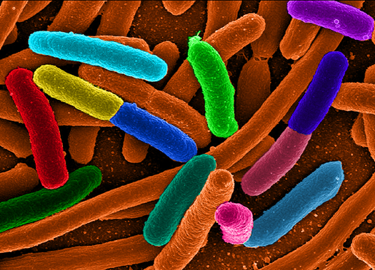
Remember my previous post, in which I gave you a free slide show on the role of the appendix in keeping the gut microbiome happy?  Well, to sort of "prove the point" of the importance of a healthy gut microbiome, we've been hearing all about that weird Escherichia coli (E. coli) epidemic in Europe.

If you're like me, you'll want to take the opportunity to emphasize concepts learned in class by applying them to "real life" events reported in the news.  If you're like me, you may want to check out these journal articles:

Deadly bugs: Toxin-producing E. coli strain causes outbreak in Germany
Tina Hesman Saey
Science News web edition : Tuesday, June 7th, 2011

my-ap.us/lfMeYE
[Brief, highly readable introduction to the current outbreak in Europe.  Plus a cool photo!]

Bacterial infections: new and emerging enteric pathogens
Sherman, P et al.
Current Opinion in Gastroenterology:January 2010 - Volume 26 - Issue 1 - p 1-4
doi: 10.1097/MOG.0b013e328333d73b
my-ap.us/lJv8PC
[from the abstract: "The aim of this review is to highlight recent advances in knowledge of bacterial enteric infections. We focus on understanding of enterohemorrhagic Escherichia coli O157:H7 and Campylobacter jejuni infections, and to link these acute events with long-term consequences in a susceptible host, including irritable bowel syndrome and chronic inflammatory bowel diseases."]

Clinical Relevance of Shiga Toxin Concentrations in the Blood of Patients With Hemolytic Uremic Syndrome
Brigotti, Maurizio et al.
Pediatric Infectious Disease Journal: June 2011 - Volume 30 - Issue 6 - pp 486-490
doi: 10.1097/INF.0b013e3182074d22
my-ap.us/j72bUA

[from the abstract: "Intestinal infections with Shiga toxin-producing Escherichia coli (STEC) in children can lead to the hemolytic uremic syndrome (HUS). Shiga toxins (Stx) released in the gut by bacteria enter the blood stream and target the kidney causing endothelial injury. Free toxins have never been detected in the blood of HUS patients, but they have been found on the surface of polymorphonuclear leukocytes (PMN)."]

Infectious colitis
Navaneethan, Udayakumar and Giannella, Ralph A
Current Opinion in Gastroenterology: January 2011 - Volume 27 - Issue 1 - p 66–71
doi: 10.1097/MOG.0b013e3283400755

my-ap.us/mTOlpN
[from the abstract: "The incidence of gastrointestinal infections continues to increase and infectious colitis contributes to significant morbidity and mortality worldwide. The purpose of this review is to highlight the recent advances in knowledge of pathogens causing infectious colitis. We describe the various pathogens and specifically focus on enterohemorrhagic Escherichia coli (EHEC) O157:H7, Salmonella, Shigella, Campylobacter, and Entamoeba histolytica infections, and their impact on long-term effects, including postinfectious irritable bowel syndrome and inflammatory bowel disease."]

An inside job: subversion of the host secretory pathway by intestinal pathogens
Sharp, Tyler M and Estes, Mary K
Current Opinion in Infectious Diseases: October 2010 - Volume 23 - Issue 5 - p 464–469
doi: 10.1097/QCO.0b013e32833dcebd

my-ap.us/leb7Le
[from the abstract: "The cellular secretory pathway, composed of the endoplasmic reticulum, Golgi apparatus, and cellular vesicles, mediates the intracellular trafficking of proteins and lipids. Gastrointestinal pathogens frequently affect the functions of enterocytes, the differentiated cells involved in secretion and absorption of extracellular molecules. Microbial pathogenesis can be enhanced by altering the trafficking of key molecules such as brush border enzymes, soluble immune mediators such as cytokines and chemokines, and MHC Class I molecules, all of which rely on the secretory pathway for their appropriate cellular localization. This review focuses on our current understanding of the distinct mechanisms employed by enteric pathogens to antagonize the secretory pathway."]


Probiotics: progress toward novel therapies for intestinal diseases
Yan et al.
Current Opinion in Gastroenterology: March 2010 - Volume 26 - Issue 2 - p 95–101
doi: 10.1097/MOG.0b013e328335239a

my-ap.us/iH93qP
[from the abstract "As the beneficial effects of probiotics on health and disease prevention and treatment have been well recognized, the demand for probiotics in clinical applications and as functional foods has significantly increased in spite of limited understanding of the mechanisms. This review focuses on the most recent advances in probiotic research from genetics to biological consequences regulated by probiotics and probiotic-derived factors."]

For a really cool, copyright-free image to use in your course, go to my-ap.us/lVEg69

Sunday, April 3, 2011

Plaque-fighting bacteria

Have you noticed that the microbiome of the human body has taken off as one of the hottest areas?  Each month, new concepts of how our microbial partners keeps us healthy are revealed.  Last week, we were discussing teeth in my A&P 2 course and I wish I'd had this new tidbit to share with my students:

Researchers recently found that Streptococcus salivarius, one of the microbes in our mouth, can help fight the buildup of plaque on our teeth.  It does so by producing the enzyme FruA, which breaks down carbohydrates in our mouth more efficiently than can the bacteria that form plaque biofilms.  Thus, the plaque-forming bacteria are robbed of their nutrients.

I'll bet S. salivarius will become popular as an oral probiotic.  And its discovery may help us find better ways to manage our mouth's ecosystem to promote good health.

Want to know more?

Inhibition of Streptococcus mutans Biofilm Formation by Streptococcus salivarius FruA 
A. Ogawa, et al.
Applied and Environmental Microbiology Vol. 77, March 2011, p. 1572 doi:10.1128/AEM.02066-10, published online January 14, 2011
[Original research article]
http://my-ap.us/h7RuaA

Bacterial fight dental plaque
Tina Hesman Saey
Science News Published online April 1, 2011
[Brief summary of the discovery]
http://my-ap.us/hsj1nb
Click the image above to access a FREE animation of tooth decay you can use in your course as you explain the process.

Sunday, November 8, 2009

Bacterial microbiomes on human skin

Nearly a year ago, I shared results of a study of the bacteria that live on human skin, including these fun facts:
  • Females have a higher diversity of bacteria on their hands than males . . . perhaps due to a slightly higher skin pH in women, or perhaps the mix of sebum, sweat, and lotions, or maybe even hormonal differences . . . they couldn't really say for sure at this point
  • Females have more bacteria living under the surface film of skin than males
  • 4, 742 different species of bacteria were found in the whole group of subjects
  • The species each of has on our hands is a rather unique mix--only 5 (out of 4,742) species were found on every hand in the group
  • Most of the 150 or so different species of bacteria found on skin of an individual hand are beneficial or harmless . . . only a small minority are pathogenic
  • The diversity of bacteria differs between a person's right hand and left hand
  • Hand washing (as practiced in this group) did not remove many of the bacteria (or the populations recovered rapidly after washing)
Recently, another study was published that gives us an even more complete picture of the micro-ecology of human skin.  The report, published online a few days ago by the journal Science, provides an inventory of what organisms live where on the human skin.

A few fun facts about the bacteria, viruses, and fungi of the human skin gleaned from the new study:
  • Microbes on the skin outnumber human cells by at least 10 times (about 100 trillion microbial symbionts)

  • Microbial community composition is determined primarily by habitat (well, of course!)

  • The composition of microbial communities varies widely from one person to another

  • The compostion of microbial communities for an individual human do not vary much over time

  • Some locations of the skin harbor more diverse communities than even the mouth or gut
Want to know more?
Bacterial Community Variation in Human Body Habitats Across Space and Time.
Elizabeth K. Costello, et al. 
Science Express, 5 November 2009, online .
doi: 10.1126/science.1177486
[Recent study on human flora]

Bacteria Flourish in Favorite Ecosystems on the Human Body
Laura Sanders
Science News November 5, 2009
[Nice summary of the study's importance and implications]

Variation In Bacterial Populations From Person To Person Surprises Researchers

C. Paddock
Medical News Today 6 November 2009
[Press release about the new study]

Skin Ecology
K. Patton
The A&P Professor 18 November 2008
[My previous article on the topic.  Includes links to other articles.]

Monday, August 24, 2009

New "old" news about the appendix


A recent article in ScienceDaily discusses a new article in the Journal of Evolutionary Biology regarding the evolution of the human appendix.

As you know, Darwin thought that the vermiform appendix was a vestigial, nonfunctional structure "left over" from our evolutionary past.

And if you been using my A&P textbooks for the last ten years or so, you also know that biologists have long suspected (and recently confirmed) that the appendix is no such thing. Its function is to incubate intestinal flora (gut bacteria)—which helps keep the ecosystem of the gut in a healthy balance by repopulating the colon with beneficial bacteria after an illness or other disturbance.

The new journal article, written by some of the same researchers involved in the most recent confirmation of the "incubator model of the appendix," tackles the evolutionary aspects of the appendix.

Want to know more?

Evolution Of The Human Appendix: A Biological 'Remnant' No More.
Duke University Medical Center
ScienceDaily Duke University Medical Center. 21 August 2009.
[Summary of the journal article and its context]
Comparative anatomy and phylogenetic distribution of the mammalian cecal appendix
Smith, et al.Journal of Evolutionary Biology. published online 12 August 2009
[Abstract of the journal article, published in advance of the print version]


Click here for a FREE surgical photo of a human vermiform appendix.

Tuesday, May 19, 2009

What is life?

A video I saw on the web recently reminded me of the meaning of life. OK, that sounds a LOT more profound than I mean it to.

What I mean is . . . one of the most interesting questions in science
that I deal with is the most fundamental question, and one that comes right
at the beginning of my A&P course. The question is, what does it
mean for a human to be alive? What constitutes a living human organism?

Unbelievably, most courses set aside the core of that question and deal solely with the superficial aspects:
  • defining living processes such as regulation, respiration, digestion, reproduction, etc.
  • outlining the levels of organization with a human body: chemicals, cells, tissues, organs, systems
  • describing the homeostatic nature of healthy human function
OK, I know it's only an beginning undergraduate course. I know that this is science and not philosophy (although I'm not certain of the distinction, really). I know we don't have much time in the course. I know the students don't really care about that right now (and quite possibly, never).

What really got me thinking about how central this question is to understanding the human organism was the book What is Life? by Lynn Margulis. Lynn, you recall, was the agent behind the serial endosymbiosis theory (SET) . . . which got us all thinking about how we can imagine ourselves more as a cooperative symbiotic community of organelles and cells than a distinct and fully integrated unit.

If the mitochondria in my cells can be thought of as bacteria-like symbionts, then am I not a collection of organisms rather than one organism? Well, no. By definition, a useful definition, my mitochondria ARE me and not a separate species living in me.

But we now know that I cannot survive without the proper functioning of the flora living on my skin, in my gut . . . just about everywhere my body makes direct or indirect contact with the outside world. Are these creatures, which are by definition NOT me, really a part of the organism? I wonder if our definition might change someday soon.

What got me thinking about this recently was a web video from a recent TED Conference and features a talk by Bonnie Bassler in which she really hones in on our concept of human life and relates it to the ability of some bacteria to regulate each other in much the same way that our cells communicate with and regulate each other.


[If you don't see the video viewer in your newsletter or feed version of this article, please go to The A&P Professor blog site to view it. Want to learn how to embed YouTube videos in your blog, website, or PowerPoint? Check it out at The A&P Professor website.]

Considering our rapidly growing understanding of our body as a set of interrelated functioning units­--perhaps even including the creatures that live on and in us--the day is upon us that we really need an understanding of what a single human organism truly is in a biological sense.

We really need such an understanding at a beginning, undergraduate level so that we can use it as a framework for building the kind of solid understanding that will inform our later studies . . . and our professional work as scientists and healers.


Tuesday, November 18, 2008

Skin ecology


You'll likely be wanting to wash your hands after reading this.

I continue to be fascinated with the fact that we do not walk through this world alone . . . we have a host of bacteria and other tiny organisms living in us and on us. The ecological balance of these diminutive communities is crucial to our good health. In my opinion, the body's management of microbial flora is an important part of our defensive strategy against infection.

The general public is slowly becoming aware of the importance of an ecologically balanced flora in and on the body. Witness the ongoing campaigns marketing the various health benefits of the bacterial colonies in yogurt.

A new study published in the Proceedings of the National Academy of Sciences focuses on the ecology of human skin. Researchers surveyed the DNA of bacteria present on the hands of 51 male and female subjects and come up with some interesting results . . .

Here are a few interesting data discovered by the researchers:
  • Females have a higher diversity of bacteria on their hands than males . . . perhaps due to a slightly higher skin pH in women, or perhaps the mix of sebum, sweat, and lotions, or maybe even hormonal differences . . . they couldn't really say for sure at this point
  • Females have more bacteria living under the surface film of skin than males
  • 4, 742 different species of bacteria were found in the whole group of subjects
  • The species each of has on our hands is a rather unique mix--only 5 (out of 4,742) species were found on every hand in the group
  • Most of the 150 or so different species of bacteria found on skin of an individual hand are beneficial or harmless . . . only a small minority are pathogenic
  • The diversity of bacteria differs between a person's right hand and left hand
  • Hand washing (as practiced in this group) did not remove many of the bacteria (or the populations recovered rapidly after washing)
Want to know more?


The influence of sex, handedness, and washing on the diversity of hand surface bacteria
Noah Fierer, Micah Hamady, Christian L. Lauber, and Rob Knight
published 12 November 2008, 10.1073/pnas.0807920105

[This is the original article]

Hands down, women lead in diversity of bacteria
Randolph E. Schmid
The Seattle Times (online). November 4, 2008.
[Summarizes some of the results of the study.]


The Bacterial Flora of Humans
Kenneth Todar
Todar's Online Textbook of Bacteriology. University of Wisconsin-Madison. Accesseed 5 November 2008.


Scientists work at recruiting "good bugs"

Robert S. Boyd
The Seattle Times (online). November 5, 2008.
[Summarizes current research with engineered "probiotic" beneficial bacteria to treat or prevent disease; cool image of MRSA bacteria]


Click this thumbnail for a FREE image of the skin structure that you can use in your course!