Monday, November 9, 2015

Are Learning Styles Real?

Years ago, I posted an article here entitled Learning Styles that pointed to and summarized advice to A&P students that I gave at my blog The A&P Student. A recent article in a science magazine purporting to debunk the notion of learning styles prompts me to revisit this concept. There have been many, many articles claiming to debunk the concept of learning styles, but this most recent one concisely states the concern.

The myth-busting article claims that teaching strategies tailored to student learning styles do not improve learning, and implies that learning styles are thus useless. Perhaps I'm overstating that implication, but considering that readers are enticed with this headline 10 Lies About the Brain, on the magazine cover, I don't think so.

Many A&P study activities are
fun for visual and kinesthetic learners.
I think the article correctly interprets what we know from experimentation when it concludes that students, as a group, can learn just as well with strategies that do not match their learning styles. Perhaps we have a lot more experimentation to do before we get too comfortable with that idea, though. One issue is the variety of descriptions of what a "learning style" even is. Are we talking left-brain/right-brain? Or the VARK styles based on sensory preferences? Or perhaps big-picture/little-pieces learning preferences? Or is it active-passive, or maybe quiet-loud? Really, there are all kinds of learning style preferences that could be examined.

Another issue to consider is how experimental results apply to individuals—rather than giving us a group's average measure of performance impacts of teaching to specific learning styles. Have we sufficiently eliminated the possibility that some students are more style-dependent than others?

Questions that occur to me include how and when "learning" is measured for such experiments. Could learning be quicker when targeting teaching to learning styles? Could some kinds of learning benefit from a learning-style approach to teaching, but not other kinds of learning?

Regardless of the potential pitfalls of research that has failed to prove that teaching strategies keyed to specific learning strategies improves learning, there are some additional aspects to consider. One is that learning-style targeted teaching has not proven to do any harm. Perhaps there are other benefits to providing multiple types of learning experiences to our students besides the "ability to learn" the concepts. Do students enjoy learning more when it better fits their personal style?

Another issue not addressed by the recent article, nor any of the similar articles that I've seen, is that it doesn't seem to consider that most of us teachers don't much rely on style-specific teaching strategies, anyway.

What we most often do is suggest that students make their own choices about study strategies based on their learning style preferences. We do that because we know that each student has their own way of thinking and learning. Why not help them do a bit of metacognition and analyze which study strategies work best for them? Or at least help them figure out which strategies are more comfortable—or less painful?

A&P students can tailor
personal study to their learning styles
To use an analogy, let's compare learning styles to clothing styles. Students in my face-to-face classes can tell you my clothing "uniform" worn every day for teaching: sneakers, jeans or khakis, pocket-T shirt or mock turtleneck, and sport coat. Each of these items is worn mainly for comfort. It seems easier and more enjoyable for me to teach in this kind of outfit. But I've also presented information in suit-and-tie and even in academic regalia (believe it or not). And I think I've been no less effective as a teacher—even if I'm not at all comfortable in formal outfits. My point is that for me, clothing style does not affect my ability to teach—but it does affect my comfort and joy of teaching. And I think perhaps that can potentially affect the comfort and joy of learning among my students.

Likewise, I could learn all the features of the human skull by reading a narrative description—but it's a heck of a lot more fun to instead learn them by exploring an actual human skull held in my hands. Perhaps that's because I'm a kinesthetic learner and my comfort and joy of learning blossoms when I can play with specimens.

Doesn't comfort and joy of learning—perhaps enhanced by making personalized choices in study strategies—have some value alongside the value of learning outcomes measured on an exam?

Perhaps it's not very useful to have a black-and-white approach to learning research that implies that because we have not proven a link between learning-style teaching strategies and learning outcomes, such strategies are therefore useless. Let's consider a more shades-of-gray approach that allows the possibility that suggesting learning-style-specific study strategies to our students may make learning more enjoyable for them—and possibly more efficient.


What can we use from this in teaching undergraduate A&P?


  • Learning research has not confirmed a significant impact on learning when we provide learning activities matched to the learning styles of our students, so perhaps we should avoid focusing too much on trying to do that.

  • Consider the value of metacognition—thinking about how we think and learn—for our students when they are planning their own study strategies. Perhaps suggesting to them a personalized approach based on individual learning styles can be helpful.

  • Learning styles should not be seen as hard barriers to learning, but instead seen as areas where the process of learning can be adjusted for ease and comfort of learning.

  • When students insist that you must deliver content to match their learning styles, or "I can't learn," suggest to them the more reasonable assertion that they can still learn effectively no matter the style of presentation. Then consider helping them tailor their own study strategies to their preferred learning style.

  • Although a learning-style approach should never limit the strategies we can use in learning, adapting personal study strategies to learning-style preferences may enhance the experience of learning.


Want to know more?


Brain Myths Busted (10 Lies About the Brain)

  • M. Scudellari. Popular Science. November 2015. p.49-53.
  • One of many articles that in recent years claiming to debunk the entire concept of learning styles by refuting their value in designing lesson plans.
  • my-ap.us/1QJBDPU


VARK: A Guide to Learning Styles

  • VARK-learn.com. Accessed 8 November 2015.
  • Website explaining the VARK system of learning styles, including its origin and research related to VARK.
  • http://my-ap.us/1PADLLu


The VARK Questionnaire

  • VARK: A Guide to Learning Styles. VARK-learn.com. Accessed 8 November 2015.
  • A self-quiz that helps any student determine their own learning style in the VARK system (see above).
  • my-ap.us/1XYc7L6


Learning Styles Online

  • A website that outlines yet another system of learning styles.
  • my-ap.us/1XYcKEt


Learning Styles

  • Kevin Patton. Lion Den. Last updated January 2015.
  • One of my A&P Study Tips & Tools pages from the Lion Den website. Your students can link to this page, get a brief intro to learning styles, then link to various tools to help them use them.
  • lionden.com/learning_styles.htm


Survival Guide for Anatomy & Physiology

  • Kevin Patton. Elsevier Publishing. 2nd edition, 2014.
  • My compact study tool for all A&P students, includes information for students on using learning styles to improve their success in the A&P course.
  • my-ap.us/APsurvival


The A&P Student


Top photo: melodi2
Bottom photo: picaland


Monday, October 5, 2015

Water Intoxication Case in Hiker

While emphasis is often placed on keeping athletes and outdoor enthusiasts properly hydrated, too much water can be just as dangerous. Exercise-associated hyponatremia (EAH), a form of water intoxication, results in an extreme, and potentially fatal, sodium imbalance.

In the latest issue of Wilderness & Environmental Medicine, investigators detail the case of a hiker who died as an illustration of the potential danger of endurance exercise and excessive water intake lowering serum sodium to a dangerous level. There have been several deaths from EAH associated with various sports. This is one of few reported fatalities due to EAH in a wilderness setting.

The study details the death of a 47-year-old female hiker who passed out after a 10 km hike on the South Kaibab Trail in Grand Canyon National Park in 2008. Over a five-hour period, the woman consumed a large amount of water but ate very little food. She collapsed approximately an hour after finishing the hike. Despite the efforts of emergency medical personnel, the patient was pronounced dead 19 hours later from cerebral edema brought on by water intoxication.

As outdoor endurance recreation grows in popularity, so does the risk of EAH. Early symptoms of EAH often mimic dehydration and include nausea, vomiting, malaise, and headache. If left untreated, the condition can lead to an altered mental status, seizures, and death. Unfortunately, many practitioners have difficulty recognizing the condition and treating it appropriately.

“EAH results from overhydration, and avoiding additional fluid overload is critically important in treatment,” explained co-investigator Martin D. Hoffman, MD. “Because dehydration is often a reflex diagnosis for a symptomatic exerciser in a hot environment, the usual treatment of oral or IV isotonic or hypotonic fluids needs to be resisted if there is a strong likelihood that the underlying condition is EAH.”

What can we use from this in teaching undergraduate A&P?


  • When discussing fluid/electrolyte balance, we can introduce the concepts of water intoxication and EAH

  • This particular case can be discussed as an example of water intoxication

  • This particular case could be adapted into a case-study activity in your course—or a case-study problem on a test.

  • The fact that early symptoms of EAH mimic dehydration, creates a clinical issue.  Class discussions regarding this—including suggested solutions from the research article—could be fruitful in increasing the understanding of basic principles of fluid/electrolyte balance in the body—as well as critical thinking skills.

  • This is a good example of practical applications of fluid/electrolyte balance, thus answering the common question, "why do we have to learn all this?!"

  • Everybody should know this, right?


Quick points about water intoxication (EAH)


EAH symptoms
  • Impaired exercise performance
  • Nausea and vomiting
  • Headache
  • Altered level of consciousness
  • Seizure (convulsion)
  • Muscle cell breakdown (rhabdomyolysis) with the development of acute kidney failure
NOT symptoms of neither EAH nor dehydration
  • Dizziness and fainting
  • Muscle cramping
  • Wheezy breathing
Risk factors of EAH
  • Overdrinking water, sports drinks, and other hypotonic beverages
  • Weight gain during exercise
  • Exercise duration of more than 4 hours
  • Event inexperience or inadequate training
  • Slow performance pace
  • High or low body mass index
  • Readily available fluids


Want to know more?


Hiker Fatality From Severe Hyponatremia in Grand Canyon National Park
  • Thomas M. Myers, MD, and Martin D. Hoffman, MD. Wilderness & Environmental Medicine, Volume 26, Issue 3 (September 2015) DOI: http://dx.doi.org/10.1016/j.wem.2015.03.001
  • Research article outlining the case, including causes, outcomes, and suggestions for successful diagnosis and treatment of EAH.
  • my-ap.us/1h7J3Qy

Seven clear symptoms of Exercise-Associated Hyponatremia
  • Timothy Noakes. Human Kinetics. Accessed 28 Sep 2015. 
  • Excerpt from the book Waterlogged: The Serious Problem of Overhydration in Endurance Sports (ISBN-13: 9781450424974) Source of the symptoms "quick points" listed above.
  • my-ap.us/1h7JeeJ

Guidelines Released for Exercise-Associated Hyponatremia
  • Diana Phillips. Medscape Medical News. July 03, 2015
  • Summary of recent guidelines published in a statement developed at this year's Third International Exercise-Associated Hyponatremia Consensus Development Conference. Source of the risk factors in the "quick points" listed above.
  • my-ap.us/1P41yTN


Some text adapted from an Elsevier press release
Top photo: Sanja Gjeero
Bottom photo: Sara Hammeraback

Thursday, September 24, 2015

Noisy A&P Classrooms and Labs

I'll never forget the feedback a dean gave me a few years ago when he visited my (then) new A&P supplement course. The feedback was memorable because it seemed to miss the whole point (learning outcomes) of the course!

Should A&P students always be quiet?
I was very happy with the way that day's class went. Meant to be an informal coaching and collaboration course to provide supplemental support for my human anatomy and physiology students, we had more fun than usual that day. Held in a teaching lab with about 22 students, we started with questions submitted on index cards as the students entered the room. They freely commented "Yeah!" to concerns about a topic being hard to understand. They piped up to give advice to the whole group about what worked for them.

Then we moved on to an active learning session with students working collaboratively in small groups. But not quietly. At all. Some occasionally "shouted" a remark to another group. Once or twice members of one group talked across the aisle to another group about their progress or to get "unstuck" on a concept. And few shouted, "Kevin! We need you over here!"

After that, we wrapped up with the usual "need for speed" activity where I'd put up slides of lab specimens—it was either bone markings or tissue types that day—and they'd have five or ten seconds to give the correct response with their clickers. A bit like a game show, my students are not shy about expressing their enthusiasm loudly. They groan when most of them get one wrong.  They cheer when most of them get it right. They sometimes, yell out a "No way that's hyaline cartilage!" and we have a chance to work out the "differential diagnosis" in a way that helps them all get better at identifying tissue types.

In fact, my students love the chaotic need-for-speed session so much that they complained that our class was just too short. So we eventually changed the schedule to give more time in class for this activity.

That classroom-visit day was particularly chaotic and raucous. And I walked out on cloud nine, as did my students. How often does such enthusiastic and effective learning take place?

So when I later met with my dean after the classroom visit, I was eager for his feedback. Being a fellow A&P teacher himself, known for his informal and playful approach to both teaching and college administration, I thought he'd have enjoyed the experience. And he did. But he expressed concern about the potential of the class being "so close" to getting "out of control."

Yikes. The aspect of the class that I had thought went particularly well, was seen by him as a cause for concern. And this guy is a bright, insightful educator who is usually very constructive in any criticism he gives. Being the self-analytical dude that I am, I spent many months thinking about this. I even discussed it with teaching colleagues and former students—was I doing it all wrong?

I (and others) eventually concluded that my dean wasn't fully appreciating the "big picture" of what was really going on that day—the way he usually does.

As a former lion tamer who presided over many raucous—but highly productive—training sessions, I can appreciate that anyone "outside the pride" may not understand what they are witnessing. It easily could seem more like a dangerous, chaotic, mess than a particularly wonderful day inside the lion den. They don't know the personalities of the animals (and people) involved, how their comfortable (and loud) relationship developed, and how the loudness was an indicator of enthusiasm rather than distraction (or menace).

So I was particularly heartened to read a blog post from the Center for Teaching Quality today on this very topic—learning is loud. Have a read yourself and see if there's anything you can glean from it that may fit your own teaching style—or that may want to make part of your teaching style.

There are some other related resources listed below, too!

Want to know more? 


Learning is Loud
  • J. Keigan. Center for Teaching Quality-Teaching in the Present Tense. 20 Sep 2015.
  • Blog post that explores the value of loudness in pre-college classrooms has lessons for college teachers, too. It was this post that dredged up the memory described above.
  • my-ap.us/1FwFv5R

When Is a Good Day Teaching a Bad Thing?
  • T. Slater. The Physics Teacher. 41, 437 (2003); doi: 10.1119/1.1616492
  • Brief article that contrasts the "hidden contract" of students keeping quiet may be at odds with successful teaching and learning.
  • my-ap.us/1FwJdMS

How ‘Deprogramming’ Kids From How to ‘Do School’ Could Improve Learning
  • K. Schwartz. KQED News - Big Ideas 15 Dec 2014
  • Brief article that explores the idea the many classrooms do not resemble what we know about how people learn best.
  • my-ap.us/1FwJGhW


Using Supplemental Courses to Reinforce Concepts and Promote Learning Skills
  • K. Patton. The A&P Professor. Accessed 24 Sep 2015.
  • Want to know more about the A&P Supplement course I referred to in this post? This online presentation explores a case study outline the use of supplemental courses to helps student prepare effectively for their first A&P course, and to improve their learning skills as they begin their first A&P course.
  • my-ap.us/fmemoT

Photo credits: click image

Tuesday, July 14, 2015

Dissolving Microneedle Vaccinations

Researchers recently demonstrated that a flu vaccine delivered using microneedles that dissolve in the skin can protect people against infection even better than the standard needle-delivered vaccine.


The new microneedle patch is made of dissolvable material, eliminating needle-related risks. Not to mention the sea change it may mean for patients with severe needle anxiety!  I suspect this approach may also be more tolerable for many patients than oral and nasal vaccination methods. It is also easy to use without the need for trained medical personnel—making it ideal for use where healthcare resources are limited.

“Our novel transcutaneous vaccination using a dissolving microneedle patch is the only application vaccination system that is readily adaptable for widespread practical use,” said Professor Shinsaku Nakagawa, one of the authors of the study from Osaka University. “Because the new patch is so easy to use, we believe it will be particularly effective in supporting vaccination in developing countries.”

The new microneedle patch – MicroHyala – is dissolvable in water. The tiny needles are made of hyaluronic acid, a naturally occurring substance in tissue matrix and the synovial fluid that cushions the joints. When the patch is applied sort of like a Band-Aid, the needles pierce the epidermis of skin and dissolve into the body, taking the vaccine with them.

The researchers compared the new system to traditional needle delivery by vaccinating two groups of people against three strains of influenza: A/H1N1, A/H3N2 and B. None of the subjects had a bad reaction to the vaccine, showing that it is safe to use in humans. The patch was also effective: people given the vaccine using the microneedles had an immune reaction that was equal to or stronger than those given the vaccine by injection.

“We were excited to see that our new microneedle patch is just as effective as the needle-delivered flu vaccines, and in some cases even more effective,” said Professor Nakagawa.

Previous research has evaluated the use of microneedles made of silicon or metal, but they were not shown to be safe. Microneedles made from these materials also run the risk of breaking off in the skin, leaving tiny fragments behind. The new dissolvable patch eliminates this risk because the microneedles are designed to dissolve in the skin.

What can we use from this in teaching undergraduate A&P?


  • Consider mentioning this advance when discussing the layers of the skin, this giving a clinical application to pique student interest.

  • When discussing immunity and vaccination, consider mentioning this discovery.

  • If you discuss hyaluronic acid when covering histology, this information may help students realize the importance of knowing such details because of clinical applications of materials science.

Want to know more?


  • Clinical study and stability assessment of a novel transcutaneous influenza vaccination using a dissolving microneedle patch.
    • Sachiko Hirobe, et al. Biomaterials. Vol 57 (July 2015), Elsevier. doi: 10.1016/j.biomaterials.2015.04.007
    • The original research article.
    • my-ap.us/1eXzAud


Microneedle image courtesy of S. Nakagawa
Some content adapted from an Elsevier newsroom release

Friday, June 26, 2015

There May Not Be a Single Language Comprehension Center in the Brain

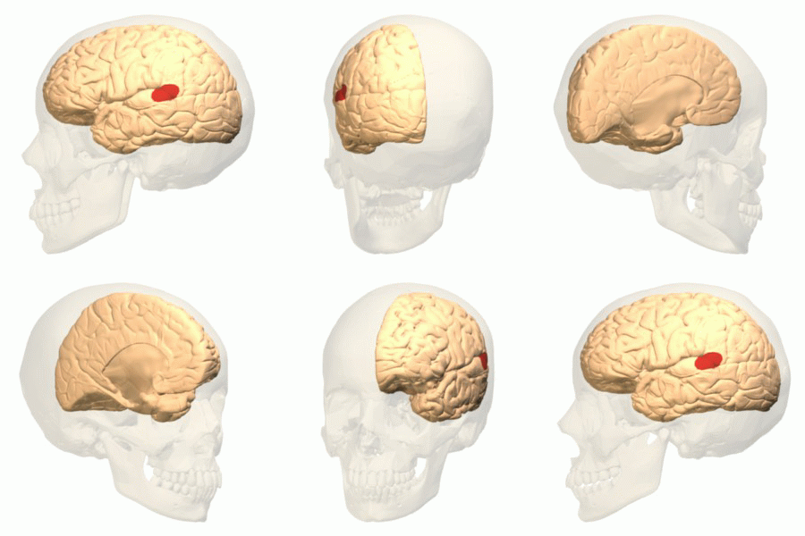
A recent paper in the journal Brain, a journal of neurology, challenges the long-held notion that the sensory speech area of the left temporal lobe of the cerebrum—often called the Wernicke area—acts as the center of language comprehension.

Wernicke area
The report describes research that leads one to the conclusion that although the Wernicke area (pictured) apparently has a role in understanding individual words, the task of sentence comprehension is accomplished by a complex network of diverse areas of the cerebrum. These include, "temporoparietal components of Wernicke’s area, Broca’s area, and [the] dorsal premotor cortex."


What can we use from this in teaching undergraduate A&P?


  • When discussing the sensory speech (Wernicke) area, one might bring up this discovery and how it may modify our description of its main function.

  • This discovery is a good example of how science works—how theories are formed and how they are modified as new information is discovered.  And how new research must be confirmed before it becomes widely established as fact.

  • When discussing cerebral localization of function, this story may be helpful in emphasizing that complex cerebral functions often involve integration of diverse cortical areas.

  • This story is a good example of our rapidly advancing knowledge of complex brain function.

  • You'll sound very "with it" when you can drop in casual asides about the latest brain research in your classroom discussions!

Want to know more?


New Human Brain Language Map | Researchers find that Wernicke’s area, thought to be the seat of language comprehension in the human brain for more than a century, is not.
  • Bob Grant. The Scientist  Published online June 26, 2015
  • Brief, plain-English article summarizing the recent findings.
  • my-ap.us/1QTTN5z 

The Wernicke conundrum and the anatomy of language comprehension in primary progressive aphasia 
  • M-Marsel Mesulam , et al. Brain. DOI: http://dx.doi.org/10.1093/brain/awv154 First published online: 25 June 2015
  • The original research article.
  • my-ap.us/1QTWPXw

Scientists redraw traditional brain map of language comprehension
  • Northwestern University press release. Published online June 25, 2015
  • my-ap.us/1QTYpsl


Cortical Areas Involved in Speech Processing


Image credits: Database Center for Life Science (Wernicke area)
Leuthardt, et al. (cortical speech areas)

Monday, June 15, 2015

Virtual Cardiology Lab


Looking for a supplemental hands-on activity with cardiology in your course?

Try the FREE online interactive Cardiology Virtual Lab from the Howard Hughes Medical Institute.

It covers these concepts:
  • Symptoms of a selection of heart diseases, to serve as examples of what kinds of things can go wrong with the heart. 
    • How are symptoms detected and why?
  • Tools and techniques used for diagnosis. 
    • What can the different techniques detect and how do they work?
  • Principles of pedigree analysis.


What can we use from this in teaching undergraduate A&P?


  • Link to this virtual lab activity from your online syllabus, course web page, or LMS (or in an email  or tweet to students)
  • If you want to give course points for the lab, consider an online quiz or lab report submitted through your learning management system (LMS) or emailed to you.
  • Gives students a "real life" clinical lab application for the concepts they are learning in A&P.
  • Provides a cardiology lab option for online/hybrid courses or wet labs that don't have funding for cardiology experiments.
  • Helps integrate principles of genetics with cardiology, so it can be used in your genetics unit.


Want to know more?


Cardiology Virtual Lab

  • BioInteractive. Howard Hughes Medical Institute. Accessed 16 Sep 2014.
  • This virtual lab will familiarize you with heritable diseases of the heart. Learn about the diagnostic tools used to examine and diagnose patients.
  • my-ap.us/1qKWiK7

BioInteractive Virtual Labs

  • BioInteractive. Howard Hughes Medical Institute. Accessed 16 Sep 2014.
  • List of all the FREE virtual labs offered by HHMI's BioInteractive project.
  • my-ap.us/1wAX92q

Cardiovascular Topics

  • Kevin Patton. The A&P Professor. Various dates.
  • List of previous blog posts on cardiovascular topics
  • my-ap.us/1uEGngu

Wednesday, June 3, 2015

A Brain-Lymphatic Connection

Existing dogma in neuroscience states that the brain does not possess the classical lymphatic drainage system found in other parts of body. However, a recent letter in the journal Nature reports the discovery of lymphatic vessels lining the dural sinuses in mice. These were shown to drain immune cells and cerebrospinal fluid (CSF) into the deep cervical lymph nodes.

Although more work is yet to be done in humans, this discovery will cause neuroscientists to revisit a number of concepts related to CSF and lymphatic drainage, as well as immune functions in the brain.

For example, do these new data truly challenge the notion of immune privilege in the nervous tissue of the central nervous system—or do they apply to the brain as an organ and allow for lymphatic drainage of tissues outside the nervous tissue of the brain?

What can we use from this in teaching undergraduate A&P?

  • When discussing CSF drainage, consider mentioning the possibility that filtration of CSF directly into dural sinuses may be augmented by the newly discovered lymphatic drainage.

  • If you discuss the dogma of the "immune privilege" of the brain, consider mentioning this possible challenge to the concept. This may trigger a great discussion of whether these newly discovered lymphatic vessels are truly "in the brain."

  • If you discuss disorders involving altered immunity, such as multiple sclerosis, consider mentioning this discovery.

  • Bringing up this new information may be useful in discussions related to the process of science—how existing concepts are sometimes challenged by new information, for example. Perhaps a discussion of the need for more investigation would stimulate students to think about what future steps can be taken to map out a possible lymphatic network in or around the brain.
Current concept of lymphatic drainage (left) compared to updated version to reflect new data (right).

Want to know more?


Brain Drain | The brain contains lymphatic vessels similar to those found elsewhere in the body, a mouse study shows.
  • Ashley P. Taylor, The Scientist, June 1, 2015 (online)
  • Plain-English article summarizing the study and its significance.
  • my-ap.us/1IezLLF
Structural and functional features of central nervous system lymphatic vessels,
  • A. Louveau et al., Nature, June 1, 2015.doi:10.1038/nature14432, 
  • The original research article. Includes images and video.
  • my-ap.us/1KNFPy6
Missing link found between brain, immune system; major disease implications
  • University of Virginia Health System, Science Daily, June 1, 2015
  • Illustrated press release describing the research.
  • my-ap.us/1HQys2J

Images: DBCLS (top)
Univ Va Health System (bottom)

Tuesday, May 19, 2015

Testing as a Learning Tool | UPDATE

I radically changed my approach to testing my A&P students about 13 years ago. It was was one of those experiments that had me questioning my sanity leading up to its roll-out. That's because in some fundamental ways, it challenged my experience of how I and others had always thought about the role of testing in the learning process.

But when it did finally roll out in the first set classes in which I tried it, my new method of using tests as learning tools was a phenomenal success. The class averages on my exams went up by a whole letter grade! Students were working harder—but grumbling less. And student attitudes, which I'm grateful have always been pretty positive anyway, suddenly became downright sunny!

About six years ago, I published an article about online testing as a method of teaching in my blog The Electronic Professor. In the article, I shared my experience in using frequent online tests in my anatomy & physiology courses as a way to get students engaged with the material on an ongoing basis. It outlines how they used testing as a way of learning—promoting construction of a cognitive framework of concepts that helped them prepare for their in-class exams.

Almost a year later, research published in Science further supported this idea. Not that I needed the support . . . my own experience over many years has confirmed for me that it works. In fact, it works VERY well in enhancing student learning. But as a scientist, a variety of independent confirmations of a topic is always valuable and appreciated.

Of course, the concept of frequent, online formative testing (as opposed to summative testing) is not at all new. But like a lot of breakthroughs in teaching and learning, it hasn't caught on with many professors "out in the trenches" yet. But it's really worth taking a look at--and trying it out yourself.

First, check out an updated version of a seminar that I've given on this topic several times over the years.
Seminar: Testing as Teaching
  • Kevin Patton The A&P Professor.
  • Narrated presentation outlining a method to produce randomzed formative tests for A&P.
  • my-ap.us/qtAclX
Next, check out my article from 2009 to get my take on some reactions I've gotten from other professors.
Teaching as Testing
  • Kevin Patton. The Electronic Professor. 27 Feb 2009.
  • Article outlining my use of online, randomized formative tests in teaching A&P.
  • my-ap.us/p3rM6B
After that, take a look at the research published in Science a year ago.
To Really Learn, Quit Studying and Take a Test
  • Pam Belluck. The New York Times. 20 January 2011.
  • Brief summary of the research, including a graph of the results.
  • my-ap.us/yP6jZ0 
Retrieval Practice Produces More Learning than Elaborative Studying with Concept Mapping
  • J.D. Karpicke, J. R. Blunt. Science. Published Online January 20 2011. DOI: 10.1126/science.1199327
  • Original research mentioned above.
  • my-ap.us/yTr2b7
Next, read these brief articles on a few of the many adaptive learning tools that are now available to accompany many of the major A&P textbooks. Oh, if only I'd had something like this back then!
Students Have Fun and Learn Quickly with EAL Adaptive Learning
  • Kevin Patton. Anatomy & Physiology 18 March 2015
  • Article outlining a self-quizzing tool based on the popular Cerego system.
  • my-ap.us/1zgNLEg
Adaptive Quizzing Helps Students Get Ready for Tests and Exams
  • Kevin Patton. Anatomy & Physiology 25 March 2015
  • Brief article outlining a quizzing system that helps students prepare for their exams.
  • my-ap.us/1xzwB3W
Lastly, just take the plunge and try it. Come on—the water's fine and you'll have a blast!

Content updated from a
previous post in
The A&P Professor 
Photo: M. Bowden

Wednesday, April 15, 2015

Mechanism of Cracking Knuckles


It never fails—somebody always asks about what's going on when knuckles crack when we get to the topic of articulations.

Recently, researchers used modern MRI techniques to demonstrate exactly what is happening—a process first described in the 1940s.  As joint surfaces separate, the changing tension in the synovial fluid causes a vapor pocket or "air bubble" to form in a process called cavitation.

The recent research proved that the popping sound typical of cracking a knuckle is produced as the vapor pocket forms. It also disproved the theory that the popping occurs when the bubble collapses.

What can we use from this in teaching undergraduate A&P?

  • You'll now always be ready for that inevitable "what causes knuckles to crack?" question when discussing joints in your A&P course.

    • Consider having a slide containing the media offered below to show students.

  • Discussing joint-cracking mechanisms engages students in a subject that may not immediately interest them.

  • Joint-cracking allows us to reinforce concepts of fluid dynamics in the context of synovial joints.

  • Consider stimulating some critical thinking by asking "what could cause a cracking sound in a cartilaginous or fibrous joint?"   (Hint: think "fracture" as cavitation would not occur where there are no fluids)
  • Do a demonstration by hitting a glass bottle full of water with a rubber mallet (see the embedded video below)
    • I've done this with an unopened glass bottle of ketchup by upturning it and hitting the base with my palm

Want to know more?

  • Real-Time Visualization of Joint Cavitation
    • GN Kawchuk, et al. PLoS|ONE. April 15, 2015 DOI: 10.1371/journal.pone.0119470
    • The original journal article. Includes downloadable images and PowerPoint slides, plus a link to a video showing the MRI results.
  • Researchers pull fingers to solve why knuckles crack | Finding bursts bubble of popular theory
    • Tina Hesman Saey Science News April 15, 2015
    • A plain-English summary of the new work on knuckle cracking with an embedded video (see below)
    • my-ap.us/1J4cGKP
Check out this video from the research article


Here's a video of the glass bottle demo of cavitation


Photo credit: Kawchuk et al.

Monday, February 9, 2015

Cytotoxic T Cell Horror Flick


Liven up your A&P class with a great video showing a gruesome attack by a killer T cell on a cancer cell. It's a fantastic bit of video microscopy produced by Cambridge University.

Okay, with the oddly soothing music score instead of a more appropriate score for the graphic violence shown in this video, it's not much of a horror flick.  Especially when you consider that it's the "bad guy" cell getting whacked.  But it is graphic and dramatic and impressive.

Just the thing to liven up a discussion of adaptive immunity, which (let's face it) can often cause a catatonic state in many students.

It's a free resource available on YouTube.

Monday, February 2, 2015

Virtual Immunology Lab


Looking for a supplemental hands-on activity with the immune system in your course?

Try the FREE online interactive Immunology Virtual Lab from the Howard Hughes Medical Institute.

It covers these concepts:

  • The basis of humoral immunity 
  • The foundation for ELISA (enzyme-linked immunosorbent assay)
  • Potential errors in conducting an ELISA
  • Sensitivity and specificity of a diagnostic test


What can we use from this in teaching undergraduate A&P?


  • Link to this virtual lab activity from your online syllabus, course web page, or LMS (or in an email  or tweet to students)
  • If you want to give course points for the lab, consider an online quiz or lab report submitted through your learning management system (LMS) or emailed to you.
  • Gives students a "real life" clinical lab application for the concepts they are learning in A&P.
  • Provides an immunology lab option for online/hybrid courses or wet labs that don't have funding for immunology experiments.


Want to know more?


Immunology Virtual Lab

  • BioInteractive. Howard Hughes Medical Institute. Accessed 16 Sep 2014.
  • This virtual lab teaches the procedures of performing an ELISA test to determine whether a particular antibody is present in a patient's blood sample.
  • my-ap.us/YPWv3f
ELISA
  • MedlinePlus. National Library of Medicine. Accessed 16 Sep 2014
  • Brief overview of what ELISA is.  You can link your students to this as a brief intro to the virtual lab.
  • my-ap.us/1o1dVjg

BioInteractive Virtual Labs

  • BioInteractive. Howard Hughes Medical Institute. Accessed 16 Sep 2014.
  • List of all the FREE virtual labs offered by HHMI's BioInteractive project.
  • my-ap.us/1wAX92q

Immunity Topics

  • Kevin Patton. The A&P Professor. Various dates.
  • List of previous blog posts on the topic of immunity.
  • my-ap.us/XdWVyO


Tuesday, January 6, 2015

Rhinovirus Replicates Best in the Nasal Cavity


It's that time of year, eh?  Cold and flu season.  And this week we have news from researchers giving us a bit more insight into the rhinoviruses that cause the common cold.

The unsurprising new discovery is that rhinoviruses replicate more efficiently—and therefore cause colds more effectively—in the nasal cavity than in the lungs because of a temperature difference.  In mice, the animals used in the recent study, the immune mechanisms that fight off rhinoviruses work better in the warmer environment of the lung than in the cooler environment of the nasal cavity.

This phenomenon may be why a cold generally doesn't wreak the same havoc in lungs as do other respiratory viruses like influenza viruses.  Rhinoviruses, as their name implies, generally remain limited to the nose.

What can we use from this in teaching undergraduate A&P?

  • When discussing the nasal cavity's vascularity and air-warming functions, we may want to point out that understanding the temperature gradient between the nose and lower respiratory tract has practical clinical applications.  Such as why some pathogens are limited to the nose. And why cold weather may contribute to rhinvirus infection.

  • When discussing immunology, we may want to mention that body temperature—and sometimes organ temperature—can have an impact on how efficiently our immune mechanisms fight infection.

  • One could consider steering a conversation about "there is still no cure for the common cold" to a conversation about the fact that scientists are still working on understanding rhinoviruses—"and, oh, did you hear the latest .....?"

Want to know more?


Where Rhinovirus Replicates Best
  • Tracy Vence,  The Scientist.  Online. January 6, 2015.
  • Plain-English summary of the discovery.
  • my-ap.us/1FgWKr7

Temperature-dependent innate defense against the common cold virus limits viral replication at warm temperature in mouse airway cells
  • Ellen F. Foxman, et al.  Proceedings of the National Academy of Science. Online. January 5, 2015. doi: 10.1073/pnas.1411030112 
  • Original research paper.
  • my-ap.us/1xA4v6m
Photo credit: mcfarlandmo


Friday, January 2, 2015

Fat Cells in Skin Kill Bacteria


Scientists reported today that adipocytes in mouse and human skin produce an antimicrobial peptide (AMP) called cathelicidin is response to Staph aureus infections, including MRSA. Experimental animals that were deficient in the AMP were more susceptible to skin infections.

Adipocytes may recognize S. aureus by detecting bacterial peptides with toll-like receptors (TLRs), but more work is needed to fully understand the mechanisms.

This finding adds more to our understanding of human skin as a vital part of our body's defenses against infection. It also opens the door to understanding how diabetes, metabolic syndrome, and other conditions can reduce resistance to skin infections by altering the availability of AMPs in the fat associated with skin.

All of this may eventually lead to additional—perhaps more effective—strategies in preventing or curing serious skin infections such as MRSA.

I realize that we generally think of fat cells as belonging to the hypodermis, not the dermis, as described in the research. However, recent evidence shows the presence of adipocytes in the dermis that are distinct from those in the hypodermis. These adipocytes derive from a common precursor cell that produces both dermal fibroblasts and intradermal adipocytes. These dermal adipocytes have been shown to have a role in wound healing and the regeneration of hair follicles. And the research summarized here suggests that they also have a role in immunity.


What can we use from this in teaching undergraduate A&P?

  • Mention this discovery when discussing the roles of adipose tissue and adipocytes in your coverage of tissues of the body.

    • Consider clarifying that dermal adipocytes are distinct from fat cells in the hypdermis. And perhaps mention that it's a detail often left out of introductory discussions of skin.

  • This is a good point to mention when discussing the protective functions of the skin when covering the integumentary system.

  • When discussing the immune system, this concept helps illustrate several important principles:

    • The role of the skin as the first line of defense against infection

      • The variety of mechanisms available in the skin to act defensively

    • The role of TLRs and pattern recognition in immunity

    • The fact that immunity is a role for many tissues—not just lymphocytes and other WBCs

  • Take a moment NOW to add this to your course notes!

Want to know more?


Killer Fat
  • J. Alcorn and J. Kolls. Science 2 January 2015: Science Vol. 347 no. 6217 pp. 26-27 DOI: 10.1126/science.aaa4567
  • Editorial summary of the research in plain English. Includes a really nice, simple illustration of the concept (includes FREE teaching slide)
  • my-ap.us/1vBWbNP

Dermal adipocytes protect against invasive Staphylococcus aureus skin infection
  • L. Zhang1, et al. Science 2 January 2015: Vol. 347 no. 6217 pp. 67-71 DOI: 10.1126/science.126097
  • Original research article. Additional images available here, including some nice micrographs showing increase in adipocytes in response to S. aureus infection
  • my-ap.us/1xePNS4

Defining dermal adipose tissue.
  • Driskell RR, et al. Exp Dermatol. 2014. Exp Dermatol. 2014 Sep;23(9):629-31. doi: 10.1111/exd.12450.
  • Review article describing dermal adipocytes.
  • my-ap.us/1GuH3bL

FREE teaching slide
Click the image
to download a


Adipose image credit: my-ap.us/13MYGWO
This post was updated 6 OCT 2015

Monday, December 8, 2014

5 Useful Features of The A&P Professor Blog That You May Have Missed


Okay, the title is a bit misleading because a some of the five features you may have missed are new, so it may just be too early to have noticed them yet.  But there are some that have been around a while and you may not have given them much thought until now.

  1. Top menu bar.  

    I recently added a menu bar to the top of the blog page, just under the blog description.   You can link to the Home Page, Site Map, Subscription and various off-blog resources.  Explore each one to see if it's useful to you.

  2. Site Map.

    This new feature is a table of contents that lists blog posts in alphabetical lists by topic.  It's pretty big because I've been doing this blog for many years, and some posts are listed under more than one topic.  But if you are "into" the teaching of A&P, this might be a fun treasure hunt to find things you may have missed—or have simply forgotten about.

  3. Email Newsletter.

    The email newsletter for this blog, powered by Feedblitz, is a way to subscribe to this blog and be notified by email each time a new article is posted in blog.  It's free and you can safely unsubscribe at any time.  Then re-subscribe later when you find that you missed getting updates!  You may not realize that by subscribing, you also get additional content.  For example, once a month, I send out a Throwback Thursday newsletter reprising a popular article from the past that remain relevant.  I occasionally send out other "extra issues" that do not appear in the blog itself.

  4. Social Media.

    As more and more of you join the rest of the world on Facebook, Twitter, and other social media networks, you may want to follow The A&P Professor there, too.  As with the email newsletters, my Twitter and Facebook followers get additional content not found on the blog or other channels.  You can even follow my YouTube channel, where I post videos with study tips for A&P that you can share with your students.

  5. Family of Blogs and Websites.

    I have other blogs that are focused on particular topics that overlap your interest in teaching A&P.  For example, The A&P Student blog is directed at, er, A&P students.  Teachers who follow that blog pick up ideas for sharing with students who are having difficulty in your class, or link to blog posts in their course pages or syllabus.  My o-log-y blog relates specifically to the terminology of human science and medicine.  There, you may find tips for helping your students learn the language of A&P or you may find information about some of the tricky aspects of terminology that you may not already know. Check out each one to see if any of them interest you.

    Oh, and don't forget the separate website theAPprofessor.org, where you can find my free image library, sign up to get free bookmarks for your students, participate in teaching seminars, and gain access to my free library of teaching slides.

Play around the edges of this blog and see what other helpful little tools you may have missed.

Monday, November 17, 2014

Your Textbook is a Mitten, Not a Glove


How well does the textbook you use in your course fit?  What I mean by that is:

  • Have you looked at whether the reading style is geared toward the students you typically encounter? Is the text written for beginners or experts?

  • Have you compared your actual expectations (objectives & expected outcomes) to the content available in the textbook?

  • Is the textbook organized in way that facilitates learning by both reading and raiding?

Of course, no textbook is going to fit you, your students, and your course like a glove.  Even if you create your own, or edit a custom version of existing text material, the fit will change as you and your course evolves as you gain teaching skills, adjust your techniques, and your students' needs change.

Textbooks fit like a mitten, not a glove. Because the best they can do is approximately fit course objectives and content, student learning needs, and your teaching approach.

But mittens are great.  They cover your hand and give you some wiggle room.

As a "mitten" your course textbook should cover what you need it to cover.  It's okay if it covers a bit more than you need.  That extra coverage will be used later for reference and future learning in professional and clinical courses.  Extra content also helps fill in prior learning gaps, which allows students to "get it" on their own as they read or raid.  And extra content may help students better see the context and importance of the required content.  Satisfying natural curiosity is not a bad thing, either.

Although well-fitting mittens cover a bit more than they need to, a mitten should not be too big.  So having so much content in a textbook that it's hard to stay focused mainly on the required content is not good for learning. Conversely, a mitten should not be so small that you can't get it around your big hand or wiggle your fingers a bit.

Some mittens have special features like a slit through which you can extend your fingers when you need more precise movements than your mitten will allow—as when opening your flask of hot tea.  If your students would benefit from a supplemental atlas, an audio glossary, a list of word parts for each term, or other special learning features of textbooks, then consider those carefully, too.

The key is to think of a textbook as an off-the-shelf item that will never fit precisely.  It's up to you to make sure it fits well enough.  And the only way to get that fit is to try on all the mittens.  Don't just glance at them, really put your hand in and see what they're like.

Plaid mittens are the best.  That has nothing to do with the analogy I'm making here—I just like plaid mittens.


Photo credits: Mélanie, Bin im Garten


Tuesday, November 11, 2014

Shepherd's Apps for Teaching Physiology


Here are some computer-based activities that help students learn specific concepts of physiology produced by veteran professor Pete Shepherd.  Dr. Shepherd has been developing over the course of many years based on his extensive teaching experience.

A while back, I told you about the Life Science Teaching Resource Community and its archive of free teaching and learning resources.  Here's an example of a collection of resources from that archive that your students can use to "play around with" physiological conditions to see what changes occur in the body as a result.  All of them can either be used in the classroom/lab setting or can be used individually by students for self-learning.

Included in the collection are these apps, most of which are available on multiple platforms:

Capillary Pressure, which illustrates the vascular control of capillary hydrostatic pressure.

Alveolar Gas, which lets you study some of the physiological factors that affect the composition of alveolar and expired gases. Such factors include dead space, tidal volume, the frequency of breathing, and the rates of oxygen consumption and carbon dioxide production. A worksheet is included.

Blood Oxygen, which enables the user to change variables like the PO2, hemoglobin concentration, and hemoglobin's affinity for oxygen and calculate the concentrations of oxygen in the form of oxyhemoglobin and dissolved oxygen. Simulations include anemia, polycythemia, comparing the effects of oxygen inhalation in a pulmonary "patient" with a normal person, carbon monoxide poisoning, hyperbaria, etc., as well as the concepts of the Fick Principle and the arteriovenous oxygen difference.

Pulse Pressure, which simulates the arterial pressure pulse. The user explores factors like heart rate, stroke volume, arterial compliance, and arterial resistance and see how they affect the arterial pulse pressure.

Sat Curves, which allows the user to demonstrate the effects of pH, PCO2, DPG, and temperature on the oxyhemoglobin dissociation curve. Two graphs are displayed so that one can serve as a control and compared with the other. Cursors on both graphs can be manipulated to obtain exact readings of oxyhemoglobin saturation as specified PO2s.


If you want to check out these amazing resources, please access them in the Life Science Teaching Resource Community at


Image credit: Shepherd

Monday, November 3, 2014

Protein Mechanism in Neurodegenerative Diseases

Huntingtin

Huntingtin, the abnormal protein that produces clumps characteristic of Huntington disease (HD), can spread from one neuron to another. That's what a recent study has uncovered. Because such protein clumping is observed in other neurodegenerative disorders such as Alzheimer disease (AD) and Parkinson disease (PD), some scientists hope that understanding this newly discovered mechanism of transmission within brain tissue may lead to possible treatments or preventive strategies.

If you want to read more about it, check out the resources I've provided below.

What can we use from this in teaching undergraduate A&P?

  • This information can help us answer those pesky "why do we need to know all this if I'm going to be a [insert health profession here]?" challenges when covering the details of protein structure.  The sequence of amino acids and the complex folded structure of proteins really does have real-world clinical implications.  And is already becoming necessary to understand disease mechanisms and treatment strategies.  In real life!
  • Discussing the basic idea of this discovery provides a starting platform from which we can jump into discussions of 
    • Degeneration of tissues in general and neurodegeneration in particular
    • Why neurodegeneration in specific brain locations produces specific neural deficits
    • Prions and their possible roles in various disorders
    • The possible roles of genetic mechanisms in neurodegenerative disorders
    • The need to know details about protein structure (see item above)
    • Current directions in medical research—that proteins are hot!

Want to know more?


Neurodegeneration’s Spread

  • Ashley P. Taylor. The Scientist. August 4, 2014
  • Plain-English article describing the new research showing that pathogenic protein aggregates that accumulate within neurons and are a hallmark of Huntington’s disease can propagate from cell to cell.
  • my-ap.us/1kDnJnk


Transneuronal propagation of mutant huntingtin contributes to non-cell autonomous pathology in neurons

  • E. Pecho-Vrieseling et al., Nature Neuroscience, 13 July 2014, doi:10.1038/nn.3761, 2014.
  • The original research article.
  • my-ap.us/YKuauI


Huntington disease

  • PubMed Health. last reviewed 28 May 2013
  • Basic information about HD.
  • my-ap.us/1tAVHbM


Why Bother with Protein Folding?

  • Kevin Patton. The A&P Professor. 22 May 2012.
  • Further discussion of why protein folding is important in the undergrad A&P course; includes links to other resources.
  • my-ap.us/1vtd9Ad


Want to Fold Some Proteins?

  • Kevin Patton. The A&P Professor. 13 June 2012.
  • Introduction of the Foldit game that allows anyone to participate in protein-folding research by playing an online folding game.
  • my-ap.us/1l1PYMz


Monday, October 13, 2014

RNA Interference. Again.


Five years ago, I extolled the virtues of teaching a little bit about RNA interference (RNAi) in undergraduate A&P courses.  But for a while it looked like the promise of RNAi in basic and clinical research might be sputtering.  However, a recent article by Eric Bender called The Second Coming of RNAi shows that RNAi "the gene-silencing technique [now] begins to fulfill some of its promises."

I recommend reading the entire article at my-ap.us/1BbxvB9  Before you read it, allow me to reprise my reasons of five years ago supporting my proposal to include RNAi in your course.

What can we use from this in teaching undergraduate A&P?


  • RNAi plays a role in defending our cells against viruses by stopping viral genetic code from being translated in host cells

  • RNAi likely plays a role in regulating gene activity in a cell by preventing translation of the gene product(s)

  • RNAi is increasingly used as method for "knocking out" a particular gene's effects in research animals in order to study the gene's functions

  • RNAi is being used to treat genetic disease. . . an application that will likely expand greatly over the next few decades
I'll add two more items to my previous list:
  • RNA interference is a mechanism of human disease, as has been demonstrated in some cases of inherited progressive hearing loss (for example).

  • Learning about RNAi helps clarify a general understanding of the many roles played by RNA in our lives—some perhaps still undiscovered.

I'm not sure that it's useful to expect beginning undergraduate students to learn the nitty-gritty details of RNAi mechanisms.  But I do think it's valuable to be exposed to the general concept of RNA interference and gene silencing.  A&P students are going to run up against these eventually as they learn about and then administer RNAi-based therapies, after all.  And perhaps we should prepare them.

Want to know more?


The Second Coming of RNAi
  • Eric Bender. The Scientist. September 1, 2014
  • Article mentioned above. In plain English, it shows that clinical progress in RNAi therapy against liver diseases, the gene-silencing technique begins to fulfill some of its promises. Includes useful illustrations and links to other resources.
  • my-ap.us/1BbxvB9

Why do we need to know about RNA interference?
  • Kevin Patton. The A&P Professor. 14 April 2009
  • My first article promoting the idea of teaching RNAi in the A&P course.  It links to an expanded article with additional teaching resources.
  • my-ap.us/1xbq4v6

RNA interference revisited
  • Kevin Patton. The A&P Professor. 9 June 2009
  • Brief follow-up article that references the role of RNA interference as a mechanism of human disease.  Links to other resources.
  • my-ap.us/1oDflzw

RNA Interference Animation and Slideshow
  • Nature Reviews Genetics. Accessed 3 September 2014
  • FREE animation, slideshow, and poster on RNAi, as well as a link to more details.
  • my-ap.us/1roNGYm

RNA Interference BioInteractive
  • Howard Hughes Medical Institute. Accessed 3 September 2014
  • FREE slideshow with worksheet that students fill out as they view the slideshow.  Links to FREE DVD from HMMI called The Double Life of RNA.
  • my-ap.us/1pJ9r5V


Wednesday, October 8, 2014

Nobel Prize 2014: Super-resolved fluorescence microscopy


The Royal Swedish Academy of Sciences has decided to award the Nobel Prize in Chemistry for 2014 to

Eric Betzig
Janelia Farm Research Campus, Howard Hughes Medical Institute, Ashburn, VA, USA,

Stefan W. Hell
Max Planck Institute for Biophysical Chemistry, Göttingen, and German Cancer Research Center, Heidelberg, Germany

and

William E. Moerner
Stanford University, Stanford, CA, USA

“for the development of
super-resolved fluorescence microscopy”


Surpassing the limitations of the light microscope


For a long time optical microscopy was held back by a presumed limitation: that it would never obtain a better resolution than half the wavelength of light. Helped by fluorescent molecules the Nobel Laureates in Chemistry 2014 ingeniously circumvented this limitation. Their ground-breaking work has brought optical microscopy into the nanodimension.

In what has become known as nanoscopy, scientists visualize the pathways of individual molecules inside living cells. They can see how molecules create synapses between nerve cells in the brain; they can track proteins involved in Parkinson’s, Alzheimer’s and Huntington’s diseases as they aggregate; they follow individual proteins in fertilized eggs as these divide into embryos.

It was all but obvious that scientists should ever be able to study living cells in the tiniest molecular detail. In 1873, the microscopist Ernst Abbe stipulated a physical limit for the maximum resolution of traditional optical microscopy: it could never become better than 0.2 micrometres. Eric Betzig, Stefan W. Hell and William E. Moerner are awarded the Nobel Prize in Chemistry 2014 for having bypassed this limit. Due to their achievements the optical microscope can now peer into the nanoworld.

Two separate principles are rewarded. 


One enables the method stimulated emission depletion (STED) microscopy, developed by Stefan Hell in 2000. Two laser beams are utilized; one stimulates fluorescent molecules to glow, another cancels out all fluorescence except for that in a nanometre-sized volume. Scanning over the sample, nanometre for nanometre, yields an image with a resolution better than Abbe’s stipulated limit.

Eric Betzig and William Moerner, working separately, laid the foundation for the second method, single-molecule microscopy. The method relies upon the possibility to turn the fluorescence of individual molecules on and off. Scientists image the same area multiple times, letting just a few interspersed molecules glow each time. Superimposing these images yields a dense super-image resolved at the nanolevel. In 2006 Eric Betzig utilized this method for the first time.

Today, nanoscopy is used world-wide and new knowledge of greatest benefit to mankind is produced on a daily basis.


This video is a brief animation of how STED works and how it improves resolution of individual particles.


This video is a longer, more detailed presentation by one of the Nobel laureates (Hell).


What can we use from this in teaching undergraduate A&P?

  • Discuss how this technology has enabled us to better visualize the chemicals and structures within our cells, enabling scientists to better understand the structure and function of cell, organelles, microbiome constituents, and other structures of the human body.

  • If you do a brief run-through of the theory of microscopy—perhaps in your A&P lab—you can add a mention of this technology.  

  • Your textbook or other learning resource may already have an example of this type of microscopy.

  • A discussion of this  Nobel Prize could evolve into a meaningful example of how science works, including how incremental improvements in classical tools for observation expand the number of questions that can be answered.

  • Use the links below (and images above) to use for a handout and/or teaching slides.


Want to know more?


Resources from Nobelprize.org

  • Popular Information 
  • Scientific Background
    • Handout: More detailed information includes references to original research articles
    • my-ap.us/ZdLJ69
  • Advanced Information
  • Images
  • Biographies
    • Eric Betzig, 
      • U.S. citizen. Born 1960 in Ann Arbor, MI, USA. Ph.D. 1988 from Cornell University, Ithaca, NY, USA. Group Leader at Janelia Farm Research Campus, Howard Hughes Medical Institute, Ashburn, VA, USA.
      • http://janelia.org/lab/betzig-lab
    • Stefan W. Hell, German citizen. 
      • Born 1962 in Arad, Romania. Ph.D. 1990 from the University of Heidelberg, Germany. Director at the Max Planck Institute for Biophysical Chemistry, Göttingen, and Division head at the German Cancer Research Center, Heidelberg, Germany.
      • http://www3.mpibpc.mpg.de/groups/hell
    • William E. Moerner, U.S. citizen. 
      • Born 1953 in Pleasanton, CA, USA. Ph.D. 1982 from Cornell University, Ithaca, NY, USA. Harry S. Mosher Professor in Chemistry and Professor, by courtesy, of Applied Physics at Stanford University, Stanford, CA, USA.
      • http://web.stanford.edu/group/moerner


Diagrram cretit: Ganbaatar
Micrograph credit: Tesselkaffee
Text adapted from press release from Nobel Media

Monday, October 6, 2014

Nobel Prize 2014: The Brain's Positioning System


The Nobel Assembly at Karolinska Institutet has today decided to award

The 2014 Nobel Prize in Physiology or Medicine

with one half to

John O´Keefe

and the other half jointly to

May-Britt Moser and Edvard I. Moser

for their discoveries of cells that constitute a positioning 
system in the brain.


How do we know where we are? How can we find the way from one place to another? And how can we store this information in such a way that we can immediately find the way the next time we trace the same path? This year´s Nobel Laureates have discovered a positioning system, an “inner GPS” in the brain that makes it possible to orient ourselves in space, demonstrating a cellular basis for higher cognitive function.

In 1971, John O´Keefe discovered the first component of this positioning system. He found that a type of nerve cell in an area of the brain called the hippocampus that was always activated when a rat was at a certain place in a room. Other nerve cells were activated when the rat was at other places. O´Keefe concluded that these “place cells” formed a map of the room.

More than three decades later, in 2005, May-Britt and Edvard Moser discovered another key component of the brain’s positioning system. They identified another type of nerve cell, which they called “grid cells”, that generate a coordinate system and allow for precise positioning and pathfinding.

Their subsequent research showed how place and grid cells make it possible to determine position and to navigate.

The discoveries of John O´Keefe, May-Britt Moser and Edvard Moser have solved a problem that has occupied philosophers and scientists for centuries – how does the brain create a map of the space surrounding us and how can we navigate our way through a complex environment?


How do we experience our environment?


The sense of place and the ability to navigate are fundamental to our existence. The sense of place gives a perception of position in the environment. During navigation, it is interlinked with a sense of distance that is based on motion and knowledge of previous positions.

Questions about place and navigation have engaged philosophers and scientists for a long time. More than 200 years ago, the German philosopher Immanuel Kant argued that some mental abilities exist as a priori knowledge, independent of experience. He considered the concept of space as an inbuilt principle of the mind, one through which the world is and must be perceived. With the advent of behavioural psychology in the mid-20th century, these questions could be addressed experimentally. When Edward Tolman examined rats moving through labyrinths, he found that they could learn how to navigate, and proposed that a “cognitive map” formed in the brain allowed them to find their way. But questions still lingered - how would such a map be represented in the brain?


John O´Keefe and the place in space


John O´Keefe was fascinated by the problem of how the brain controls behaviour and decided, in the late 1960s, to attack this question with neurophysiological methods. When recording signals from individual nerve cells in a part of the brain called the hippocampus, in rats moving freely in a room, O’Keefe discovered that certain nerve cells were activated when the animal assumed a particular place in the environment (Figure 1). He could demonstrate that these “place cells” were not merely registering visual input, but were building up an inner map of the environment. O’Keefe concluded that the hippocampus generates numerous maps, represented by the collective activity of place cells that are activated in different environments. Therefore, the memory of an environment can be stored as a specific combination of place cell activities in the hippocampus.


May-Britt and Edvard Moser find the coordinates


May-Britt and Edvard Moser were mapping the connections to the hippocampus in rats moving in a room when they discovered an astonishing pattern of activity in a nearby part of the brain called the entorhinal cortex. Here, certain cells were activated when the rat passed multiple locations arranged in a hexagonal grid (Figure 2). Each of these cells was activated in a unique spatial pattern and collectively these “grid cells” constitute a coordinate system that allows for spatial navigation. Together with other cells of the entorhinal cortex that recognize the direction of the head and the border of the room, they form circuits with the place cells in the hippocampus. This circuitry constitutes a comprehensive positioning system, an inner GPS, in the brain (Figure 3).


A place for maps in the human brain


Recent investigations with brain imaging techniques, as well as studies of patients undergoing neurosurgery, have provided evidence that place and grid cells exist also in humans. In patients with Alzheimer´s disease, the hippocampus and entorhinal cortex are frequently affected at an early stage, and these individuals often lose their way and cannot recognize the environment. Knowledge about the brain´s positioning system may, therefore, help us understand the mechanism underpinning the devastating spatial memory loss that affects people with this disease.

The discovery of the brain’s positioning system represents a paradigm shift in our understanding of how ensembles of specialized cells work together to execute higher cognitive functions. It has opened new avenues for understanding other cognitive processes, such as memory, thinking and planning.


What can we use from this in teaching undergraduate A&P?

  • The role of the hippocampus and it's place cells could be briefly discussed as you explore the cognitive functions of the brain.

  • A discussion of this year's Nobel Prize could evolve into a meaningful example of how science works, including the use of discoveries in animals that can later be applied to learning more about human structure and function.

  • Information revealed by this discovery could be discussed when discussing human disorders, such as Alzheimer disease, that involve impairments of spatial orientation and/or place memory.


Want to know more?

Here's a video in which May-Britt Moser and Edvard I. Moser explain their research, which you can use in your course discussion.


2014 Nobel Prize announcement

  • Official announcement, which includes photos of the laureates and links to related information.
  • my-ap.us/1oJH82y


Scientific Background: The Brain’s Navigational Place and Grid Cell System

  • Nobelprize.org accessed 6 October 2014
  • Plain English hand-out that can be used with your students.  Illustrated with clear diagrams.
  • my-ap.us/1pGLVlm

Nobel Prize in Medicine Is Awarded for Discovery of Brain’s ‘Inner GPS’

  • L. Altman The New York Times. OCT. 6, 2014
  • Plain English article summarizing the discoveries.
  • my-ap.us/10ELy5h

The hippocampus as a spatial map. Preliminary evidence from unit activity in the freely‐moving rat.

  • O'Keefe, J., and Dostrovsky, J. (1971). Brain Research 34, 171-175.
  • Original research paper describing key discoveries for which this prize is given.
  • my-ap.us/1s4fi5Z


Place units in the hippocampus of the freely moving rat.

  • O´Keefe, J. (1976). Experimental Neurology 51, 78-109.
  • Original research paper describing key discoveries for which this prize is given.
  • my-ap.us/1s2NYGd


Spatial representation in the entorhinal cortex.

  • Fyhn, M., Molden, S., Witter, M.P., Moser, E.I., Moser, M.B. (2004)  Science 305, 1258-1264.
  • Original research paper describing key discoveries for which this prize is given.
  • my-ap.us/1BIvdI4


Microstructure of spatial map in the entorhinal cortex.

  • Hafting, T., Fyhn, M., Molden, S., Moser, M.B., and Moser, E.I. (2005). Nature 436, 801-806.
  • Original research paper describing key discoveries for which this prize is given.
  • my-ap.us/1nX0jLe


Article adapted from Nobel Media press release