Wednesday, October 10, 2012
G-protein coupled receptors
The Royal Swedish Academy of Sciences has decided to award the
Nobel Prize in Chemistry for 2012
to
Robert J. Lefkowitz
Howard Hughes Medical Institute and Duke University Medical Center, Durham, NC, USA
and
Brian K. Kobilka
Stanford University School of Medicine, Stanford, CA, USA
"for studies of G-protein–coupled receptors"
Smart receptors on cell surfaces
Your body is a fine-tuned system of interactions between billions of cells. Each cell has tiny receptors that enable it to sense its environment, so it can adapt to new situtations. Robert Lefkowitz and Brian Kobilka are awarded the 2012 Nobel Prize in Chemistry for groundbreaking discoveries that reveal the inner workings of an important family of such receptors: G-protein–coupled receptors (GPCRs).
For a long time, it remained a mystery how cells could sense their environment. Scientists knew that hormones such as adrenalin had powerful effects: increasing blood pressure and making the heart beat faster. They suspected that cell surfaces contained some kind of recipient for hormones. But what these receptors actually consisted of and how they worked remained obscured for most of the 20th Century.
Lefkowitz started to use radioactivity in 1968 in order to trace cells' receptors. He attached an iodine isotope to various hormones, and thanks to the radiation, he managed to unveil several receptors, among those a receptor for adrenalin: β-adrenergic receptor. His team of researchers extracted the receptor from its hiding place in the cell wall and gained an initial understanding of how it works.
The team achieved its next big step during the 1980s. The newly recruited Kobilka accepted the challenge to isolate the gene that codes for the β-adrenergic receptor from the gigantic human genome. His creative approach allowed him to attain his goal. When the researchers analyzed the gene, they discovered that the receptor was similar to one in the eye that captures light. They realized that there is a whole family of receptors that look alike and function in the same manner.
Today this family is referred to as G-protein–coupled receptors. About a thousand genes code for such receptors, for example, for light, flavour, odour, adrenalin, histamine, dopamine and serotonin. About half of all medications achieve their effect through G-protein–coupled receptors.
The studies by Lefkowitz and Kobilka are crucial for understanding how G-protein–coupled receptors function. Furthermore, in 2011, Kobilka achieved another break-through; he and his research team captured an image of the β-adrenergic receptor at the exact moment that it is activated by a hormone and sends a signal into the cell. This image is a molecular masterpiece--the result of decades of research.
Laureate's Links
Robert J. Lefkowitz, U.S. citizen. Born 1943 in New York, NY, USA. M.D. 1966 from Columbia University, New York, NY, USA.Investigator, Howard Hughes Medical Institute. James B. Duke Professor of Medicine, and Professor of Biochemistry, Duke University Medical Center, Durham, NC, USA.
lefkolab.org
Brian K. Kobilka, U.S. citizen. Born 1955 in Little Falls, MN, USA. M.D. 1981 from Yale University School of Medicine, New Haven, CT, USA. Professor of Medicine, and Professor of Molecular and Cellular Physiology, Stanford University School of Medicine, Stanford, CA, USA.
med.stanford.edu/kobilkalab
Want to know more?
Popular Information
[A great synopsis, entitled "Cells and Sensibility," written for general reader and nicely illustrated. This would make a great handout or reading assignment for your A&P class.]
my-ap.us/SLShCF
Scientific Background
[A more technical treatment of the discovery, but still accessible to advanced A&P students as well as A&P professors. Includes extensive references. Illustrated.]
my-ap.us/PnK4Fz
Adapted from my-ap.us/TwmIyd
Monday, October 8, 2012
Reprogramming cells to be pluripotent
The Nobel Assembly at Karolinska Institutet has today decided to award
The Nobel Prize in Physiology or Medicine 2012
jointly to
John B. Gurdon and Shinya Yamanaka
for the discovery that mature cells can be reprogrammed to become pluripotent
Summary
The Nobel Prize recognizes two scientists who discovered that mature, specialised cells can be reprogrammed to become immature cells capable of developing into all tissues of the body. Their findings have revolutionised our understanding of how cells and organisms develop.
John B. Gurdon discovered in 1962 that the specialisation of cells is reversible. In a classic experiment, he replaced the immature cell nucleus in an egg cell of a frog with the nucleus from a mature intestinal cell. This modified egg cell developed into a normal tadpole. The DNA of the mature cell still had all the information needed to develop all cells in the frog.
Shinya Yamanaka discovered more than 40 years later, in 2006, how intact mature cells in mice could be reprogrammed to become immature stem cells. Surprisingly, by introducing only a few genes, he could reprogram mature cells to become pluripotent stem cells, i.e. immature cells that are able to develop into all types of cells in the body.
These groundbreaking discoveries have completely changed our view of the development and cellular specialisation. We now understand that the mature cell does not have to be confined forever to its specialised state. Textbooks have been rewritten and new research fields have been established. By reprogramming human cells, scientists have created new opportunities to study diseases and develop methods for diagnosis and therapy.
Life – a journey towards increasing specialisation
All of us developed from fertilized egg cells. During the first days after conception, the embryo consists of immature cells, each of which is capable of developing into all the cell types that form the adult organism. Such cells are called pluripotent stem cells. With further development of the embryo, these cells give rise to nerve cells, muscle cells, liver cells and all other cell types - each of them specialised to carry out a specific task in the adult body. This journey from immature to specialised cell was previously considered to be unidirectional. It was thought that the cell changes in such a way during maturation that it would no longer be possible for it to return to an immature, pluripotent stage.
Frogs jump backwards in development
John B. Gurdon challenged the dogma that the specialised cell is irreversibly committed to its fate. He hypothesised that its genome might still contain all the information needed to drive its development into all the different cell types of an organism. In 1962, he tested this hypothesis by replacing the cell nucleus of a frog's egg cell with a nucleus from a mature, specialised cell derived from the intestine of a tadpole. The egg developed into a fully functional, cloned tadpole and subsequent repeats of the experiment yielded adult frogs. The nucleus of the mature cell had not lost its capacity to drive development to a fully functional organism.
Gurdon's landmark discovery was initially met with scepticism but became accepted when it had been confirmed by other scientists. It initiated intense research and the technique was further developed, leading eventually to the cloning of mammals. Gurdon's research taught us that the nucleus of a mature, specialized cell can be returned to an immature, pluripotent state. But his experiment involved the removal of cell nuclei with pipettes followed by their introduction into other cells. Would it ever be possible to turn an intact cell back into a pluripotent stem cell?
A roundtrip journey – mature cells return to a stem cell state
Shinya Yamanaka was able to answer this question in a scientific breakthrough more than 40 years after Gurdon´s discovery. His research concerned embryonal stem cells, i.e. pluripotent stem cells that are isolated from the embryo and cultured in the laboratory. Such stem cells were initially isolated from mice by Martin Evans (Nobel Prize 2007) and Yamanaka tried to find the genes that kept them immature. When several of these genes had been identified, he tested whether any of them could reprogram mature cells to become pluripotent stem cells.
Yamanaka and his co-workers introduced these genes, in different combinations, into mature cells from connective tissue, fibroblasts, and examined the results under the microscope. They finally found a combination that worked, and the recipe was surprisingly simple. By introducing four genes together, they could reprogram their fibroblasts into immature stem cells!
The resulting induced pluripotent stem cells (iPS cells) could develop into mature cell types such as fibroblasts, nerve cells and gut cells. The discovery that intact, mature cells could be reprogrammed into pluripotent stem cells was published in 2006 and was immediately considered a major breakthrough.
From surprising discovery to medical use
The discoveries of Gurdon and Yamanaka have shown that specialised cells can turn back the developmental clock under certain circumstances. Although their genome undergoes modifications during development, these modifications are not irreversible. We have obtained a new view of the development of cells and organisms.
Research during recent years has shown that iPS cells can give rise to all the different cell types of the body. These discoveries have also provided new tools for scientists around the world and led to remarkable progress in many areas of medicine. iPS cells can also be prepared from human cells.
For instance, skin cells can be obtained from patients with various diseases, reprogrammed, and examined in the laboratory to determine how they differ from cells of healthy individuals. Such cells constitute invaluable tools for understanding disease mechanisms and so provide new opportunities to develop medical therapies.
Sir John B. Gurdon was born in 1933 in Dippenhall, UK. He received his Doctorate from the University of Oxford in 1960 and was a postdoctoral fellow at California Institute of Technology. He joined Cambridge University, UK, in 1972 and has served as Professor of Cell Biology and Master of Magdalene College. Gurdon is currently at the Gurdon Institute in Cambridge.
Shinya Yamanaka was born in Osaka, Japan in 1962. He obtained his MD in 1987 at Kobe University and trained as an orthopaedic surgeon before switching to basic research. Yamanaka received his PhD at Osaka City University in 1993, after which he worked at the Gladstone Institute in San Francisco and Nara Institute of Science and Technology in Japan. Yamanaka is currently Professor at Kyoto University and also affiliated with the Gladstone Institute.
Key publications:
Gurdon, J.B. (1962). The developmental capacity of nuclei taken from intestinal epithelium cells of feeding tadpoles. Journal of Embryology and Experimental Morphology 10:622-640.
Takahashi, K., Yamanaka, S. (2006). Induction of pluripotent stem cells from mouse embryonic and adult fibroblast cultures by defined factors. Cell 126:663-676.
Link to a FREE handout you can use in your A&P class: my-ap.us/UNtV9i
Want to know more? Try my-ap.us/TbNCWG
Adapted from my-ap.us/TlWoqC
Thursday, September 6, 2012
That junk is valuable!
During spring cleaning in my household, I'm often heard lamenting that yet another of my treasures has been deemed "junk" and hurled into the "it's outta here" box. As we've been discovering in science, so-called "junk DNA" is also truly a treasure.
As scientists have been outlining for years, even before the start of the ENCODE project to explore the genome "within and between the genes," the noncoding regions of DNA contain important information that allows cells to regulate the activity of genes.
The ENCODE (Encyclopedia of DNA Elements) just announced the publication of 30 linked papers in Nature and other journals that give us the clearest picture yet of the critical roles played by noncoding DNA.
The journal Nature has a great site that links many resources about these new discoveries in one nifty "dashboard." Besides the 30 linked papers, you can access podcasts, news, comments, quick summaries of the ENCODE project, and more. It's a great place to get up to speed on what's going on, so that we can be more informed about the current state of knowledge as we weave the genomic story into our A&P courses.
You might even find some resources that you can use directly in your course . . . or as supplemental activities.
Explore Nature's ENCODE site at my-ap.us/NatureENCODE
As scientists have been outlining for years, even before the start of the ENCODE project to explore the genome "within and between the genes," the noncoding regions of DNA contain important information that allows cells to regulate the activity of genes.
The ENCODE (Encyclopedia of DNA Elements) just announced the publication of 30 linked papers in Nature and other journals that give us the clearest picture yet of the critical roles played by noncoding DNA.
The journal Nature has a great site that links many resources about these new discoveries in one nifty "dashboard." Besides the 30 linked papers, you can access podcasts, news, comments, quick summaries of the ENCODE project, and more. It's a great place to get up to speed on what's going on, so that we can be more informed about the current state of knowledge as we weave the genomic story into our A&P courses.
You might even find some resources that you can use directly in your course . . . or as supplemental activities.
Explore Nature's ENCODE site at my-ap.us/NatureENCODE
Friday, July 27, 2012
Doping. Again.
Once again, we're hearing doping stories in the news. On the heels of continuing scandals in the sport of cycling, we are now hearing reports of doping in Olympic athletes.
As many longtime readers of this blog know, besides several blog articles, I have a resource page on doping for A&P teachers at my companion website The A&P Professor.
There is also a recent article in The Scientist
Want to know more?
Anti-Doping Research Gets Creative: Scientists work hard to keep up with ever-evolving performance enhancement techniques that go undetected by existing tests.
Sabrina RichardsThe Scientist Online July 26, 2012
[Comprehensive, easy-to-follow article on the latest in anti-doping strategies. Might be good assigned reading for your course.]
my-ap.us/SXKHU0
Kevin's blog articles on doping
. . . and how to use doping to illustrate A&P in your classroom
my-ap.us/aa3AjM
Resource page on doping at The A&P Professor website
my-ap.us/942vMZ
Saturday, July 21, 2012
Andrew Huxley
A few weeks ago, science lost one of its greats . . . Andrew Fielding Huxley.
As explained in my textbooks, "The British physiologist Andrew F. Huxley (born 1917) is largely responsible for explaining how muscle fibers contract. After making pioneering discoveries in how nerves conduct impulses, a feat for which he shared the 1963 Nobel Prize in Medicine or Physiology, Huxley turned his attention to muscle fibers. It was he who in the 1950s proposed the sliding filament model, along with its mechanical explanation of muscle contraction."
Sometimes, our students don't fully appreciate that much of what we know about basic functions of the body have been discovered only within the last few decades. They may not realize that people alive during their lifetimes were the ones who discovered central concepts of human structure and function, such as how nerves conduct action potentials and how muscle fibers contract.
The reason I include stories of Huxley and others in both my A&P textbooks and in my classroom discussions is that I think the story of science is important in gaining deep understanding of the concepts learned in A&P. Learning "just the facts" devoid of their context and without any understanding of how we learned what we know does not give our students what they need to navigate the ongoing evolution of our scientific understanding of human A&P.
I also like to include stories of the people who helped shape our current understanding of the body's structure and function because it reveals the diversity of backgrounds, approaches, ethnic/national origins, gender, and age of the folks who have made striking discoveries and provided critical insights. I think that helps students understand that they, too, can play a role in the progress of science.
If you want to brush up on Huxley's role in the progress of science--so that you can perhaps drop it during your classroom discussions of nerve impulses and muscle contraction--check out the resources I have provided.
Want to know more?
As explained in my textbooks, "The British physiologist Andrew F. Huxley (born 1917) is largely responsible for explaining how muscle fibers contract. After making pioneering discoveries in how nerves conduct impulses, a feat for which he shared the 1963 Nobel Prize in Medicine or Physiology, Huxley turned his attention to muscle fibers. It was he who in the 1950s proposed the sliding filament model, along with its mechanical explanation of muscle contraction."
Sometimes, our students don't fully appreciate that much of what we know about basic functions of the body have been discovered only within the last few decades. They may not realize that people alive during their lifetimes were the ones who discovered central concepts of human structure and function, such as how nerves conduct action potentials and how muscle fibers contract.
The reason I include stories of Huxley and others in both my A&P textbooks and in my classroom discussions is that I think the story of science is important in gaining deep understanding of the concepts learned in A&P. Learning "just the facts" devoid of their context and without any understanding of how we learned what we know does not give our students what they need to navigate the ongoing evolution of our scientific understanding of human A&P.
I also like to include stories of the people who helped shape our current understanding of the body's structure and function because it reveals the diversity of backgrounds, approaches, ethnic/national origins, gender, and age of the folks who have made striking discoveries and provided critical insights. I think that helps students understand that they, too, can play a role in the progress of science.
If you want to brush up on Huxley's role in the progress of science--so that you can perhaps drop it during your classroom discussions of nerve impulses and muscle contraction--check out the resources I have provided.
Want to know more?
Sir Andrew Huxley obituary: He shared the Nobel prize for unravelling the mechanism of the nerve impulseClick here for a photo you can use in your course.
Anthony Tucker
guardian.co.uk, Thursday 31 May 2012 13.05 EDT
[Nice article summarizing the life and contributions of A. Huxley]
my-ap.us/O6jMUL
Andrew Huxley, Nobel-Winning Physiologist, Dies at 94
By DENISE GELLENE
New York Times (online) June 4, 2012
[Another comprehensive obituary of Huxley]
my-ap.us/O7flW7
Tuesday, July 10, 2012
Stem cells in the ovary
Beginning about 8 years ago, scientists began providing evidence that apparently overturns the dogma that adult ovaries do not contain stem cells capable of producing oocytes. Jonathon Tilly found such cells first in mice, then later in middle-aged women. Other labs have replicated such findings.
However, a study by members of Kui Liu's lab published today disputes that the stem cells actually produce oocytes. Needless to say, this is stirring up quite a bit of controversy. Tilly believes Liu was looking at oocytes, not stem cells. Liu doesn't agree.
What does this mean for the undergraduate A&P class? I discuss the recent discoveries of stem cells in adult ovaries in my course. I use it as an opportunity to point out that there is still much to learn about how the human body works . . . that we are continually surprised by new research findings. This is part of a year-long subtext of "how science works." I tell my A&P students that I'm telling them "the last, best story" about the human body. But that my story changes from year to year as scientists tease out more information . . . and thus revise "the story."
How does this fit in? Well, isn't this how science is supposed to work? Wouldn't it seem logical that it is in the best interest of everyone to have vigorous debate and extensive re-examination before we throw out the last, best story in favor of a new version?
The first article below briefly summarizes the issues involved in today's publication.
Want to know more?
The image above is copyright free and can be used in your course!
However, a study by members of Kui Liu's lab published today disputes that the stem cells actually produce oocytes. Needless to say, this is stirring up quite a bit of controversy. Tilly believes Liu was looking at oocytes, not stem cells. Liu doesn't agree.
What does this mean for the undergraduate A&P class? I discuss the recent discoveries of stem cells in adult ovaries in my course. I use it as an opportunity to point out that there is still much to learn about how the human body works . . . that we are continually surprised by new research findings. This is part of a year-long subtext of "how science works." I tell my A&P students that I'm telling them "the last, best story" about the human body. But that my story changes from year to year as scientists tease out more information . . . and thus revise "the story."
How does this fit in? Well, isn't this how science is supposed to work? Wouldn't it seem logical that it is in the best interest of everyone to have vigorous debate and extensive re-examination before we throw out the last, best story in favor of a new version?
The first article below briefly summarizes the issues involved in today's publication.
Want to know more?
Ovarian Stem Cell Debate
Ed Yong
The Scientist (online) July 9, 2012
[Brief article discusses that opinion is divided on a new paper showing that adult ovaries do not contain egg-making stem cells, contrary to two recent studies that appeared to overturn longstanding dogma.]
my-ap.us/Mf1Y5w
Experimental evidence showing that no mitotically active female germline progenitors exist in postnatal mouse ovaries
H. Zhang et al.
Proceedings of the National Academy of Sciences doi:10.1073/pnas.1206600109
[Recent paper disputing presence of stem cells in adult ovaries]
my-ap.us/RX4McG
Ovarian Stem Cells in Humans?
Sabrina Richards
The Scientist (online) February 27, 2012
[Brief summary of recent paper by Tilly's lab stating that adult human ovaries contain a population of stem cells capable of generating immature egg cells.]
my-ap.us/OWGY8G
Oocyte formation by mitotically active germ cells purified from ovaries of reproductive-age women
Y.A.R. White et al.
Nature Medicine 18:413–21, 2012.
[Original research paper by Tilly's lab showing presence of stem cells in human ovaries.]
my-ap.us/LFaYqX
The image above is copyright free and can be used in your course!
Wednesday, June 13, 2012
Want to fold some proteins?
In a previous post, I proposed that A&P students should be aware of the basic elements of protein folding. To follow up, I'd like to mention a interesting phenomenon related to protein folding and "citizen science" using an online game called Foldit.
The Foldit game is an online puzzle game in which anybody can try their hand and finding which way a given protein folds most efficiently. Interestingly, this has proven to yield useful results for biochemists not obtainable by traditional methods.
You may want to mention the Foldit game to students. I've already posted it at my blog The A&P Student.
Want to know more?
The Foldit game is an online puzzle game in which anybody can try their hand and finding which way a given protein folds most efficiently. Interestingly, this has proven to yield useful results for biochemists not obtainable by traditional methods.
You may want to mention the Foldit game to students. I've already posted it at my blog The A&P Student.
Want to know more?
Online Gamers Achieve First Crowd-Sourced Redesign of Protein
Jessica Marshall & Nature magazine
Scientific American Online January 22, 2012
[Brief article about recent redesign of a protein by online gamers using Foldit. Original paper published in Nature Biotechnology]
my-ap.us/wRK2bV
Foldit Online Protein Puzzle
Scientific American Citizen Science accessed 23 January 2012
[Brief description of the online game Foldit and the goals of the project.]
my-ap.us/zIV75F
Foldit - Solve Puzzles for Science
[Direct link to portal for the game Foldit]
my-ap.us/wfRQPF
Tuesday, May 22, 2012
Why bother with protein folding?
For those of you who use (or refer to) my textbooks, you may notice that I've been gradually adding more and more coverage of protein folding to most of them. My newest text (due out in March) adds a bit more to the story. Why bother? Isn't that way more than beginning students need to know for an A&P course preparing students for health careers?
I submit that beginning A&P students should know a bit about protein folding.
Knowing the very basic principles of protein folding help students visualize the complex shape of proteins. That, in turn, helps them understand that "it's all about shape" when trying to understand how proteins like enzymes, receptors, and most other proteins work—proteins that they'll encounter many times throughout their A&P course and beyond.
Besides that, protein folding has become a key concept in understanding not only how the body functions, but how to intervene therapeutically in important diseases. If a class of therapy based on protein folding is now being developed, a class of therapy that many of our students will likely encounter in their professions, don't we owe it to them to cover the basic ideas of protein folding?
This latest idea was brought up at a recent meeting of the American Society of Cell Biology (ASCB). You may want to read the article below, which briefly summarizes some current work being done in developing drugs that affect protein folding systems. None of the specific information in the article would be appropriate for A&P students to learn. But reading it will give the A&P professor better insights about why the concept of protein folding is important for students to learn. And it gives you a chance to say, "I was just reading about how scientists are now developing drugs based on protein folding . . . " to get their attention in class!
Want to know more?
I submit that beginning A&P students should know a bit about protein folding.
Knowing the very basic principles of protein folding help students visualize the complex shape of proteins. That, in turn, helps them understand that "it's all about shape" when trying to understand how proteins like enzymes, receptors, and most other proteins work—proteins that they'll encounter many times throughout their A&P course and beyond.
Besides that, protein folding has become a key concept in understanding not only how the body functions, but how to intervene therapeutically in important diseases. If a class of therapy based on protein folding is now being developed, a class of therapy that many of our students will likely encounter in their professions, don't we owe it to them to cover the basic ideas of protein folding?
This latest idea was brought up at a recent meeting of the American Society of Cell Biology (ASCB). You may want to read the article below, which briefly summarizes some current work being done in developing drugs that affect protein folding systems. None of the specific information in the article would be appropriate for A&P students to learn. But reading it will give the A&P professor better insights about why the concept of protein folding is important for students to learn. And it gives you a chance to say, "I was just reading about how scientists are now developing drugs based on protein folding . . . " to get their attention in class!
Want to know more?
Protein Folding and Disease: The Path from Bench to Bedside
V. Glaser
Genetic Engineering & Biotechnology News 15 Jan 2012. Vol. 32, No. 2
[Brief, illustrated article that clearly summarizes some recent work in applying principles of protein folding to drug therapies.]
my-ap.us/wFh6F2
Protein folding animation
[Interesting Quicktime animation that shows a protein folding]
my-ap.us/xzWnW4
GCSF Protein Folding Illustration Movie
[Another animation, a bit more complex than the previous one. Clearly shows different types of models used in most A&P textbooks.]
my-ap.us/xpSisS
The Three-Dimensional Structure of Proteins
[Narrated animation showing four levels of protein structure, including visualization of protein folding.]
my-ap.us/yAS2fr
Saturday, February 11, 2012
Free book on heart function!
Besides free advice, The A&P Professor is also a big fan of free resources and references. And here's a set of free resources that will help give you a new perspective on cardiovascular function, particularly the factors that affect cardiac output.
I was recently contacted by Doug Anderson, a relative of the late cardiac surgeon, educator, and inventor Robert M. Anderson. Doug told me about their family's efforts to make Dr. Anderson's contributions to understanding cardiovascular function widely available to the educational community.
Besides a FREE downloadable textbook outlining an approach to understanding cardiovascular function that is different than what you might be used to, there is also a FREE video that summarizes Anderson's concepts.
The video features Anderson himself walking the viewer through the operation of an elegant pump that he designed and built for teaching purposes. For a deeper understanding of the fluid dynamics behind cardiac function, you should consider watching the video.
By the way, this textbook has a Creative Commons license that allows you to use all or part of it FREE in your course!
If you are looking for a FREE "medical school lesson" on the factors that influence blood flow, then check out these resources:
Want to know more?
Free textbook
Free video
Free website
I was recently contacted by Doug Anderson, a relative of the late cardiac surgeon, educator, and inventor Robert M. Anderson. Doug told me about their family's efforts to make Dr. Anderson's contributions to understanding cardiovascular function widely available to the educational community.
Besides a FREE downloadable textbook outlining an approach to understanding cardiovascular function that is different than what you might be used to, there is also a FREE video that summarizes Anderson's concepts.
The video features Anderson himself walking the viewer through the operation of an elegant pump that he designed and built for teaching purposes. For a deeper understanding of the fluid dynamics behind cardiac function, you should consider watching the video.
By the way, this textbook has a Creative Commons license that allows you to use all or part of it FREE in your course!
| Anderson with his circulation model |
Want to know more?
Free textbook
Gross Physiology of the Cardiovascular System
Robert M. Anderson
PDF (printable) my-ap.us/zGtqOa
Kindle and ePub formats coming soon!
Free video
The Determinants of Cardiac Output
University of Arizona Health Sciences Center with Robert M. Anderson et. al.
Video my-ap.us/xT5dx9
Illustrated transcript my-ap.us/ywWcgL
Free website
Gross Physiology of the Cardiovascular SystemAnderson's approach is a bit different than what many of us are used to. Tell me what you think!
Includes additional resources
cardiac-output.info
Saturday, January 21, 2012
Testing as a Learning Tool
Almost two years ago, I published an article about testing as a method of teaching in my blog The Electronic Professor. In the article, I shared my experience in using frequent online tests in my anatomy & physiology courses as a way to get students engaged with the material on an ongoing basis.
Almost a year later, research published in Science further supported this idea. Not that I needed the support . . . my own experience over several years has confirmed for me that it works. In fact, it works VERY well in enhancing student learning. But as a scientist, a variety of independent confirmations of a topic is appreciated.
Of course, the concept of frequent, online formative testing (as opposed to summative testing) is not at all new. But like a lot of breakthroughs in teaching and learning, it hasn't caught on with many professors "out in the trenches" yet. But it's really worth taking a look at.
First, check out my article from 2009 to get an idea of what I'm talking about.
After that, take a look at the research published in Science a year ago.
Almost a year later, research published in Science further supported this idea. Not that I needed the support . . . my own experience over several years has confirmed for me that it works. In fact, it works VERY well in enhancing student learning. But as a scientist, a variety of independent confirmations of a topic is appreciated.
Of course, the concept of frequent, online formative testing (as opposed to summative testing) is not at all new. But like a lot of breakthroughs in teaching and learning, it hasn't caught on with many professors "out in the trenches" yet. But it's really worth taking a look at.
First, check out my article from 2009 to get an idea of what I'm talking about.
Teaching as TestingThen check out the seminar that I gave on this topic a few years ago.
Kevin Patton
The Electronic Professor. 27 Feb 2009.
[Article outlining my use of online, randomized formative tests in teaching A&P.]
my-ap.us/p3rM6B
Seminar: Testing as Teaching
Kevin Patton
The A&P Professor. Accessed Jan 2012.
[Narrated presentation outlining a method to produce randomzed formative tests for A&P.]
my-ap.us/qtAclX
After that, take a look at the research published in Science a year ago.
To Really Learn, Quit Studying and Take a Test
Pam Belluck
The New York Times. 20 January 2011.
[Brief summary of the research, including a graph of the results.]
my-ap.us/yP6jZ0
Retrieval Practice Produces More Learning than Elaborative Studying with Concept Mapping
J.D. Karpicke, J. R. Blunt
Science. Published Online January 20 2011. DOI: 10.1126/science.1199327
[Original research mentioned above.]
my-ap.us/yTr2b7
Saturday, January 7, 2012
Academic integrity in the A&P course
One of my favorite "teaching" books is What the Best College Teachers Do
That revelation changed the way I look at cheating in my courses. Rather than working hard at developing complex anti-cheating strategies, I work hard at educating my students about the value of academic integrity. Although one can never be absolutely certain of the extent of cheating in one's courses, the tools I do have available tell me that cheating is not a significant problem in my courses.
Of course, I do pay attention to setting things up in ways that discourage cheating, but I don't go overboard . . . and I don't worry about it.
How, exactly, do I promote academic integrity? And what are some of the specific methods that I use to discourage cheating? Those answers and more can be found in the resources below:
Want to know more?
Why be honest?
Kevin Patton
The A&P Student 5 January 2012
[Brief article for students. Explains why they should want to be honest. You can link to this in your syllabus or course website.]
my-ap.us/zHHd7H
Academic Integrity
Kevin Patton
The A&P Professor accessed 5 January 2012
[Extended version of this article. It also gives specific tips and examples, as well as free resources such as handouts, syllabus example, and PowerPoint slides.]
my-ap.us/xSDoxP
Tuesday, January 3, 2012
Free bookmarks for your students!
As you begin another term of A&P, don't forget to stock up on those FREE eyeball bookmarks for your students!
These unique "anatomically correct" first-day-of-class gifts for your students include information for your students on how to access my blog The A&P Student. This blog has a continuously updated library of study tips for A&P, shortcuts, links to learning resources, and more.
These bookmarks are available in packs of 50 to qualified A&P instructors. And if you act now, you'll also get some fun freebies for yourself!
Get your free bookmarks here: my-ap.us/99NNTx
These unique "anatomically correct" first-day-of-class gifts for your students include information for your students on how to access my blog The A&P Student. This blog has a continuously updated library of study tips for A&P, shortcuts, links to learning resources, and more.
These bookmarks are available in packs of 50 to qualified A&P instructors. And if you act now, you'll also get some fun freebies for yourself!
Get your free bookmarks here: my-ap.us/99NNTx
Tuesday, November 29, 2011
Caffeine strengthens synapses
Can caffeine help us learn A&P (or anything else)? Can it improve our memory?
Recent research published in Nature Neuroscience this week suggests that the answer may be yes. In animal studies, caffeine strengthened synaptic connections in the hippocampus of the brain. That's not enough to demonstrate that caffeine will be an effective learning enhancer . . . but is does suggest the possibility.
We've been talking about synapses and memory in our A&P 1 course recently, so I thought you might also. And perhaps want to drop in this tidbit about the latest research.
Next time our students ask how they can possibly remember everything in the A&P course, perhaps a trip to the campus coffee shop may be in order, eh?
Want to know more?
Recent research published in Nature Neuroscience this week suggests that the answer may be yes. In animal studies, caffeine strengthened synaptic connections in the hippocampus of the brain. That's not enough to demonstrate that caffeine will be an effective learning enhancer . . . but is does suggest the possibility.
We've been talking about synapses and memory in our A&P 1 course recently, so I thought you might also. And perhaps want to drop in this tidbit about the latest research.
Next time our students ask how they can possibly remember everything in the A&P course, perhaps a trip to the campus coffee shop may be in order, eh?
Want to know more?
Coffee delivers jolt deep in the brain
Laura Sanders
Science News Web edition : Monday, November 21st, 2011
[A brief synopsis discussing the discovery.]
my-ap.us/upTvh2
Caffeine-induced synaptic potentiation in hippocampal CA2 neurons
Stephen B Simons, et. al.
Nature Neuroscience (2011) Published online 20 November 2011 doi:10.1038/nn.2962
[The original research article]
my-ap.us/sbedTE
| Caffeine |
Monday, November 21, 2011
Touch sense is enhanced by deafness gene
| Voltage-gated K+ channel protein | KCNQ4 |
Well, today in Nature Neuroscience researchers reveal that a gene for a protein in voltage-gated potassium channels in sensory cells that is mutated in a form of progressive deafness is also responsible for helping us sense vibration in the skin. When the gene is mutated, it limits hearing. But the mutation heightens touch sensitivity in the skin.
So folks with this form of deafness lose hearing but gain touch sensitivity.
However, there doesn't seem to be any measurable advantage to the increased touch sensitivity. Probably, there is a disadvantage.
The important thing here, I think, is that it shows us something about how this particular potassium ion channel, which inhibits neuron excitability, can be used to adjust the sensitivity of sensory neurons for touch.
Studies such as this help us understand that certain genes can be expressed in different cells and have similar functions--but different roles to play. It also underscores the tendency of the human body to make more than one use of a particular process.
If our A&P students start looking for the same mechanisms that appear in different parts of the body, then they'll gain a deeper understanding of human structure and function.
Want to know more?
Deafness Gene Heightens Touch
Tia Ghose
TheScientist November 20, 2011
my-ap.us/s5OP8n
[Brief news article summarizing the discovery]
KCNQ4 K+ channels tune mechanoreceptors for normal touch sensation in mouse and man
M. Heidenreich, et. al.
Nature Neuroscience 20 November , 2011
doi:10.1038/nn.298
my-ap.us/sNhC08
[Original research article]
Monday, October 3, 2011
Dendritic cell pioneers win Nobel Prize
The Nobel Assembly at Karolinska Institutet has today decided that
The Nobel Prize in Physiology or Medicine 2011
shall be divided, with one half jointly to
Bruce A. Beutler and Jules A. Hoffmann
for their discoveries concerning the activation of innate immunity
and the other half to
Ralph M. Steinman
for his discovery of the dendritic cell and its role in adaptive immunity
Summary
This year's Nobel Laureates have revolutionized our understanding of the immune system by discovering key principles for its activation.Scientists have long been searching for the gatekeepers of the immune response by which man and other animals defend themselves against attack by bacteria and other microorganisms. Bruce Beutler and Jules Hoffmann discovered receptor proteins that can recognize such microorganisms and activate innate immunity, the first step in the body's immune response. Ralph Steinman discovered the dendritic cells of the immune system and their unique capacity to activate and regulate adaptive immunity, the later stage of the immune response during which microorganisms are cleared from the body.
The discoveries of the three Nobel Laureates have revealed how the innate and adaptive phases of the immune response are activated and thereby provided novel insights into disease mechanisms. Their work has opened up new avenues for the development of prevention and therapy against infections, cancer, and inflammatory diseases.
Two lines of defense in the immune system
We live in a dangerous world. Pathogenic microorganisms (bacteria, virus, fungi, and parasites) threaten us continuously but we are equipped with powerful defense mechanisms (please see image below). The first line of defense, innate immunity, can destroy invading microorganisms and trigger inflammation that contributes to blocking their assault. If microorganisms break through this defense line, adaptive immunity is called into action. With its T and B cells, it produces antibodies and killer cells that destroy infected cells. After successfully combating the infectious assault, our adaptive immune system maintains an immunologic memory that allows a more rapid and powerful mobilization of defense forces next time the same microorganism attacks. These two defense lines of the immune system provide good protection against infections but they also pose a risk. If the activation threshold is too low, or if endogenous molecules can activate the system, inflammatory disease may follow.The components of the immune system have been identified step by step during the 20th century. Thanks to a series of discoveries awarded the Nobel Prize, we know, for instance, how antibodies are constructed and how T cells recognize foreign substances. However, until the work of Beutler, Hoffmann and Steinman, the mechanisms triggering the activation of innate immunity and mediating the communication between innate and adaptive immunity remained enigmatic.
Discovering the sensors of innate immunity
Jules Hoffmann made his pioneering discovery in 1996, when he and his co-workers investigated how fruit flies combat infections. They had access to flies with mutations in several different genes including Toll, a gene previously found to be involved in embryonal development by Christiane Nüsslein-Volhard (Nobel Prize 1995). When Hoffmann infected his fruit flies with bacteria or fungi, he discovered that Toll mutants died because they could not mount an effective defense. He was also able to conclude that the product of the Toll gene was involved in sensing pathogenic microorganisms and Toll activation was needed for successful defense against them.Bruce Beutler was searching for a receptor that could bind the bacterial product, lipopolysaccharide (LPS), which can cause septic shock, a life threatening condition that involves overstimulation of the immune system. In 1998, Beutler and his colleagues discovered that mice resistant to LPS had a mutation in a gene that was quite similar to the Toll gene of the fruit fly. This Toll-like receptor (TLR) turned out to be the elusive LPS receptor. When it binds LPS, signals are activated that cause inflammation and, when LPS doses are excessive, septic shock. These findings showed that mammals and fruit flies use similar molecules to activate innate immunity when encountering pathogenic microorganisms. The sensors of innate immunity had finally been discovered.
The discoveries of Hoffmann and Beutler triggered an explosion of research in innate immunity. Around a dozen different TLRs have now been identified in humans and mice. Each one of them recognizes certain types of molecules common in microorganisms. Individuals with certain mutations in these receptors carry an increased risk of infections while other genetic variants of TLR are associated with an increased risk for chronic inflammatory diseases.
A new cell type that controls adaptive immunity
Ralph Steinman discovered, in 1973, a new cell type that he called the dendritic cell. He speculated that it could be important in the immune system and went on to test whether dendritic cells could activate T cells, a cell type that has a key role in adaptive immunity and develops an immunologic memory against many different substances. In cell culture experiments, he showed that the presence of dendritic cells resulted in vivid responses of T cells to such substances. These findings were initially met with skepticism but subsequent work by Steinman demonstrated that dendritic cells have a unique capacity to activate T cells.Further studies by Steinman and other scientists went on to address the question of how the adaptive immune system decides whether or not it should be activated when encountering various substances. Signals arising from the innate immune response and sensed by dendritic cells were shown to control T cell activation. This makes it possible for the immune system to react towards pathogenic microorganisms while avoiding an attack on the body's own endogenous molecules.
From fundamental research to medical use
The discoveries that are awarded the 2011 Nobel Prize have provided novel insights into the activation and regulation of our immune system. They have made possible the development of new methods for preventing and treating disease, for instance with improved vaccines against infections and in attempts to stimulate the immune system to attack tumors. These discoveries also help us understand why the immune system can attack our own tissues, thus providing clues for novel treatment of inflammatory diseases.Bruce A. Beutler was born in 1957 in Chicago, USA. He received his MD from the University of Chicago in 1981 and worked as a scientist at Rockefeller University in New York and the University of Texas in Dallas, where he discovered the LPS receptor. Since 2000 he has been professor of genetics and immunology at The Scripps Research Institute, La Jolla, USA.
Jules A. Hoffmann was born in Echternach, Luxembourg in 1941. He studied at the University of Strasbourg in France, where he obtained his PhD in 1969. After postdoctoral training at the University of Marburg, Germany, he returned to Strasbourg, where he headed a research laboratory from 1974 to 2009. He has also served as director of the Institute for Molecular Cell Biology in Strasbourg and during 2007-2008 as President of the French National Academy of Sciences.
Ralph M. Steinman was born in 1943 in Montreal, Canada, where he studied biology and chemistry at McGill University. After studying medicine at Harvard Medical School in Boston, MA, USA, he received his MD in 1968. He has been affiliated with Rockefeller University in New York since 1970, has been professor of immunology at this institution since 1988, and is also director of its Center for Immunology and Immune Diseases.
Key publications: |
| Poltorak A, He X, Smirnova I, Liu MY, Van Huffel C, Du X, Birdwell D, Alejos E, Silva M, Galanos C, Freudenberg M, Ricciardi-Castagnoli P, Layton B, Beutler B. Defective LPS signaling in C3H/HeJ and C57BL/10ScCr mice: Mutations in Tlr4 gene. Science 1998;282:2085-2088. |
| Lemaitre B, Nicolas E, Michaut L, Reichhart JM, Hoffmann JA. The dorsoventral regulatory gene cassette spätzle/Toll/cactus controls the potent antifungal response in drosophila adults. Cell 1996;86:973-983. |
| Steinman RM, Cohn ZA. Identification of a novel cell type in peripheral lymphoid organs of mice. J Exp Med 1973;137:1142-1162. |
| Steinman RM, Witmer MD. Lymphoid dendritic cells are potent stimulators of the primary mixed leukocyte reaction in mice. Proc Natl Acad Sci USA 1978;75:5132-5136. |
| Schuler G, Steinman RM. Murine epidermal Langerhans cells mature into potent immunostimulatory dendritic cells in vitro. J Exp Med 1985;161:526-546. |
The Nobel Assembly, consisting of 50 professors at Karolinska Institutet, awards the Nobel Prize in Physiology or Medicine. Its Nobel Committee evaluates the nominations. Since 1901 the Nobel Prize has been awarded to scientists who have made the most important discoveries for the benefit of mankind.
Nobel Prize® is the registered trademark of the Nobel Foundation
The information above is taken directly from
The 2011 Nobel Prize in Physiology or Medicine - Press Release
Nobelprize.org. 3 Oct 2011 my-ap.us/pE7zzC
Want to know more?
Immune Responses[An animated activity from the Nobel Prize folks.]
Find a brief explanation of dendritic cells in these textbooks:
- Anatomy & Physiology
(Chapter 21) Patton & Thibodeau
- Essentials of Anatomy and Physiology (Chapter 19) Patton, Thibodeau, & Douglas
Find FREE images and videos you can use in your course
Dendritic cells
http://my-ap.us/pkQycM
Watch a brief video on dendritic cells.
Sunday, August 7, 2011
Bookmark time again!
As you begin another term of A&P, don't forget to stock up on those FREE eyeball bookmarks for your students!
These bizarre "first day of class" gifts for your students include information for your students on how to access my blog The A&P Student. This blog has a continuously updated library of study tips for A&P, shortcuts, links to learning resources, and more.
These bookmarks are available in packs of 50 to qualified A&P instructors. And if you act now, you'll also get some fun freebies for yourself!
Get your free bookmarks here: my-ap.us/99NNTx
These bizarre "first day of class" gifts for your students include information for your students on how to access my blog The A&P Student. This blog has a continuously updated library of study tips for A&P, shortcuts, links to learning resources, and more.
These bookmarks are available in packs of 50 to qualified A&P instructors. And if you act now, you'll also get some fun freebies for yourself!
Get your free bookmarks here: my-ap.us/99NNTx
Wednesday, June 8, 2011
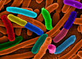
That weird E. coli epidemic
Remember my previous post, in which I gave you a free slide show on the role of the appendix in keeping the gut microbiome happy? Well, to sort of "prove the point" of the importance of a healthy gut microbiome, we've been hearing all about that weird Escherichia coli (E. coli) epidemic in Europe.
If you're like me, you'll want to take the opportunity to emphasize concepts learned in class by applying them to "real life" events reported in the news. If you're like me, you may want to check out these journal articles:
For a really cool, copyright-free image to use in your course, go to my-ap.us/lVEg69
If you're like me, you'll want to take the opportunity to emphasize concepts learned in class by applying them to "real life" events reported in the news. If you're like me, you may want to check out these journal articles:
Deadly bugs: Toxin-producing E. coli strain causes outbreak in Germany
Tina Hesman Saey
Science News web edition : Tuesday, June 7th, 2011
my-ap.us/lfMeYE
[Brief, highly readable introduction to the current outbreak in Europe. Plus a cool photo!]
Bacterial infections: new and emerging enteric pathogens
Sherman, P et al.
Current Opinion in Gastroenterology:January 2010 - Volume 26 - Issue 1 - p 1-4
doi: 10.1097/MOG.0b013e328333d73b
my-ap.us/lJv8PC
[from the abstract: "The aim of this review is to highlight recent advances in knowledge of bacterial enteric infections. We focus on understanding of enterohemorrhagic Escherichia coli O157:H7 and Campylobacter jejuni infections, and to link these acute events with long-term consequences in a susceptible host, including irritable bowel syndrome and chronic inflammatory bowel diseases."]
Clinical Relevance of Shiga Toxin Concentrations in the Blood of Patients With Hemolytic Uremic Syndrome
Brigotti, Maurizio et al.
Pediatric Infectious Disease Journal: June 2011 - Volume 30 - Issue 6 - pp 486-490
doi: 10.1097/INF.0b013e3182074d22
my-ap.us/j72bUA
[from the abstract: "Intestinal infections with Shiga toxin-producing Escherichia coli (STEC) in children can lead to the hemolytic uremic syndrome (HUS). Shiga toxins (Stx) released in the gut by bacteria enter the blood stream and target the kidney causing endothelial injury. Free toxins have never been detected in the blood of HUS patients, but they have been found on the surface of polymorphonuclear leukocytes (PMN)."]
Infectious colitis
Navaneethan, Udayakumar and Giannella, Ralph A
Current Opinion in Gastroenterology: January 2011 - Volume 27 - Issue 1 - p 66–71
doi: 10.1097/MOG.0b013e3283400755
my-ap.us/mTOlpN
[from the abstract: "The incidence of gastrointestinal infections continues to increase and infectious colitis contributes to significant morbidity and mortality worldwide. The purpose of this review is to highlight the recent advances in knowledge of pathogens causing infectious colitis. We describe the various pathogens and specifically focus on enterohemorrhagic Escherichia coli (EHEC) O157:H7, Salmonella, Shigella, Campylobacter, and Entamoeba histolytica infections, and their impact on long-term effects, including postinfectious irritable bowel syndrome and inflammatory bowel disease."]
An inside job: subversion of the host secretory pathway by intestinal pathogens
Sharp, Tyler M and Estes, Mary K
Current Opinion in Infectious Diseases: October 2010 - Volume 23 - Issue 5 - p 464–469
doi: 10.1097/QCO.0b013e32833dcebd
my-ap.us/leb7Le
[from the abstract: "The cellular secretory pathway, composed of the endoplasmic reticulum, Golgi apparatus, and cellular vesicles, mediates the intracellular trafficking of proteins and lipids. Gastrointestinal pathogens frequently affect the functions of enterocytes, the differentiated cells involved in secretion and absorption of extracellular molecules. Microbial pathogenesis can be enhanced by altering the trafficking of key molecules such as brush border enzymes, soluble immune mediators such as cytokines and chemokines, and MHC Class I molecules, all of which rely on the secretory pathway for their appropriate cellular localization. This review focuses on our current understanding of the distinct mechanisms employed by enteric pathogens to antagonize the secretory pathway."]
Probiotics: progress toward novel therapies for intestinal diseases
Yan et al.
Current Opinion in Gastroenterology: March 2010 - Volume 26 - Issue 2 - p 95–101
doi: 10.1097/MOG.0b013e328335239a
my-ap.us/iH93qP
[from the abstract "As the beneficial effects of probiotics on health and disease prevention and treatment have been well recognized, the demand for probiotics in clinical applications and as functional foods has significantly increased in spite of limited understanding of the mechanisms. This review focuses on the most recent advances in probiotic research from genetics to biological consequences regulated by probiotics and probiotic-derived factors."]
For a really cool, copyright-free image to use in your course, go to my-ap.us/lVEg69
Monday, May 9, 2011
FREE animated function of the appendix
Being a visual learner by preference, and therefore preferentially a visual teacher, I find any concept easier to teach if I can draw a picture of it. Even better if I can put that picture into motion.
I recently uploaded a new set of animated PowerPoint slides that anyone can use to explain the immune function of the appendix.
To download and view the FREE set of slides, go to http://www.mediafire.com/file/jqgizrv1xr8s6kd/AppendixFunction-LionDen-KPatton.pptx
To access the file, you'll need a password. If you don't already have a current password to the Lion Den Slide Collection, you can get one by filling out the form at http://my-ap.us/eOtyVq
Feel free to use it in your classroom or website (or both).
I'll be adding a narrated version to my YouTube channel soon at youtube.com/user/kevintpatton
Want to know more?
New "old" news about the appendix
K. Patton
The A&P Professor 24 Aug 2009
[From the archive of this blog, includes some interesting comments from our readers plus links to journal articles]
http://my-ap.us/eIHUKI
The Cecal Appendix: One More Immune Component With a Function Disturbed By Post-Industrial Culture
Michel Laurin et al.
The Anatomical Record. Article first published online: 2 MAR 2011 DOI: 10.1002/ar.21357
[Recent review article that outlines an interesting perspective on the function of the appendix.]
http://my-ap.us/fUeGPX
Monday, May 2, 2011
Video: Neutrophils to the Rescue
Have you seen this video from Science Videolab that shows fluorescent-stained neutrophils rushing toward the site of a tissue injury?
The clip actually strings together several videos showing bright green neutrophils rushing toward damaged cells in liver tissue (seen as bright red areas). The narrator explains in simple terms what is going on and what it means in understanding what happens when tissue damage occurs.
This is a great FREE video to show your class when discussing any or all of these topics:
The clip actually strings together several videos showing bright green neutrophils rushing toward damaged cells in liver tissue (seen as bright red areas). The narrator explains in simple terms what is going on and what it means in understanding what happens when tissue damage occurs.
This is a great FREE video to show your class when discussing any or all of these topics:
- WBCs in general
- Neutrophils
- Immune response
- Inflammation
- Chemotaxis
http://my-ap.us/fh1ExmWant to know more?
Intravascular Danger Signals Guide Neutrophils to Sites of Sterile Inflammation
Braedon McDonald et al.
Science 15 October 2010: Vol. 330 no. 6002 pp. 362-366 DOI: 10.1126/science.1195491
[Research article that summarizes the discovery about how neutrophils use a multistep process to navigate toward noninfectious sites of tissue injury. ]
http://my-ap.us/f5Fua6
EDITORS' CHOICE: Immunology Inflammation Response in Living Color
Kristen L. Mueller
Sci. Signal., 19 October 2010 Vol. 3, Issue 144, p. ec324 DOI: 10.1126/scisignal.3144ec324
[Editor's summary of the processes described above]
http://my-ap.us/fFfwOq
Monday, April 25, 2011
Supplementary courses help A&P students succeed
A few years ago, we brainstormed about what else we could do as A&P professors to help our students succeed. We realized that the two most common things holding our students back from reaching their full potential were:
To address the lack of preparation, we have a prerequisite of "C or better in high school biology or its equivalent within the last five years." That's the best we could manage given the constraints of our institution and its programs. But even with the most stringent prerequisites, it's rare that students really walk into an A&P class ready with a comfortable foundation in biological chemistry and cell biology.
So I developed a refresher course that incoming A&P students could take just before entering their A&P 1 course. Foundations in Science for Health Careers is a developmental level, one-hour course that is offered in a completely online self-paced format. We offer it only during the short mini-mesters and half-semesters.
The Foundations course covers the basic chemistry and biology concepts students need as they begin A&P.
To address the lack of study skills, I developed a one-credit course for our A&P 1 students to take along with A&P 1. Having been given the idea of a supplemental course by my friend Mari Hopper at Southern Indiana University, we began offering A&P 1 Supplement at our institution.
This course parallels the A&P 1 course, giving students how-to tips on specific study skills useful in A&P. Students also have the opportunity to bring their sticking points to the class to get help in getting them unstuck.
The Foundations course is the refresher course and the Supplement course is the shortcut course.
Want to know more?
Check out my video . . .
Then check out the handout and helpful links at The A&P Professor website:
- Lack of adequate preparation to begin A&P on a solid footing
- Lack of basic learning and study skills
To address the lack of preparation, we have a prerequisite of "C or better in high school biology or its equivalent within the last five years." That's the best we could manage given the constraints of our institution and its programs. But even with the most stringent prerequisites, it's rare that students really walk into an A&P class ready with a comfortable foundation in biological chemistry and cell biology.
So I developed a refresher course that incoming A&P students could take just before entering their A&P 1 course. Foundations in Science for Health Careers is a developmental level, one-hour course that is offered in a completely online self-paced format. We offer it only during the short mini-mesters and half-semesters.
The Foundations course covers the basic chemistry and biology concepts students need as they begin A&P.
To address the lack of study skills, I developed a one-credit course for our A&P 1 students to take along with A&P 1. Having been given the idea of a supplemental course by my friend Mari Hopper at Southern Indiana University, we began offering A&P 1 Supplement at our institution.
This course parallels the A&P 1 course, giving students how-to tips on specific study skills useful in A&P. Students also have the opportunity to bring their sticking points to the class to get help in getting them unstuck.
The Foundations course is the refresher course and the Supplement course is the shortcut course.
Want to know more?
Check out my video . . .
Then check out the handout and helpful links at The A&P Professor website:
SEMINAR: Helping Students Succeed
Do these courses work? We're still working on the statistics, but as the above presentations tell you, student feedback from anonymous surveys show that students are happy with what they are getting from these courses. When we get some statistical analysis done, I'll let you know!
[NOTE: If your students would like to take our online pre-A&P refresher course (BIO 095 Foundations in Science for Health Careers) prior to taking your A&P course, they can enroll at St. Charles Community College during either of two 5-wk summer sessions or during either of two 1-wk pre-fall sessions . . . or beyond.]
Monday, April 18, 2011
First human brain map unveiled
Today NewScientist reported that the world's first computerized map of the brain was unveiled last week by neuroscientists at the Allen Institute for Brain Science.
The FREE interactive brain map must be downloaded and installed on your computer at http://my-ap.us/f8Rabf It's fun . . . you should try it!
You can see both of two brains used to produce the maps and check which parts of the brain you want to see. Each is shown in a different color and you check and uncheck brain parts as you explore. For example, you can visualize just the cerebral nuclei, then add in the cerebral cortex. You can also click on each part of the cortex and it will highlight (and name) the particular gyrus or region that you are on.
There are far more features than I've had the time to explore . . . and far more than I'll need to use in the classroom to help my students visualize the brain's structure.
All these richer features are available because it's meant as a research tool rather than a teaching tool. The new map can show the biochemistry and gene expression at various sites based on in depth studies done on two human brains, for example. But you don't have to use any of the richer features.
One of many interesting and useful tidbits of information that has come out of the research end of the project is that there is a 94% similarity in the biochemistry of the two human brains used int he study.
Another interesting fact is that at least 82% of all human genes are expressed in the human brain. (Except perhaps in mine, especially on Fridays.)
While exploring the website at Allen Institute for Brain Science I also stumbled upon a nifty, interactive tool that I'll also probably use in my A&P course. This FREE tool allows you to view different planes of the brain simultaneously while navigating around the brain. I imagine that this tool would be fun to use in class to visualize anatomical relationships of the brain as students themselves navigate around and answer their own questions about the general nature of brain structure.
Want to know more?
The FREE interactive brain map must be downloaded and installed on your computer at http://my-ap.us/f8Rabf It's fun . . . you should try it!
You can see both of two brains used to produce the maps and check which parts of the brain you want to see. Each is shown in a different color and you check and uncheck brain parts as you explore. For example, you can visualize just the cerebral nuclei, then add in the cerebral cortex. You can also click on each part of the cortex and it will highlight (and name) the particular gyrus or region that you are on.
There are far more features than I've had the time to explore . . . and far more than I'll need to use in the classroom to help my students visualize the brain's structure.
All these richer features are available because it's meant as a research tool rather than a teaching tool. The new map can show the biochemistry and gene expression at various sites based on in depth studies done on two human brains, for example. But you don't have to use any of the richer features.
One of many interesting and useful tidbits of information that has come out of the research end of the project is that there is a 94% similarity in the biochemistry of the two human brains used int he study.
Another interesting fact is that at least 82% of all human genes are expressed in the human brain. (Except perhaps in mine, especially on Fridays.)
While exploring the website at Allen Institute for Brain Science I also stumbled upon a nifty, interactive tool that I'll also probably use in my A&P course. This FREE tool allows you to view different planes of the brain simultaneously while navigating around the brain. I imagine that this tool would be fun to use in class to visualize anatomical relationships of the brain as students themselves navigate around and answer their own questions about the general nature of brain structure.
Want to know more?
World's first human brain map unveiled
H. Crawford
NewScientist published online 15 April 2011
[Brief news synopsis with images of applications of the new brain map]
http://my-ap.us/dRY1Qy
Allen Institute's online MRI explorer
[FREE interactive tool that allows you to explore a human brain MRI to visualize brain structure at different levels that you control.]
http://my-ap.us/hjGa7c
Allen Institute's download page for Brain Explorer 2
[FREE interactive tool that allows researchers to locate biochemistry and/or gene expression at specific brain locations.]
http://my-ap.us/fDppYG
Monday, April 11, 2011
Looking for a new one-semester A&P textbook?
I'm excited about the recent publication of my latest textbook for A&P students! Essentials of Anatomy & Physiology is designed for use in one-semester A&P courses.
Coauthors Gary Thibodeau and Matt Douglas worked closely with me and a very talented team of creative editors and scientific illustrators to produce a textbook that students will love to use.
What? A text book that students will actually use?! How can that be?
Let me summarize just two of the many reasons:
Want to know more?
If you go to the electronic brochure, you can view a sample chapter, get a list of available ancillaries, learn about the complete online course available with the textbook, and request a FREE examination copy.
Coauthors Gary Thibodeau and Matt Douglas worked closely with me and a very talented team of creative editors and scientific illustrators to produce a textbook that students will love to use.
What? A text book that students will actually use?! How can that be?
Let me summarize just two of the many reasons:
- This book is the most visually oriented textbook in its niche.
- There are more illustrations than in most other one-semester books, providing students with additional visual help in mastering concepts.
- Each illustration is carefully designed for maximum learning effectiveness.
- Most figures include a detailed "walk through" that explains the meaning of image, rather than merely providing a perfunctory title.
- It includes the Clear View of the Human Body, a bound-in set of transparency overlays that provide a virtual dissection experience for readers as they peel away (or add) layers of the body from either an anterior view or a posterior view. This experience allows readers to develop a sense of anatomical relationships among body structures.
- Numerous summary tables act as graphic organizers to help students see relationships among concepts.
- We worked hard to get the images and tables close to related text. This kind of visual integration not as easy at is sounds, requiring several passes at the layout to "get it right" and creatively fit everything together.
- This text is carefully constructed to be easy to read and easy to raid. Polls conducted with my students show that most students who use a textbook use some combination of reading chapter sections straight through and simply raiding parts of chapter sections when they need to find something.
- Even strong readers have some difficulty reading highly technical scientific texts. We use straightforward, conversational language to communicate difficult terminology and difficult concepts.
- This text breaks the material down into smaller chapters so that readers do not get overwhelmed and get so discouraged they won't read the book.
- Our page design uses many levels of bold headings to help students understand the organization of concepts as they read and to find specific concepts when they raid.
- I worked with reading specialists and ESL teachers to find ways to make the book more accessible to all readers. For example:
- We include a significantly larger glossary than most texts in this market.
- In-chapter pronunciation guides for all boldface terms used in each chapter.help students master the language of A&P.
- We provide the meanings of word parts for all boldface terms so that students can start building their skills in understanding scientific terminology
- A comprehensive outline summary at the end of each chapter visually organizes concepts so that readers can solidify their comprehension of the chapter.
- Downloadable audio chapter summaries (included in the included online resources) can be used along with the printed chapter summaries to strengthen understanding even more.
Want to know more?
If you go to the electronic brochure, you can view a sample chapter, get a list of available ancillaries, learn about the complete online course available with the textbook, and request a FREE examination copy.
Click the link: http://my-ap.us/gcH7Jr
Sunday, April 3, 2011
Plaque-fighting bacteria
Have you noticed that the microbiome of the human body has taken off as one of the hottest areas? Each month, new concepts of how our microbial partners keeps us healthy are revealed. Last week, we were discussing teeth in my A&P 2 course and I wish I'd had this new tidbit to share with my students:
Researchers recently found that Streptococcus salivarius, one of the microbes in our mouth, can help fight the buildup of plaque on our teeth. It does so by producing the enzyme FruA, which breaks down carbohydrates in our mouth more efficiently than can the bacteria that form plaque biofilms. Thus, the plaque-forming bacteria are robbed of their nutrients.
I'll bet S. salivarius will become popular as an oral probiotic. And its discovery may help us find better ways to manage our mouth's ecosystem to promote good health.
Want to know more?
Researchers recently found that Streptococcus salivarius, one of the microbes in our mouth, can help fight the buildup of plaque on our teeth. It does so by producing the enzyme FruA, which breaks down carbohydrates in our mouth more efficiently than can the bacteria that form plaque biofilms. Thus, the plaque-forming bacteria are robbed of their nutrients.
I'll bet S. salivarius will become popular as an oral probiotic. And its discovery may help us find better ways to manage our mouth's ecosystem to promote good health.
Want to know more?
Click the image above to access a FREE animation of tooth decay you can use in your course as you explain the process.
Inhibition of Streptococcus mutans Biofilm Formation by Streptococcus salivarius FruA
A. Ogawa, et al.
Applied and Environmental Microbiology Vol. 77, March 2011, p. 1572 doi:10.1128/AEM.02066-10, published online January 14, 2011
[Original research article]
http://my-ap.us/h7RuaA
Bacterial fight dental plaque
Tina Hesman Saey
Science News Published online April 1, 2011
[Brief summary of the discovery]
http://my-ap.us/hsj1nb
Friday, March 25, 2011
Progesterone's action on sperm finally solved
Two recent articles in Nature reveal a mystery that's been puzzling physiologists for a couple of decades . . . how does progesterone signal sperm cells?
The short version of the story is this . . .
Progesterone is a steroid hormone and thus usually enters its target cell and activates transcription of a gene. In this case, however, progesterone instead directly triggers a calcium channel. It's not even a second-messenger system, but a direct binding to the channel.
Secretion of progesterone by cumulus cells apparently also provides a chemical gradient that helps sperm navigate toward the ovum.
Recall also that calcium influx into the ovum triggered by contact with a sperm cell plays a role in producing changes within the ovum that result in successful fertilization.
I recommend that my students keep running concept lists on recurring themes or actors in story of human biology. Here's something they can now add to their calcium list. Now they can see that a calcium gradient is a truly multipurpose tool in the body. (For more about running concept lists see my-ap.us/hCIA9X)
Want to know more?
The short version of the story is this . . .
Progesterone is released from the cumulus cells that cling to the outside of the zona pellucida surrounding the ovum. This progesterone binds directly to calcium channels in the sperm's plasma membrane to open. Influx of calcium ions triggers an increase in flagellum activity. This increased work is needed for the sperm to get through the zona to the ovum.
Progesterone is a steroid hormone and thus usually enters its target cell and activates transcription of a gene. In this case, however, progesterone instead directly triggers a calcium channel. It's not even a second-messenger system, but a direct binding to the channel.
Secretion of progesterone by cumulus cells apparently also provides a chemical gradient that helps sperm navigate toward the ovum.
Recall also that calcium influx into the ovum triggered by contact with a sperm cell plays a role in producing changes within the ovum that result in successful fertilization.
I recommend that my students keep running concept lists on recurring themes or actors in story of human biology. Here's something they can now add to their calcium list. Now they can see that a calcium gradient is a truly multipurpose tool in the body. (For more about running concept lists see my-ap.us/hCIA9X)
Want to know more?
Sperm mystery solved: Scientists identify the channel by which progesterone activates sperm to swim toward an eggFrom The A&P Professor archive
M. Scudellari
The Scientist Published online 16th March 2011
[Nice summary of the significance of the discovery]
my-ap.us/grpcTp
Female hormone could be key to male contraceptive: Progesterone-sensing molecule that guides sperm to egg offers fertility solution.
E. Callaway
NatureNews Published online 16 March 2011 | Nature | doi:10.1038/news.2011.163
[Summary article outlining the papers published in Nature]
my-ap.us/fJNaYk
Progesterone activates the principal Ca2+ channel of human sperm
Lishko et al.
Nature 471:387–391 17 March 2011 doi:10.1038/nature09767
[Original research findings]
my-ap.us/gxcomX
The CatSper channel mediates progesterone-induced Ca2+ influx in human sperm
Strunker et al.
Nature 471:382–386 17 March 2011 doi:10.1038/nature09769
[Original research findings] my-ap.us/g14eTK
New discovery about sperm's ability to swim
K. Patton
The A&P Professor published online Feb 18, 2010
[Summary of new discovery that when sperm enter female tract, proton channels in the sperm head open and the resulting pH drop triggers influx of calcium, which gets the flagella started in the first place. That darn calcium shows up in every part of this story, eh?]
my-ap.us/dExdEK
Subscribe to:
Posts (Atom)