Friday, March 25, 2011

Progesterone's action on sperm finally solved

Two recent articles in Nature reveal a mystery that's been puzzling physiologists for a couple of decades . . . how does progesterone signal sperm cells?

The short version of the story is this . . .

Progesterone is released from the cumulus cells that cling to the outside of the zona pellucida surrounding the ovum. This progesterone binds directly to calcium channels in the sperm's plasma membrane to open.  Influx of calcium ions triggers an increase in flagellum activity.  This increased work is needed for the sperm to get through the zona to the ovum.

Progesterone is a steroid hormone and thus usually enters its target cell and activates transcription of a gene.  In this case, however, progesterone instead directly triggers a calcium channel.  It's not even a second-messenger system, but a direct binding to the channel.

Secretion of progesterone by cumulus cells apparently also provides a chemical gradient that helps sperm navigate toward the ovum.

Recall also that calcium influx into the ovum triggered by contact with a sperm cell plays a role in producing changes within the ovum that result in successful fertilization.

I recommend that my students keep running concept lists on recurring themes or actors in story of human biology.  Here's something they can now add to their calcium list.  Now they can see that a calcium gradient is a truly multipurpose tool in the body.  (For more about running concept lists see my-ap.us/hCIA9X)

Want to know more?
Sperm mystery solved: Scientists identify the channel by which progesterone activates sperm to swim toward an egg
M. Scudellari
The Scientist Published online 16th March 2011
[Nice summary of the significance of the discovery]
my-ap.us/grpcTp

Female hormone could be key to male contraceptive: Progesterone-sensing molecule that guides sperm to egg offers fertility solution.
E. Callaway
NatureNews Published online 16 March 2011 | Nature | doi:10.1038/news.2011.163
[Summary article outlining the papers published in Nature]
my-ap.us/fJNaYk

Progesterone activates the principal Ca2+ channel of human sperm
Lishko et al.
Nature  471:387–391 17 March 2011 doi:10.1038/nature09767
[Original research findings]
my-ap.us/gxcomX

The CatSper channel mediates progesterone-induced Ca2+ influx in human sperm
Strunker et al.
Nature 471:382–386 17 March 2011 doi:10.1038/nature09769
[Original research findings] my-ap.us/g14eTK
From The A&P Professor archive
New discovery about sperm's ability to swim
K. Patton
The A&P Professor published online Feb 18, 2010
[Summary of new discovery that when sperm enter female tract, proton channels in the sperm head open and the resulting pH drop triggers influx of calcium, which gets the flagella started in the first place.  That darn calcium shows up in every part of this story, eh?]
my-ap.us/dExdEK

Wednesday, March 16, 2011

What do tuft cells do?

We've known for a half-century that the lining of the small intestine has a scattering of weird little cells called tuft cells.  They are called that because they have distinctive tufts of microvilli facing into the intestinal lumen.  But what do they do?  We're finally getting to the answer!

A recent article in Journal of Cell Biology outlines confirms some recent discoveries about tuft cell function and extends our knowledge a bit further.

Apparently, tuft cells are secretory cells that produce opioids in the gut.  They are also the only epithelial cells that produce the COX (cyclooxygenase) enzymes needed to produce prostaglandins involved in inflammation and tumor formation.

Some physiologists had previously proposed a sensory role for tuft cells.  Could they be involved in tasting foods in the gut and be part of the signaling mechanism that regulates exocrine and endocrine secretion that controls digestive and metabolic processes?

As we learn more about tuft cells, we are sure to discover a role for them in normal regulation of intestinal function as well as in important pathological processes. 

Want to know more?

A fifth amendment to the intestine's constitution
Ben ShortJournal of Cell Biology 2011 192:706. Published March 7, 2011, doi:10.1083/jcb.1925iti2
[Brief synopsis of the discovery and its importance]
http://my-ap.us/ezeS9J

Distinct ATOH1 and Neurog3 requirements define tuft cells as a new secretory cell type in the intestinal epithelium.
Gerbe, F., et al.
Journal of Cell Biology 2011 Mar 7;192(5):767-80. doi:10.1083/jcb.201010127.
[Original research article.  Includes many illustrations, supplements, helps and cross references. FREE full text]
my-ap.us/eCNITF

Here's a really nice teaching image from the Gerbe et al. article, which complements Figure 25-18 in the Anatomy & Physiology 7/E textbook:  my-ap.us/hmdFub (includes downloadable PowerPoint slide)

Here are all the PowerPoints available with the Gerbe et al. article: my-ap.us/gKY7ZG

Chemosensory Perception in the Gut 
Hofer, D., et al.
Physiology February 1999 vol. 14 no. 1 18-23  
[Article proposing sensory function of tuft cells; FREE access to full text/PDF; nice images]
http://my-ap.us/g2HCxw


Saturday, March 12, 2011

More study tool slides

In a recent post, I briefly discussed some presentations that I do regarding using Running Concept Lists and Concept Maps to learn anatomy and physiology concepts and how they relate to one another.  I posted links to slides, narrated YouTube videos of brief versions of the presentation, and related study tip pages in my Lion Den website for students.  As promised, this week I'm posting links and short descriptions of four more presentations that may help your students succeed in A&P.

Want to present your own version of any of these study tips? Perhaps embed parts of them into your own presentations? You are welcome to use the slides, which can be found in the Lion Den Slide Collection.  (To use the slides, you'll need the password for downloading them.  Just fill in the form to get the password if you don't already have it.)

Don't forget . . . when using the direct links below, you need to have the super-secret, magic password ready!

In an upcoming post, I'll let you in on some secrets for creating some additional time with you students so that you can cover these study tips without sacrificing "content time" in your A&P course.

Previously described slides available in the Lion Den Slide Collection:


More Slides also available in the Lion Den Slide Collection:

Flash Cards: Reducing Your Study Time 
Some practical tips for using study cards to reduce your study time and get a solid foundation in learning any topic. This video also includes some surprising advanced techniques that show how to use flash cards to also learn higher-level thinking in any subject. Includes discussion of the Leitner system (plus Patton's adaptation of the Leitner system), color codes and symbols, using cards to learn processes and ordered structures, and using cards to build concept maps (mind maps).
Muscle Names Have Meaning
Learning muscles is hard enough without dealing with those crazy convoluted Latin names. But if you pay attention to those names, you'll find that they are actually phrases that help you find the muscles AND help you to remember them in the long term. Find out how this works . . . and where to find lists to help you figure out the meaning of common muscle names.
Exam Strategies
Proven strategies for success in taking tests and exams. What you can do before, during, and after an exam to improve performance in your anatomy and physiology course.
Learn from Your Mistakes: TEST ANALYSIS 
How do you effectively "go over" your tests or exams? Learn how to analyze your tests to see what went wrong and how to fix it.

Don't forget that your students can keep up with all these study tips (and more) on their own by subscribing to my blog The A&P Student.

I have some handy (and bizarre) bookmarks giving students information about The A&P Student blog that you can distribute FREE to your classes!  Just go to my bookmark request page to get bookmarks for your students now.

    Friday, March 11, 2011

    Antibodies work INSIDE virus-infected cells

    You already know that we use antibodies in several ways to combat infection in our immune system.  They bind to pathogens, they activate complement, you know the drill. Well, here's another bullet point to add to your antibody slide: we've found a new intracellular role for the antibody.

    Researchers have recently shown that antibodies can attach to a virus, which then enters a host cell where a molecule called TRIM21 quickly binds at Fc on the IgG antibodies.  By ubiquitin ligase activity, TRIM21 targets the virus's proteins for destruction by the proteasome.

    Click here for an awesome animation that shows all this violent destruction in a simple, dramatic way. Your students will love this animation, because the proteasomes' rapid and total destruction of the virus is so amazing to watch.  And it's a good opportunity to emphasize the importance of the proteasome in the cell. 

    Not only does this observation give us a new intracellular role for antibodies, it also highlights a new and important strategic link between innate immunity (TRIM21/proteasome action) and adaptive immunity (antibodies).

    Want to know more?

    Antibodies mediate intracellular immunity through tripartite motif-containing 21 (TRIM21)
    Donna L. Mallery, et al.
    Proceedings of the National Academy of Science November 16, 2010 vol. 107 no. 46 19985-19990
    Published online before print November 2, 2010, doi: 10.1073/pnas.1014074107
    [This is the original paper, available open access]

    Read this shortened evaluation highlighting the key findings:
    Koch D, Sawtell N: 2010. F1000.com/6381958

    Come Inside
    R. Grant
    The Scientist Volume 25 Issue 3 Page 58 2011-03-01
    [Quick and easy summary, including that awesome video]

    AntibodyBy the way, I love those movies showing miniaturized submarines exploring the inside of the body . . . and this reminds me of the SyFy thriller Antibody with Lance Henriksen and Robin Givens.  Yes, it's a hokey movie, but I like the scenes showing the immune cells attacking the miniaturized sub.

    Monday, February 28, 2011

    Prezi practice practicals

    In my blog The Electronic Professor, I recently wrote about the Prezi alternative to PowerPoint presentations.

    In a nutshell, Prezi creates large, complex sets of information that you can zoom into either at will or in a preplanned way.

    As I was exploring around the Prezi presentations that have been made public, I found a great idea . . . why not use Prezi to create practice lab practicals?

    My students always find it hard to get ready for practicals.  Obviously, part of the issue is the massive amount of content that we ask them to learn.  But, perhaps more imporatantly, it's hard for them to imagine the kinds of questions that they are likely to get. We set up "practice practicals" in our open lab when we can . . . and that helps a lot.  But wouldn't it be great to have an easy to access online place our students can practice for their praticals?

    Well, Rob Swatski at the York Campus of Harrisburg Area Community College (York, PA) has already cracked this egg!

    Take a look at his Virtual Lab Exam for muscles in A&P 1.


    You have to start clicking around and get the feel for how this practice lab practical is set up . . . but once you have the hang of it, it's amazing.  You can see how students can practice the content and also get a feel for EXACTLY the kinds of things they'll be asked to do on their practical.

    Another idea that Rob came up with is shown in this presentation that introduces the microscope.  Rob uses the particular characteristics of Prezi to full advantage to produce a resource that is useful for both teaching and learning in the A&P lab.


    Imagine how useful this sort of thing can be for
    • online tests and quizzes for labs
    • exploring large, complex anatomical structures in a lecture class
    • virtual dissections
    • student presentations
    • zooming in on tissues or bone markings while teaching histology
    Let's hear YOUR ideas!

    Thursday, February 17, 2011

    Concept Lists and Concept Maps

    I've recently posted two new videos that help students learn A&P to my YouTube channel.

    You are welcome to use them in your course by linking to them or by embedding them in your website, LMS (learning management system), or PowerPoint presentation.  Or you can just keep them in mind when students come to you looking for study tips.  Or they may just spark some ideas of your own!


    Concept Lists - A Powerful Study Strategy 
    Running concept lists are a great way to build up what you are learning layer by layer, while at the same time learning relationships between different concepts. If you run your concept lists faithfully, you will also have a handy "personal encyclopedia" of concepts and how they are related.  (Closed Captioned)


    The Concept Lists video is also embedded in one of my Lion Den Study Tips & Tools pages entitled Concept Lists.


    Concept Maps - A Learning & Study Strategy 
    Concepts maps (mind maps) help you understand relationships in human anatomy and physiology in ways that deepen understanding. This video summarizes what a concept maps is, how to make and use one, and outlines some examples of different styles of concept maps.  (Closed Captioned)


    The Concep Maps video is also embedded in one of my Lion Den Study Tips & Tools pages entitled Concept Maps


    Want to present your own version of any of these study tips? Perhaps embed parts of them into your own presentations? You are welcome to use the slides, which can be found in the Lion Den Slide Collection.  (To use the slides, you'll need the password for downloading them.  Just fill in the form to get the password if you don't already have it.)
    When using the direct links, you need to have the super-secret, magic password ready!

    Want more slides? More study tip materials to share with your students?  In an upcoming post, I'll be sharing a few more, ok? 

    Don't forget that your students can keep up with all these study tips (and more) on their own by subscribing to my blog The A&P Student.


    I have some handy (and bizarre) bookmarks giving students information about The A&P Student blog that you can distribute FREE to your classes!  Just go to my bookmark request page to get bookmarks for your students now.


    Thursday, January 6, 2011

    Lion Den Slide Collection

    As you think about getting started in a new semester, you may find that sets of FREE animated slides might help you spice up your classroom presentations.

    For the last several years, I sent a set of hundreds of slides from my Lion Den site for students to anyone who donated to keeping that site up and running.  But starting in this new year of a new decade of a (relatively new) century, I'm now opening up the "secret vault" for anyone who wants them.  And promises to use them for good and not evil.

    If you go to the Lion Den downloads page and join the Lion Den, you'll get acccess to all the slide files.

    Once you are in the download location, you can access the original set of slides in the folder marked  Lion Den Slide Collection Version 1.0. Newer and updated slide collections will be available in the folder Lion Den Slide Collection Version 2.0.  You can follow the links to the folders to see what's there, but you cannot download any of the slide sets until you fill out the form and get the super-secret password.

    You can preview some of the slides by going to the Lion Den slide page.  However, not all the slides in the Lion Den Slide Collection are posted on that website, the slides are posted individually (not in sets), and they are in "slide show" format (which may not be fully editable by you).  The slide sets in the downloadable collection are in fully editable .ppt or .pptx format.  It's much easier to go to the Lion Den download page, join the highly-classified-super-secret membership roster and download the sets of slides.

    All the slides can be freely used (and adapted) for noncommercial educational purposes (see the license embedded in the Notes section of each slide).

    As the User Manual states, these slides are not necessarily as detailed (or as simplified) as you would like to use in your own presentation.  But because they are editable, you can change that, right?  Also, some of these slides were originally intended for a particular purpose (such as introducing a topic that will be explored more fully later).

    Another thing to remember is that the slide sets are not meant to be a complete set of slides covering all topics of A&P.  In my classroom, I use a lot of images from the textbook (provided by the publisher).  The slides in the Lion Den Slide Collection are meant to be supplemental slides.

    Keep in mind that the slide sets are NOT created by a professional graphic artist . . . just old Kevin hacking away in PowerPoint.  So they may not be as slick as some slides you've seen.

    All the slides are in PowerPoint-compatible files.  The newer slides are in the newer XML PowerPoint format (.pptx).

    Tuesday, December 28, 2010

    Google Body Browser

    My editor Jeff recently pointed me to a story about Google's recently launched Body Browser.  I was amazed at this latest creation of Google Labs.

    The Body Browser is a FREE online tool that you and your students can use to explore the anatomy of the human body in a "virtual dissection" format.  Using the familiar Google Maps navigation tools, you can . . .
    • Peel (or fade) away layers of the body . . . removing the skin, then muscles, then bones, to reveal the internal organs

    • Select systems (skeletal, muscular, nervous, cardiovascular) to view

    • Click on any structure to show its label

    • Type the name of any structure in the search box to find it in the body

    • Tilt, zoom, turn the body to a variety of positions to see organs in more views that usually available in a textbook, atlas, or chart
    The Body Browser runs inside any WebGL-enabled browser, meaning that you don't have to worry about having the latest Flash or Java plugins installed.

    I think Body Browser a is a great FREE tool for A&P students to have access to an online model of the human body that can be used for a beginning study of anatomy.  Because it allows the user to type in the names of organs for which they are looking, you don't need to worry about it being too high (or too low) a level for your course.

    There are a few minor limitations of the Body Browser:
    • The only available specimen is female (that is, there is no male specimen available to complement the female specimen)

    • The specimen is partially clothed.  Although one can see some of the underlying surface structures as the "skin" layer fades back, it's not the same as seeing these structures clearly.  An odd feature that makes certain regions of the body "off limits." (I've seen some hacks to fix this, but none of them work for me using the Chrome browser)

    • Some of the organs are roughly rendered, so it's not as detailed (at least in some areas) as you may like to see

    • Only a few systems can be shown in entirety.  Some useful system views that are missing are the lymphatic system and the respiratory system

    • You cannot select or hide individual organs for display
       
    • I could find no documentation or even a help button (pretty typical of Google Labs resources)
    Even with some minor limitations, Body Browser is still a fantastic study and teaching tool.  As an A&P professor, you might use Body Browser as . . .
    • a presentation tool during a class lecture or discussion to demonstrate the location and structure of specific organs

      • you could use it live or you could record a session with Jing or similar recording tool and use the pre-recorded exploration

    • a tutoring tool with which you can send the URL of a specific view (perhaps with a label) to a student or group of students

    • a testing tool with which you can create specific views that can then be used to test from on a quiz, test, or lab practical . . . perhaps using a screen capture tool such as Jing

    • alternative lab model to use along with, or in place of, physical models in the lab

      • it may be useful as a reference to side by side with a laboratory model

      • use it in place of a laboratory model in a distance learning course
    I'll be listing some ideas for student  uses of Body Browser at my blog The A&P Student.

    Do you have some other ideas for using Body Browser in an undergraduate A&P course?  Just use the comment feature and share your ideas with us!

    Check out this video to see a demo of the currently available features of Body Browser

    Tuesday, December 14, 2010

    See you in Sarasota?

    I plan on attending the Human Anatomy and Physiology Society (HAPS) Southeast Regional Conference on January 22, 2011, in the Sarasota FL area. 

    Will I see you there?

    These regional HAPS meetings are wonderful.  Not only do you get to meet and chat with folks doing what you do from all kinds of institutions, you'll hear loads of ideas on how to better help your students succeed in A&P.

    And what better time of year to head off for coastal Florida, eh?

    Want some details?

    Go to the official meeting website my-ap.us/f7HO2K for more information on invited speakers, available workshops, travel info, etc.. . . and a handy online registration form.

    And I'll be presenting a workshop there:

    Helping A&P Students Succeed:
    Using Supplemental Courses to
    Reinforce Concepts and Promote Learning Skills
     
    Do your A&P students struggle with the whole process of learning?
    Do they seem ill prepared in their study skills and their knowledge of basic principles of biology?
    Explore a case study in which short supplemental courses provide underprepared and unskilled students
    with knowledge and skills that make them better able to succeed in the A&P course and beyond.
    You will also receive free resources to help your own students succeed.

    I hope I see you there!  I'd love to chat with you.

    Friday, December 3, 2010

    Father of Fractals

    You may have already heard the recent news of the passing of Benoit Mandelbrot, originator of the iconic Mandelbrot Set (pictured) and founder of the field of fractal geometry.  It brings to mind the deeper understanding of human structure and function that has resulted directly from applying principles of fractal geometry.  An important set of principles that I believe we A&P professors could do a better job of helping our students appreciate.

    Mandelbrot's pioneering efforts in understanding the roughness of nature led to the discovery of basic principles of fractal geometry.  A key characteristic of fractal structures is self-similarity (the parts resemble the whole).

    In human anatomy, this self-similar characteristic is observed in surfaces that have folds, which have bumps, which in turn have their own bumps, and so on . . . producing unexpectedly huge total surface areas.  For example, think of the loops of the intestines, which in turn have circular folds of mucosa, which in turn have villi, which in turn have microvilli, which in turn have membranes embedded with bumpy molecules, and so on. 

    Fractal self-similarity can also be observed in branched structures, such as the respiratory tract and the cardiovascular vessels.  These structures have branches that have branches that have branches, and so on for many levels . . . producing large numbers of pathways and huge surface areas.

    A particularly interesting characteristic of such complex fractal structures is that they are produced with relatively simple mathematical formulae.  Which means that very little genetic information is needed to produce highly complex structures like intestines, blood vessels, lymphatic vessels, bronchial trees, cerebral convolutions, etc.

    Fractal structures are also chaotic, a mathematical concept of "constrained randomness."  Put simply, chaotic structures have an element of randomness but within limits.  So when our body applies fractal geometry during development we can be certain of a particular type of structure without being certain we'll know exactly where each individual bump or branch will lie.  In other words, we can more or less be certain where the main arteries will be (with some individual variation) but not so much for the various arterioles and capillaries . . . at least not precisely.

    Principles of chaos also play out in human physiology when we observe the aperiodic (nonrhythmic) patterns of heart rate, brain waves (as in an EEG), and certain other functions.

    Mandelbrot opened up a whole new understanding of human structure and function that is only now becoming understood widely.  I've been introducing the concept of chaos and fractals in my courses, and more subtly in some of my textbooks, for several years now.  My experience is that introducing simplified principles of chaos and fractals at the beginning of A&P 1, then reinforcing them when encountered throughout both semesters of A&P, help student appreciate an intriguing and important concept of human structure and function.  A concept that is increasingly playing a central role in science's understanding of human biology.


    Want to know more?

    Benoît Mandelbrot (1924–2010)
    Ralph Gomory
    Nature Volume: 468, Page 378, Date published: 18 November 2010, doi:10.1038/468378a, Published online: 17 November 2010
    [A brief synopsis of Mandelbrot's life and contributions from the journal Nature]

    Chaos in the Human Body (Mini Lesson)
    Kevin Patton
    Lion Den http://lionden.com/chaos.htm
    [Brief outline that I use with my own students in A&P 1]

    Applications of Fractals - Human Body
    ThinkQuest
    Oracle Education Foundation. online (accessed 2 Dec 2010)
    [Brief student-produced outline of some fractal principles of the body]

    Fractal Geometry in Biological Systems: An Analytical Approach
    Philip M. Iannaccone, Mustafa Khokha
    CRC Press 1996
    [Book outlining the initial discoveries of fractals in humans.]

    Chaos: Making a New Science
    James Gleick
    Penguin 2008
    [Reprint of the classic bestseller book that outlines in simple terms the concepts of chaos and fractal geometry.  Highly recommended.  Includes some applications/examples in human biology.]

    Monday, October 4, 2010

    Nobel Prize: Test Tube Babies

    This morning, we heard the news . . . the 2010 Nobel Prize in Medicine or Physiology is awarded to Robert G. Edwards "for the development of in vitro fertilization."


    Professors and students using my Anatomy & Physiology (7th ed.) textbook can access an article on in vitro fertilization (IVF) at A&P Connect online at evolve.elsevier.com

    If you are thinking of mentioning this award in your classes this week, which I am planning to do myself, you are welcome to use the following information from the Nobel Committee, as well as the images linked to the thumbnails presented here (scroll down to the bottom for more).

    Of course, be aware that the use of IVF is condemned by some religious groups (for example, see Dignitas Personae) and thus classroom discussions may become heated.

    There is also advanced information available at the Nobel website.  This is a nice publication that summarizes the science.

    If you want a short set of slides that you can use today in your class, then use this link:


    Summary

    Robert Edwards is awarded the 2010 Nobel Prize for the development of human in vitro fertilization (IVF) therapy. His achievements have made it possible to treat infertility, a medical condition afflicting a large proportion of humanity including more than 10% of all couples worldwide.

    As early as the 1950s, Edwards had the vision that IVF could be useful as a treatment for infertility. He worked systematically to realize his goal, discovered important principles for human fertilization, and succeeded in accomplishing fertilization of human egg cells in test tubes (or more precisely, cell culture dishes). His efforts were finally crowned by success on 25 July, 1978, when the world's first "test tube baby" was born. During the following years, Edwards and his co-workers refined IVF technology and shared it with colleagues around the world.

    Approximately four million individuals have so far been born following IVF. Many of them are now adult and some have already become parents. A new field of medicine has emerged, with Robert Edwards leading the process all the way from the fundamental discoveries to the current, successful IVF therapy. His contributions represent a milestone in the development of modern medicine.

    Infertility – a medical and psychological problem

    More than 10% of all couples worldwide are infertile. For many of them, this is a great disappointment and for some causes lifelong psychological trauma. Medicine has had limited opportunities to help these individuals in the past. Today, the situation is entirely different. In vitro fertilization (IVF) is an established therapy when sperm and egg cannot meet inside the body.

    Basic research bears fruit

    The British scientist Robert Edwards began his fundamental research on the biology of fertilization in the 1950s. He soon realized that fertilization outside the body could represent a possible treatment of infertility. Other scientists had shown that egg cells from rabbits could be fertilized in test tubes when sperm was added, giving rise to offspring. Edwards decided to investigate if similar methods could be used to fertilize human egg cells.
    It turned out that human eggs have an entirely different life cycle than those of rabbits.  In a series of experimental studies conducted together with several different co-workers, Edwards made a number of fundamental discoveries. He clarified how human eggs mature, how different hormones regulate their maturation, and at which time point the eggs are susceptible to the fertilizing sperm. He also determined the conditions under which sperm is activated and has the capacity to fertilize the egg. In 1969, his efforts met with success when, for the first time, a human egg was fertilized in a test tube.
    In spite of this success, a major problem remained. The fertilized egg did not develop beyond a single cell division. Edwards suspected that eggs that had matured in the ovaries before they were removed for IVF would function better, and looked for possible ways to obtain such eggs in a safe way.

    From experiment to clinical medicine

    Edwards contacted the gynecologist Patrick Steptoe. He became the clinician who, together with Edwards, developed IVF from experiment to practical medicine. Steptoe was one of the pioneers in laparoscopy, a technique that was new and controversial at the time. It allows inspection of the ovaries through an optical instrument. Steptoe used the laparoscope to remove eggs from the ovaries and Edwards put the eggs in cell culture and added sperm. The fertilized egg cells now divided several times and formed early embryos, 8 cells in size (see figure).
    These early studies were promising but the Medical Research Council decided not to fund a continuation of the project. However, a private donation allowed the work to continue. The research also became the topic of a lively ethical debate that was initiated by Edwards himself. Several religious leaders, ethicists, and scientists demanded that the project be stopped, while others gave it their support.

    The birth of Louise Brown - an historic event

    Edwards and Steptoe could continue their research thanks to the new donation. By analyzing the patients' hormone levels, they could determine the best time point for fertilization and maximize the chances for success. In 1978, Lesley and John Brown came to the clinic after nine years of failed attempts to have a child. IVF treatment was carried out, and when the fertilized egg had developed into an embryo with 8 cells, it was returned to Mrs. Brown. A healthy baby, Louise Brown, was born through Caesarian section after a full-term pregnancy, on 25 July, 1978. IVF had moved from vision to reality and a new era in medicine had begun.

    IVF is refined and spreads around the world

    Edwards and Steptoe established the Bourn Hall Clinic in Cambridge, the world's first centre for IVF therapy. Steptoe was its medical director until his death in 1988, and Edwards was its head of research until his retirement. Gynecologists and cell biologists from all around the world trained at Bourn Hall, where the methods of IVF were continuously refined. By 1986, 1,000 children had already been born following IVF at Bourn Hall, representing approximately half of all children born after IVF in the world at that time.

    Today, IVF is an established therapy throughout the world. It has undergone several important improvements. For example, single sperm can be microinjected directly into the egg cell in the culture dish. This method has improved the treatment of male infertility by IVF. Furthermore, mature eggs suitable for IVF can be identified by ultrasound and removed with a fine syringe rather than through the laparoscope.
    IVF is a safe and effective therapy. 20-30% of fertilized eggs lead to the birth of a child. Complications include premature births but are very rare, particularly when one egg only is inserted into the mother. Long-term follow-up studies have shown that IVF children are as healthy as other children.
    Approximately four million individuals have been born thanks to IVF. Louise Brown and several other IVF children have given birth to children themselves; this is probably the best evidence for the safety and success of IVF therapy. Today, Robert Edwards' vision is a reality and brings joy to infertile people all over the world.

    Robert G. Edwards was born in 1925 in Manchester, England. After military service in the Second World War, he studied biology at the University of Wales in Bangor and at Edinburgh University in Scotland, where he received his PhD in 1955 with a Thesis on embryonal development in mice. He became a staff scientist at the National Institute for Medical Research in London in 1958 and initiated his research on the human fertilization process. From 1963, Edwards worked in Cambridge, first at its university and later at Bourn Hall Clinic, the world's first IVF centre, which he founded together with Patrick Steptoe. Edwards was its research director for many years and he was also the editor of several leading scientific journals in the area of fertilization. Robert Edwards is currently professor emeritus at the University of Cambridge.

    References:
    Edwards RG. Maturation in vitro of human ovarian oocytes. Lancet 1965; 2:926-929.
    Edwards RG, Bavister BD, Steptoe PC. Early stages of fertilization in vitro of human oocytes matured in vitro. Nature 1969; 221:632-635.
    Edwards RG, Steptoe PC, Purdy JM. Fertilization and cleavage in vitro of human oocytes matured in vivo. Nature 1970; 227:1307-1309.
    Steptoe PC, Edwards RG. Birth after the reimplantation of a human embryo. Lancet 1978; 2:366.
    Edwards RG. The bumpy road to human in vitro fertilization. Nature Med 2001; 7:1091-4.

    The preceding information is from "The 2010 Nobel Prize in Physiology or Medicine - Press Release". Nobelprize.org. 4 Oct 2010 http://nobelprize.org/nobel_prizes/medicine/laureates/2010/press.html


    Some images you may find useful (click each thumbnail)

    Sperm injection into oocyte

    Human embryos developing in vitro


    Robert G. Edwards


    PDF from the Nobel website


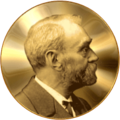
    Nobel medal

    Wednesday, September 29, 2010

    More FREE images!

    http://my-ap.us/cw3HK2
    I've recently posted some more FREE images in the collection posted at The A&P Professor website.


    Although it's just a beginning effort in a project that will take years to complete (if it really ever is "complete"), I do have quite a few images available in many different topics within A&P.

    I've been focusing mainly on images that are not likely to already be present your textbook or lab manual.  That's so you can supplement your course with these additional images.  Perhaps you could use them in PowerPoint slides or online tutorials.

    Some images are additional examples of specimens (such as tissues) where additional specimens are helpful (but would never all fit in a typical textbook).

    http://my-ap.us/dsQ5hd
    As always, if you have any specific requests for images I'll do my best to track them down for you and add them to the collection.  And if you have any images you've found that you think I should add, let me know that, too!

    And stay tuned for next week's announcement of the Nobel Prize in Medicine or Physiology!  I'll be posting links to resources you can use in your course to "bring the news home" to your students.

    I'm predicting that the discovery of the hormone leptin will be the focus of this year's prize. Let's see if I called it correctly!

    Wednesday, July 7, 2010

    Looking at cilia

     
    In a recent article in The Scientist, Peter Satir points out that the cilium was the very first distinct organelle ever directly observed by scientists--van Leeuwenhoek noted their existence in the 1660s.  But we are only now starting to fully understand these amazing and vital structures.

    Until only the last decade or so, we thought that cilia were organs of cell motility--period.  But as I've noted in recent editions of my textbooks (e.g. Anatomy & Physiology, p.82-83), we now know that cilia play a critical role in a cell's ability to sense its surroundings.

    Not only is this sensory function useful for, well, er, mediating senses such as hearing and equilibrium, it's also critical for cells to figure where to go (and when) during embryonic development.  In fact, it's been shown that situs inversus (the condition in which internal organs are flipped in their left-right orientation) is caused by a mutation affecting the structure of the primary cilium of developing cells in the embryo.

    Cilia, it turns out, are centrally involved in a lot of different cell functions.

    If you want a quick and interesting review of the history of cilia from one of the pioneers in cilia research, including answers to "why do we have to learn this stuff if I'm going to be a [insert health profession here]?," then check out this article:
    Eyelashes Up Close
    Peter Satir
    The Scientist Volume 24 | Issue 7 | Page 30 2010-07-01
    [Brief, well-illustrated review of what we know about cilia so far.  Includes great graphics and useful references.]
    For more FREE images of cilia you can use in your course, see The A&P Professor website's FREE Image Library of Cell Structures.
    .
    .

    Monday, June 7, 2010

    Sex differences in body fat distribution

    When discussing sex differences in body fat distribution in my A&P course, I off-handedly refer to the roles of sex hormones in regulating the development that leads to these differences.  But how much do we really know about how that works?  Some recent work sheds a bit of light on that.

    For example, a research review recently appearing in Obesity Reviews shows that indeed estrogen is responsible for promoting fat deposition in adult women.

    Another recent article, this one in The International Journal of Obesity, suggests that there is a huge difference between the gene activity in male fat vs. female fat.  That is, the anatomy and physiology of male fat and female fat differs far more than anyone has yet realized.

    Why Do Women Store Fat Differently From Men?.
    University of New South Wales.
    ScienceDaily 4 March 2009. 17 May 2010
    [Brief synopsis of a research review from Obesity Reviews]

    Does oestrogen allow women to store fat more efficiently? A biological advantage for fertility and gestation
    A. J. O'Sullivan
    Obesity Reviews Volume 10, Issue 2, Date: March 2009, Pages: 168-177
    [Research review]

    Belly Fat or Hip Fat: It Really Is All in Your Genes, Says Researcher.
    UT Southwestern Medical Center.
    ScienceDaily 16 May 2010. 17 May 2010

    A microarray analysis of sexual dimorphism of adipose tissues in high-fat-diet-induced obese mice. 
    K L Grove, et al.
    International Journal of Obesity, 2010; DOI: 10.1038/ijo.2010.12
    [Research article]

    Sunday, May 30, 2010

    FREE image of cerebral tracts

    I recently posted a new image to the FREE Image Library at The A&P Professor website.  This one is an amazing map of white matter tracts in the cerebrum that was made using MRI (magnetic resonance imaging) tractography.  Click on the thumbnail to see the large version of the image (and the source with copyright/use info).

    This image could be used in a PowerPoint slide in your class to make a dramatic point about the structure and function of the brain, eh?

    If you want some background on how this image was made:
    Estimating the Confidence Level of White Matter Connections Obtained with MRI Tractography.
    Gigandet X, Hagmann P, Kurant M, Cammoun L, Meuli R, et al. 
    PLoS ONE 2008 3(12): e4006. 
    doi:10.1371/journal.pone.0004006 

    Want more information on the FREE Image Library at The A&P Professor website . . . and tips on how to use it?  See my recent article in this blog.

    Thursday, May 27, 2010

    New blog feature


    This blog now has a new feature . . . AnswerTips. If you double-click any word or phrase in this blog, a floating box will appear with additional information.

    You can use this handy feature to:
    • look up the meaning of an unfamiliar term
    • find the acronym or abbreviation of a term (or, conversely, find the meaning of the acronym)
    • get a written and audio pronunciation guide for a term
    • find the word origin and word parts of a term
    • find related links
    Try it right now by clicking this term . . . erythropoietin (used in a recent blog posting).

    Now try it on ANY other word in this or any other post on this blog.

    Yes . . . I've also added this feature to The A&P Professor website that serves as a resource-rich companion to this blog.

    Oh, and another thing . . .YOU can add this feature to your online syllabus, class notes, blog, wiki, or other online resource for FREE!  It's incredibly easy. You can automatically and instantly give your students access to audio pronunciations (especially useful for ESL students) and definitions to unfamiliar terms.

    If you want to see how that works, check out this example from my course website: Learning Outline for Skin.

    And did I mention that AnswerLink is free? 

    Just go to http://my-ap.us/bwkmpX to learn more.

    .

    Saturday, May 22, 2010

    Latest in blood doping

    The subject of blood doping has come up a few times in this blog.  Recently, we heard the latest in the Floyd Landis blood doping story . . . now, after years of vigorous (and costly) denials, cycling champion Landis has now admitted that he DID dope to prepare for competitions.

    Landis states the he used EPO (erythropoietin) to increase his hematocrit to improve performance during cycling events.(EPO is pictured here.)

    He has also stated that Lance Armstrong, another champion cyclist, gave him EPO and discussed his own blood doping experiences with Landis.  Armstrong denies these claims.

    Listen to the story from NPR:

    I have an article on doping at The A&P Professor website that includes a lot of resources, as well as tips on using the topic of doping to engage students in a deeper understanding of human structure and function.
    Doping
    K. Patton
    The A&P Professor, accessed May 21, 2010
    [Tips and resources regarding doping for A&P courses.]
    .

    Thursday, May 20, 2010

    Artificial life?

    Once they "get" the basic idea of molecular genetics, my A&P students become fascinated with those teeny-weeny molecules can have such huge impacts on the structure and function of the body.  On the drive home from campus today, I heard a great story on NPR about the announcement by Craig Venter that his team has successfully created a living, reproducing cell using completely synthetic DNA.  

    They did this by using yeast cells to assemble smaller, synthesized bits of DNA and transferring it to living cells, which then reproduced the genome in offspring cells. 

    While this is a long way from the claims (and concerns) of "creating artificial life," it is a huge discovery.

    If you want to hear more about this, listen to the story yourself at Scientists Reach Milestone On Way to Artificial Life.

    .

    Monday, May 17, 2010

    Encouraging students to start their library

    Today, I posted an entry at The A&P Student blog encouraging students to begin a personal library of professional books.  I encouraged them to start with their A&P textbook.

    Many students sell their textbooks back to the bookstore as a regular thing . . . without stopping to realize that SOME textbooks should be going into their individual professional library.
     
    A professional library is the set of references that a student can begin to build NOW and continually add to throughout their professional career. Such individual libraries serve as indispensable tools to help professionals survive and excel in a health-related career.

    For health professionals, the A&P textbook will be needed for  upcoming health professions courses and clinicals/practicums. It will also serve well later, when students finally begin their careers.

    You may want to post one or more of these links to share with your students:
    [Recent blog post from The A&P Student]
    [Blog post from The A&P Student from May 2009]

    [Brief article from my Lion Den collection of Study Tips and Tools]


    Thursday, April 1, 2010

    Nature and Science to merge?

    Will it be called Natural Science or Science Nature?   You can vote on it when the esteemed journals Science and Nature combine to form a new, open-access journal.

    Want to know more?

    Science, Nature Team Up on New Journal
    John Travis
    Science NOW, 1 April 2010

    I hope you all have a crazy April 1!

    Friday, February 26, 2010

    Crazy artificial genetish

    This is just crazy.  I'm still not decided whether it's mad-scientist, what-could-they-possibly-be-thinking? crazy or it's brilliant, why-didn't-they-think-of-this-sooner, life-is-now-complete crazy.

    According to a recent post at TheScientist.com, scientists have recently created an artificial system in which a bizarre, created ribosome reads codons in mRNA that are four bases long. You read the correctly . . . instead of reading bases three at a time (like in real life), these little monsters can read a whole different form of genetic language—or genetish, as author Matt Ridley calls it.

    This breakthrough allows scientists to build a whole new system of creating proteins—one in which there could be up to 256 different possible amino acids available.  This means that instead of being limited to using only the 22 naturally-occurring amino acids currently available for playing around to produce crazy new proteins, scientists can now also use synthetically modified amino acids with a variety of chemical properties. Modified or synthetic amino acids have no 3-base codons to represent them in natural genetish.

    Just a few months ago, we were lauding the Nobel laureates who helped us figure out the structure of the ribosome.  Now we're seeing the creation of artificial ribosomes that translate artificial genetish. I guess this is a huge breakthrough for chemists hoping to synthesize new types of proteins.  It may also provide opportunities for synthetic biologists (scientists attempting to create artificial cells, tissues, and organisms).  It certainly is a great starting point for a sci-fi novel!

    Want to know more?

    Genetic coding revamp
    Jef Akst
    TheScientist.com 14 Feb 2010
    [Summary of development of a new genetic language.]

    Some background from the primary literature:

    A chemical toolkit for proteins — an expanded genetic code
    Jianming Xie et al.
    Nature Reviews Molecular Cell Biology 7, 775-782 (October 2006) doi:10.1038/nrm2005


    An evolved ribosome for genetic code expansion
    Caroline Köhrer et al.
    Nature Biotechnology 25, 745 - 746 (2007) doi:10.1038/nbt0707-745


    A network of orthogonal ribosome·mRNA pairs
    Oliver Rackham et al.
    Nature Chemical Biology 1, 159 - 166 (2005) doi:10.1038/nchembio719

    Monday, February 22, 2010

    FREE digestion images

    http://bit.ly/ciKu0T
    Although there's still a lot more to go, I have recently updated the library of images for digestion in the FREE Image Library at The A&P Professor website.


    In a previous post I outlined a few of the many ways you could use supplemental images like these.  In this batch, there are some dramatic images of gall stones, laparoscopic views of digestive organs that I may use to spice up our classroom discussions a bit.

    Thursday, February 18, 2010

    New discovery about sperm's ability to swim

    Scientists have found the trigger that gets sperm swimming in the female reproductive tract.

    Sperm cells in the testis are pretty quiet . . . they don't seem very interested in swimming.  However, after they are ejaculated into the female reproductive tract they become activated and get with the program. We already knew that the sperm cells need to raise their pH in order to kick into their swimming mode . . . but we didn't know how that is actually done.

    In an article in the journal Cell, researchers report that they have the answer . . . one-way proton (H+) channels called Hv1 that open when sperm enter the female reproductive tract.  Increasing the intracellular pH triggers the influx of calcium ions, which in turn activate the sperm flagellum.  And they're off!

    The increase in intracellular sperm pH also enables the sperm's acrosome to become activated and get ready to do its job, too.

    Hv1 may be a key to triggering the hyperactivation and capacitation of sperm necessary for male fertility.

    The researchers also found that a chemical similar to the active ingredient in marijuana inhibits the Hv1 channels and thus reduced fertility.  Perhaps this explains low fertility among males who are chronic users of marijuana.  And perhaps this opens the door to discovering chemicals that can be used to regulate the sperm fertility.

    Want to know more?
    Science News web edition : Thursday, February 4th, 2010
    [Summary article includes a cool fluorescent micrograph.]

    Acid extrusion from human spermatozoa is mediated by flagellar voltage-gated proton channel. 
    Lishko, P.V. et al.
    Cell Volume 140, Issue 3, 327-337, 5 February 2010
    [Original research article with some fabulous images in the graphical abstract and an excellent movie that features the scientists explaining their discovery. ]

    Sunday, February 14, 2010

    Any dopes in Vancouver?


    If you're like me, I mention the concept of blood doping when covering the life cycle of red blood cells (RBCs) and the homeostatic mechanisms that regulate the population numbers of RBCs.

    In 2008 and 2009,  beginning around the time of the Beijing Olympics, I wrote a series of articles on doping in this blog and an extended version at The A&P Professor website.

    I recently updated that extended doping article with a link to a recent news story from the Canadian Press service regarding the possibility of doping with the experimental anemia drug Hematide.

    The doping issue is a great way to tie an unfortunately unending series of "real life" high-profile cases to the concepts of blood physiology.

    Check out my Doping article, which includes several resources from major anti-doping agencies plus hints for incorporating doping issues in your A&P course.

    You may also be interested in the PBS video Doping for Gold, which chronicles doping in a generation of European athletes. In the 1970s, female East German athletes came from nowhere to dominate international sport. Behind their success lay a secret, state-sponsored doping program that distributed untested steroids to athletes as young as 12. Many of these girls had no knowledge that they were being doped, and now, their damaged bodies and psyches deal with the cruelty of a government that pursued international glory at the expense of its most acclaimed citizens.

    Wednesday, February 10, 2010

    Prions are our friends

    OK, let's see if I can remember what I just read about prion proteins (PrPs) . . . I think I read that they can help us store memories.  Oh yeah, that's right . . . and it turns out that they are needed to maintain the insulating myelin sheath around neurons that enables proper conduction of action potentials.

    In my Anatomy & Physiology textbook I define a prion as
    a term that is short for “proteinaceous infectious particles,” which are proteins that convert normal proteins of the nervous system into abnormal proteins, causing loss of nervous system function; the abnormal form of the protein also may be inherited; a newly discovered type of pathogen, not much is known about how the prion works; see bovine spongiform encephalopathy, variant Creutzfeldt-Jakob Disease (vCJD)


    Well, it turns out that prions are not all bad, after all.  In a recent article in Nature Neuroscience, scientists report that certain prions are needed for the axonal signaling to Schwann cells that is needed to maintain the myelin sheath (pictured) and thus maintain normal conduction of nerve impulses.

    In another finding reported in the journal Cell, scientists working with prions in sensory neurons of the sea slug found that the clumping of prions that we previously associated only with prion diseases plays a role in preserving memory.  Typically, when prions clump, they for tangles called amyloid plaques in a cell. Apparently, the clumping of certain prions at synapses increase the length of time that a memory is stored at that synapse.

    Researchers also found that the neurotransmitter serotonin promotes the formation of the memory-preserving clumps.

    More work needs to be done, of course, but these findings may lead to the discovery of a central role for prions in retaining long-term memories.

    Want to know more?
    Axonal prion protein is required for peripheral myelin maintenance. 
    Bremer, J., et al.
    Nature Neuroscience. 24 January 2010. doi:10.1038/nn.2483
    [Original research article]

    Prion protein is not all bad
    Tina Hesman Saey
    Science News February 13th, 2010; Vol.177 #4 (p. 17) 
    [Summary article describing the role of prions in maintaining the myelin sheath, as well as some general insights on the emerging new view of prions.]
    Aplysia CPEB Can Form Prion-like Multimers in Sensory Neurons that Contribute to Long-Term Facilitation
    Kausik Si, et al.
    Cell Volume 140, Issue 3, 421-435, 5 February 2010
    [Original research article included a nifty graphical summary of the central findings.]
    Click here for an audio interview with the scientist about this breakthrough

    Protein clumps like a prion, but proves crucial for long-term memory
    Tina Hesman Saey
    Science News web edition : Thursday, February 4th, 2010
    [Summary article explaining new research findings and their importance.]